
Abstract
Freeze-drying, or lyophilization, has shown great promise in addressing many of the logistical challenges of storing and preserving red blood cells (RBCs). A crucial part of any RBC lyophilization protocol is the primary drying temperature, which affects the sample drying rate and the dried cake's ability to form a stable glassy solid. Primary drying is most efficient just below the temperature at which the porous structure of the cake begins to collapse, known as the cake collapse temperature. In this short report, we utilize freeze-drying microscopy to examine the effects of human serum albumin (HSA) and hematocrit on the cake collapse temperature. Increasing the hematocrit from 0% to 20% significantly raised the cake collapse temperature from − 37.8°C to −34.8°C. Addition of 5% HSA to a 20% hematocrit RBC suspension further increased the cake collapse temperature to −20.4°C. These data provide a basis for future study of the relationship between cake collapse and overall cell survival, with the object of building a clinically-viable RBC lyophilization protocol.
Introduction
U
Materials and Methods
All chemicals were purchased from VWR. Whole human blood was collected from healthy adult volunteers with informed consent and stored at 4°C for less than 2 weeks. To prepare samples for experiments, RBCs were first isolated and washed three times using 1× Dulbecco's phosphate-buffered saline. The cells were then suspended at 30% hematocrit in a buffer containing 800 mM trehalose, 100 mOsm ADSOL, and 6.6 mM potassium phosphate, and incubated at 37°C for 7 hours to load trehalose into the cytoplasm, as described previously.2,3 After incubation, the cells were centrifuged at 2300 g for 1 min to remove the loading buffer, and a lyophilization buffer containing 100 mOsm ADSOL, 100 mM trehalose, 6.6 mM potassium phosphate, and up to 10% HSA was added to produce solutions of varying hematocrit and HSA concentration.
Freeze-drying microscopy was used to assess the effect of hematocrit and HSA concentration on cake collapse temperature. A 5 μL volume of RBC suspension was pipetted onto a glass coverslip on the temperature-controlled silver block of a Linkam FDCS 196 Cryostage mounted to a Leica DM 2500 upright microscope. A second coverslip was placed on top of the sample separated by a spacer, carefully working to avoid air bubbles, and the sample area was sealed. The sample was then cooled to −50°C at 50°C/min. After confirmation that the sample was completely frozen, a vacuum pump was turned on, which generated a pressure of about 0.07 mbar and the temperature was raised to −40°C at 4°C/min or until a drying front began to form and then raised slowly at 0.1°C/min until the cake collapsed. The temperature of overall collapse was determined as described previously.8,9 The temperature probe was confirmed to be accurate to within 0.2°C by measuring the eutectic temperature of 10% saline.
Results
The cake collapse temperature of RBC suspensions was determined from examination of microscopic images of a thin frozen sample while it was dried under vacuum using a freeze-drying microscope. Figure 1A-B shows representative images of a 20% hematocrit suspension just before and just after cake collapse. The dried cake appears as an opaque strip moving from left to right. Upon reaching the collapse temperature, which in this case is −35.2°C, the dried material loses structural integrity and translucent voids become visible.
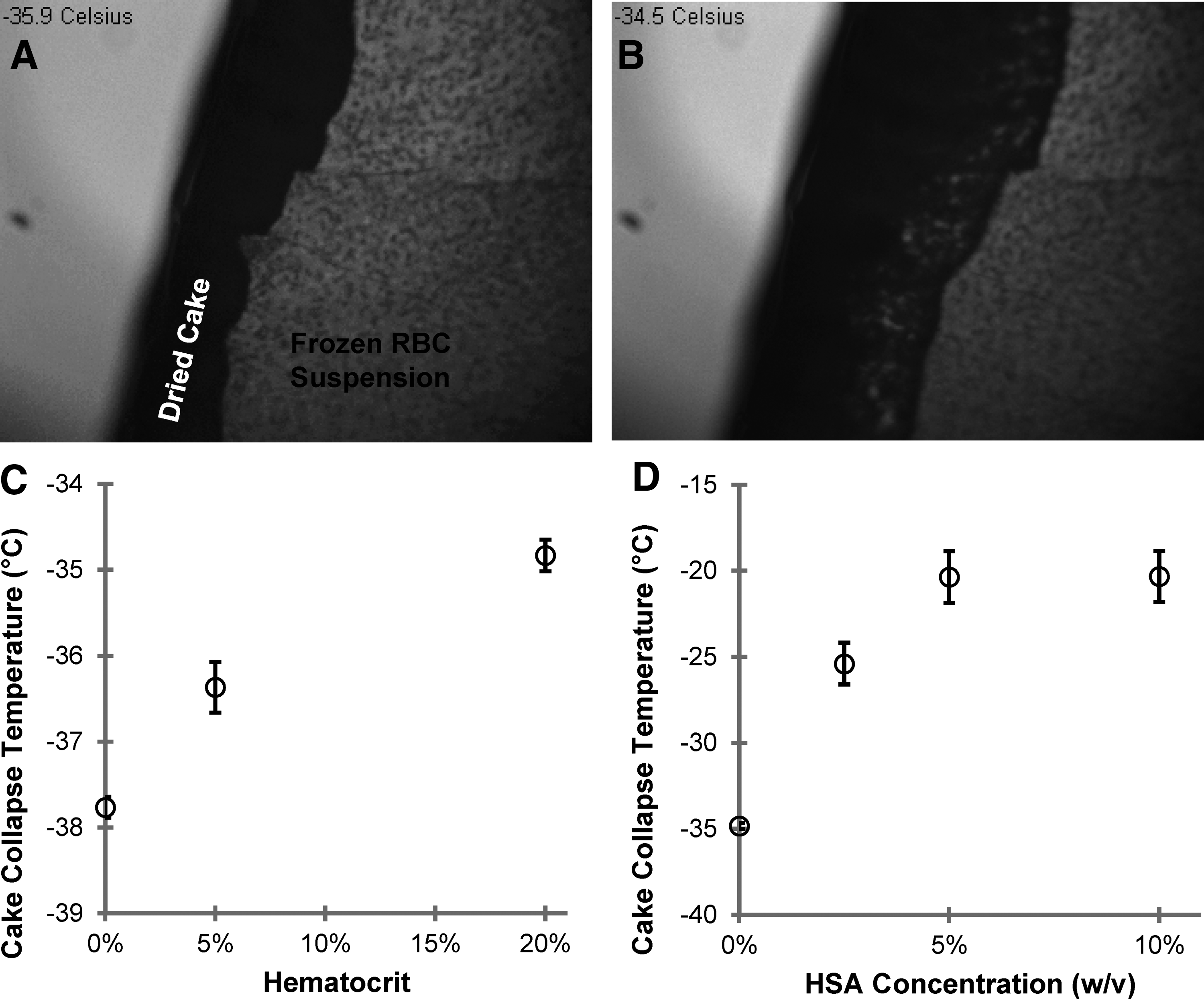
Figure 1C shows cake collapse temperatures for RBC suspensions with hematocrits ranging from 0% to 20%. Statistical analysis revealed a significant effect of hematocrit on cake collapse temperature (p=0.0002). Collapse temperatures increased from −37.8±0.1°C in cell-free lyophilization buffer to −34.8±0.2°C in the 20% hematocrit solution. We also attempted to measure the cake collapse temperature at 40% hematocrit, but this solution was too opaque to determine visually whether a cake had formed or collapsed using the current microscopy method.
Addition of HSA to the lyophilization buffer also had a statistically significant effect on the cake collapse temperature (p=0.0001), as illustrated in Figure 1D. Even adding minimal amounts of HSA significantly increased the cake collapse temperature from −34.8±0.2°C without HSA to −25.4±1.2°C with just 2.5% HSA. Further increasing the HSA concentration to 5% provided a smaller, yet significant increase to −20.4±1.5°C. However, increasing the HSA concentration to 10% did not significantly affect the overall collapse temperature.
Discussion
This study used freeze-drying microscopy to demonstrate that both HSA and hematocrit increase the cake collapse temperature of trehalose-loaded RBCs. The effect of hematocrit in particular may have implications for selection of the primary drying temperature for RBC freeze-drying. Whereas most previous studies of RBC freeze-drying have used relatively low cell densities,3–7 a clinical protocol would likely involve a hematocrit of 40% or higher. Such a high cell density would be expected to increase the cake collapse temperature by several degrees and potentially enable the use of a higher primary drying temperature.
Importantly, our results provide a framework for examining the relationship between cake collapse, primary drying temperature and RBC survival. It has previously been reported that hemolysis increases substantially when the primary drying temperature exceeds −35°C. 10 This is consistent with the cake collapse temperatures observed in the present study, suggesting a link between cake collapse and hemolysis. Future studies will enable more rigorous investigation of this idea.
Footnotes
Acknowledgments
We are grateful to the volunteer blood donors and to the Oregon State University Student Health Center for performing blood collections.
Author Disclosure Statement
No competing financial interests exist.