
Abstract
Acceleration of revival of ovarian function and maintaining of follicular reserve is mandatory after transplantation of cryopreserved ovarian tissue. In this study, hyaluronic acid hydrogel was used as a scaffold to improve restoration of ovarian estrous cycle and follicular preservation. Mature (∼8 weeks old) female Wistar rats with normal estrous cycles were divided in two groups: A: autotransplanted vitrified ovarian tissue without hyaluronic acid (HA), and B: autotransplanted vitrified ovarian tissue encapsulated with HA. Bilateral ovariectomy was performed in the diestrus stage; then ovaries were vitrified, warmed, and autotransplanted intramuscularly. Daily vaginal monitoring was performed until re-initiation of first full estrous cycle. Thereafter, follicular preservation, fibrosis, and apoptosis incidence were assessed histologically and immunohistochemically. The serum follicle stimulating hormone (FSH) levels were also accessed and compared for normal and ovariectomized rats. Re-initiation of first full cycle, atretic follicles, apoptotic index, and area of fibrosis in group A were approximately similar to group B. However, the total numbers of intact follicles were significantly lower in group B than group A. Moreover, the level of FSH in both experimental groups and normal rats was similar and in group B reduced significantly compared to the ovariectomized rats. Hyaluronic acid hydrogel did not show any negative effect on restoration of estrous cycle, but could not support follicular preservation after autotransplantation.
Introduction
O
One of the critical factors for successful ovarian transplantation is rapid angiogenesis, which is essential for follicular survival. 14 In rats, initial revascularization of ovarian autografts to ectopic sites was observed 48 hours after transplantation 15 and functional vessels have been detected after 7 days. 16 Since angiogenesis affects ovarian function, 17 a few experimental studies use scaffolds and growth factors to accelerate revascularization and ovarian function re-initiation.5,18,19 Nevertheless, improvement of the revascularization process, reduction of ischemic time, and acceleration of ovarian function retrieval require more study.
Hyaluronic acid (HA) or hyaluronan, a type of glycosaminoglycan, is typically found in the connective tissues of vertebrates. 20 Several studies have claimed that hyaluronan increases angiogenesis.21–23 In that regard, HA-rich biological glue was utilized in a study for xenograft transplantation of human ovarian tissue, and its beneficial effects were indicated when used in combination with melatonin, VEGF-A, and vitamin E. 19 Accordingly, this experiment was designed to evaluate the effects of hyaluronic acid hydrogel on estrous cycle restoration and follicular preservation following autotransplantation of vitrified rat ovary.
Material and Methods
Study design
Eight-week-old female Wistar rats (100–150 g) were purchased from the Pasteur Institute, Tehran, Iran, and maintained on a 12/12 hour light/dark cycle. One week before surgery, daily vaginal smears were taken to monitor the ovarian cycle. Animals with normal estrous cycles were selected for surgery in the diestrus stage and were randomly divided into two experimental groups (11 rats in each group): (A), autotransplanted vitrified ovarian tissue without hyaluronic acid (HA) (VT-HA group) and (B), autotransplanted vitrified ovarian tissue encapsulated in HA (VT+HA group).
In both groups, animals were ovariectomized and the right ovaries were heterotopically autotransplanted into the back muscle after vitrification and warming. Four days after transplantation, ovarian function was again monitored by daily vaginal smears and at the end of the first estrous cycle, in each group five rats were randomly sacrificed and grafted ovaries were removed, fixed in formalin, and evaluated histologically and immunohistochemically. Blood serum was also collected for measuring of the follicular stimulating hormone (FSH).
Ovariectomy
Animals were anesthetized with intraperitoneal injection of ketamine 10% and xylazine 2% (respectively, 50 mg/kg and 5 mg/kg, Alfasan, Netherlands). In a prone position, in a sterile condition, flank hair was shaved and both ovaries were removed following parasagittal incisions. Right ovaries were considered for autotransplantation, and the left ones were discarded.
Ovarian vitrification
For vitrification, the intact right ovary of each rat was equilibrated in two solutions: Solution 1 composed of 7.5% ethylene glycol (EG), 7.5% dimethyl sulfoxide (DMSO), and 10% human serum albumin (HSA) in HTCM-199)Sigma, Germany, MO, St., Louise) for 25 min, and Solution 2 composed of 20% EG, 20% DMSO, and 0.5 mol/L sucrose in HTCM-199 for 15 min. Then, each dehydrated ovary was picked up with a cryopin, 13 and immediately immersed into liquid nitrogen for 15 min.
Warming was done in three steps: first, the ovarian tissues were immersed directly in HTCM-199 supplemented with 1.0 mol/L sucrose for 1 min at 37°C. Then they were transferred to HTCM-199 supplemented with 0.5 mol/L sucrose for 5 min at room temperature, and finally, they were washed twice in HTCM-199 for 10 min before encapsulation or transplantation.
Hyaluronic acid hydrogel encapsulation
For encapsulation of vitrified-warmed ovaries in group B, Extracel™-HP hydrogel kit (Glycosan BioSystems, United States) which contains Heprasil® (a combination of thiol-modified hyaluronan, HA, and thiol-modified heparin), Gelin-S™ (thiol-modified gelatin), and Extralink® (a thiol-reactive crosslinker, polyethylene glycol diacrylate, PEGDA) was used. The ovaries were placed inside the mixture of 100 μL Heprasil, Gelin-S, and Extralink at 37°C. Gelation occurred after approximately 10 min, and the ovaries were encapsulated.
Autotransplantation
All surgeries were performed under aseptic conditions. The back areas of rats were shaved and skin and superficial muscles were incised. A space was created using scissors within the latissimus dorsi muscle, and ovaries were inserted. The skin and muscles were then sutured.
Cyclicity monitoring
From the fourth day after surgery, vaginal samples were collected daily between 7:00 and 9:00
Follicular count and histopathological evaluation
In each group, five grafted ovaries were removed following one full estrous cycle and fixed in 4% formalin for histopathological examination. The samples were embedded in paraffin and sectioned serially at 6 μm thickness. One of each six sections was stained with eosin-hematoxylin and observed under optical microscope (Nikon, Japan). Atretic and intact follicles were counted in these sections and classified according to Gougeon (1996) and Pederen's (1968) studies as follows: Primordial follicle: an oocyte surrounded by a layer of flatted granulosa cells (GCs); Transitional follicle: an oocyte surrounded by a mixture layer of flattened and cuboidal GCs; Primary follicle: an oocyte surrounded by a layer of cuboidal GCs; Preantral follicle: a growing oocyte with several layers of GCs; Early antral follicle: a follicle with small antrum or several small antral cavity within GCs; and Antral follicle: a follicle with a unit large antrum between GCs.
Apoptosis incidence
The middle section of each tissue was selected and stained with active caspase-3. Briefly, paraffin-embedded sections (6 μm) were deparaffinized and rehydrated with xylene and graded alcohol, respectively. Treatment with 3% H2O2 in methanol was used for 10 min to block endogenous peroxidase activity.
The sections were heated (98°C) in citrate buffer (pH = 6.0) for 1 h, then blocked for 10 min with protein block (Mouse and Rabbit Specific HRP/DAB (ABC) detection IHC kit, ab64264), and incubated at 4°C overnight with 1:100 dilution of active caspase-3 antibody (ab4051). Sections were washed and incubated with biotinylated goat anti-polyvalent (Mouse and Rabbit Specific HRP/DAB (ABC) detection IHC kit, ab64264) for 10 min at room temperature. They were then washed, incubated for 10 min at room temperature with streptavidin peroxidase (Mouse and Rabbit Specific HRP/DAB (ABC) detection IHC kit, ab64264), washed and finally the bound antibody was visualized after the addition of 3, 3-diaminobenzidine tetrachloride (DAB) solution (Mouse and Rabbit Specific HRP/DAB (ABC) detection IHC kit, ab64264).
After washing, the slides were counterstained with hematoxylin for facilitated counting of the total number of cells, using light microscopy. The numbers of active caspase-3-positive cells were counted with Image-J program (http://rsb.info.nih.gov/ij). Finally percentage of caspase-3-positive cells was determined by counting positive cells per total number of cells in 5 fields of view (×400) in sections as an apoptotic index. 25
Fibrosis incidence
A section adjacent to immunostaining was deparaffinized, rehydrated, and placed in hematoxylin for 4 min. Slides were washed for 6 min in running water followed by one dip in acid alcohol, then were transferred to eosin for 8 min. Thereafter, slides were washed, placed in phosphomolybdic acid (for 20 min) and stained in fast green for 3 min. They were rinsed in running water, fixed in 1% acetic acid, and the fibrotic areas were identified as green collagen fibers. Four fields per section were analyzed at ×100 magnifications with light microscopy and fibrotic areas and total section areas were measured with Image-J program (http://rsb.info.nih.gov/ij). Finally, the percentage of fibrotic areas was determined by measurement of green collagen fibers areas per total area in 4 fields of view (×100) in sections as fibrosis incidence.
Hormone measurement/hormone analysis
Blood was collected from all the rats after first full estrous cycle and allowed to clot for 10--15 minutes. Serum was separated by centrifugation (Rotofix 32A, Germany) at 3000 rpm (1509 g) for 10 min. Samples were stored at −80°C until analyzed for follicle stimulating hormone (FSH) concentration. The serum of normal rats in estrus phase (n = 5) and bilateral ovariectomized rats (n = 5) also were collected and stored at −80°C for comparison. The hormone level of serum was measured by double-antibody sandwich enzyme-linked immunosorbent assay (rat ELISA kit, bioassay technology laboratory, China). The sensitivity of the assay was 0.12 mLU/mL. Inter- and intra-assay coefficients of variance were <10% and 12%.
Briefly, standards, samples, and reagents were prepared. 50 μL of standards and 40 μL either sample with 10 μL of antibody were added to corresponding wells, followed by 50 μL Streptavidin-HRP in all wells. After 1 hour incubation at 37°C, plates were washed three times, 100 μL chromogen solutions A and B were added and incubated for 10 min at 37°C away from light. The reaction was stopped and the optical density (OD) was measured under 450 nm wavelength.
Statistical analysis
Statistical analysis was performed with the SPSS software program (Version 16; SPSS Inc., Chicago, IL, USA). Follicular count and hormone measurement were performed on five independent biological replicates in each group. The average numbers of follicles was compared by t-test between the two groups, and the mean serum FSH levels were compared by analysis of variance (ANOVA) test. The p-value <0.05 was considered to be statistically significant
Results
Cyclicity after ovarian autotransplantation
All ovarian transplantations were successful and the estrous cycle was restored in all rats (n = 22). Six–13 and 6–16 days after transplantation, cornified epithelial cells (estrous cycle re-initiation sign) were first observed in groups A and B, respectively. First full cycles were also seen 10–29 and 10–28 days after transplantation in A and B groups, respectively. There was no significant difference between groups in estrous cycle re-initiation (Fig. 1).

Estrous reinitiation after transplantation. Data are shown as mean percentage ± SE, n = 11 rats in each group. There was no significant difference between both groups. VT-HA, autotransplanted vitrified ovarian tissue without hyaluronic acid; VT+HA, autotransplanted vitrified ovarian tissue encapsulated with hyaluronic acid.
Follicular count and histopathological changes in autotransplanted vitrified ovary
A wave of follicular growth was observed in both groups and different stages of follicles (primordial, transitional, primary, preantral, early antral, and antral) were observed in all grafts. Ovarian surface epithelium (OSE) had disappeared and muscle tissue was replaced in the adjacent ovarian stroma (Fig. 2A and 2B). In group B, hyaluronic acid hydrogel remnant was occasionally seen around the ovary (Fig. 2B, 2C, and 2F) and luteinizations were seen in some follicle-like structures without oocytes (Fig. 2C and 2D). Furthermore, stromal hyperplasia and luteinization of stromal cells were observed in this group (Fig. 2E). Histologically, reduction of follicular development as a result of abnormal follicles maturation and of stromal cell condensation were evidence of ovarian atrophy. 26 In group B, ovaries showed atrophic changes, increase of fibrotic tissue, and decrease of follicular count. Further, apoptosis and necrosis changes (karyolysis, pyknosis, and karyorhexis) were observed in this group in some follicles and stromal cells (Fig. 2C and 2F).
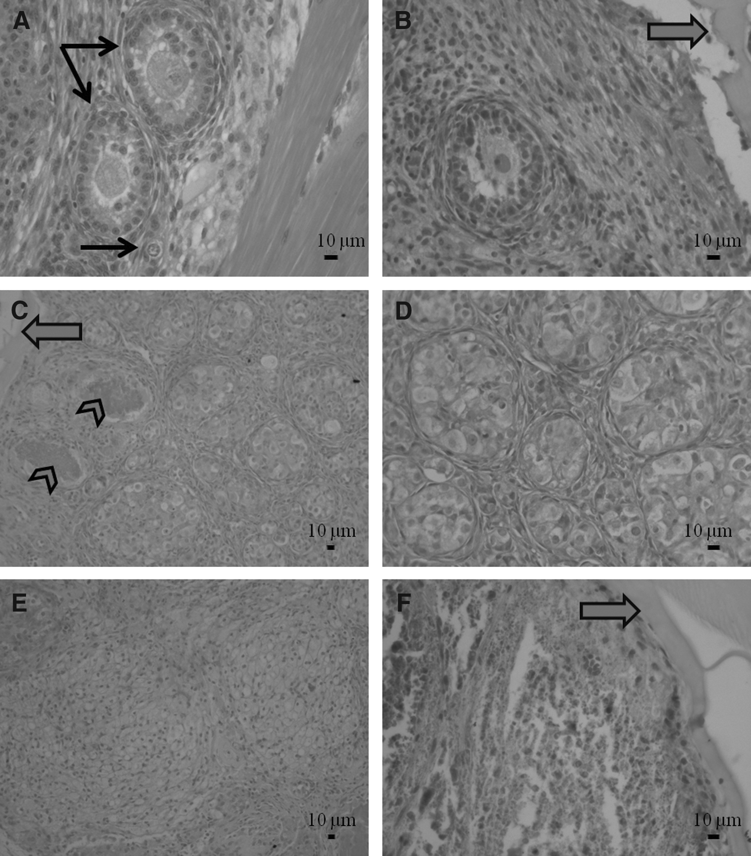
Histological sections of ovarian tissue stained with hematoxylin and eosin.
In general, the total number of follicles was significantly lower in group B than group A (p < 0.05). In group A, the mean number of intact follicles in different stages, with an exception of intact early antral follicles, was significantly higher than that of group B (p < 0.05) (Fig. 3).On the other hand, there was no significant difference in the total number of atretic follicles (Fig. 4). Although numbers of atretic preantral follicles were significantly higher in group B compared to group A, there were no significant differences in the other stages of atretic follicles between the two groups (Fig. 4). However, in group A, the percentage of total primordial follicles rather than total follicles was (35.45% ± 7.82), a little more than group B (41.03% ± 13.01), but this difference was not significant.
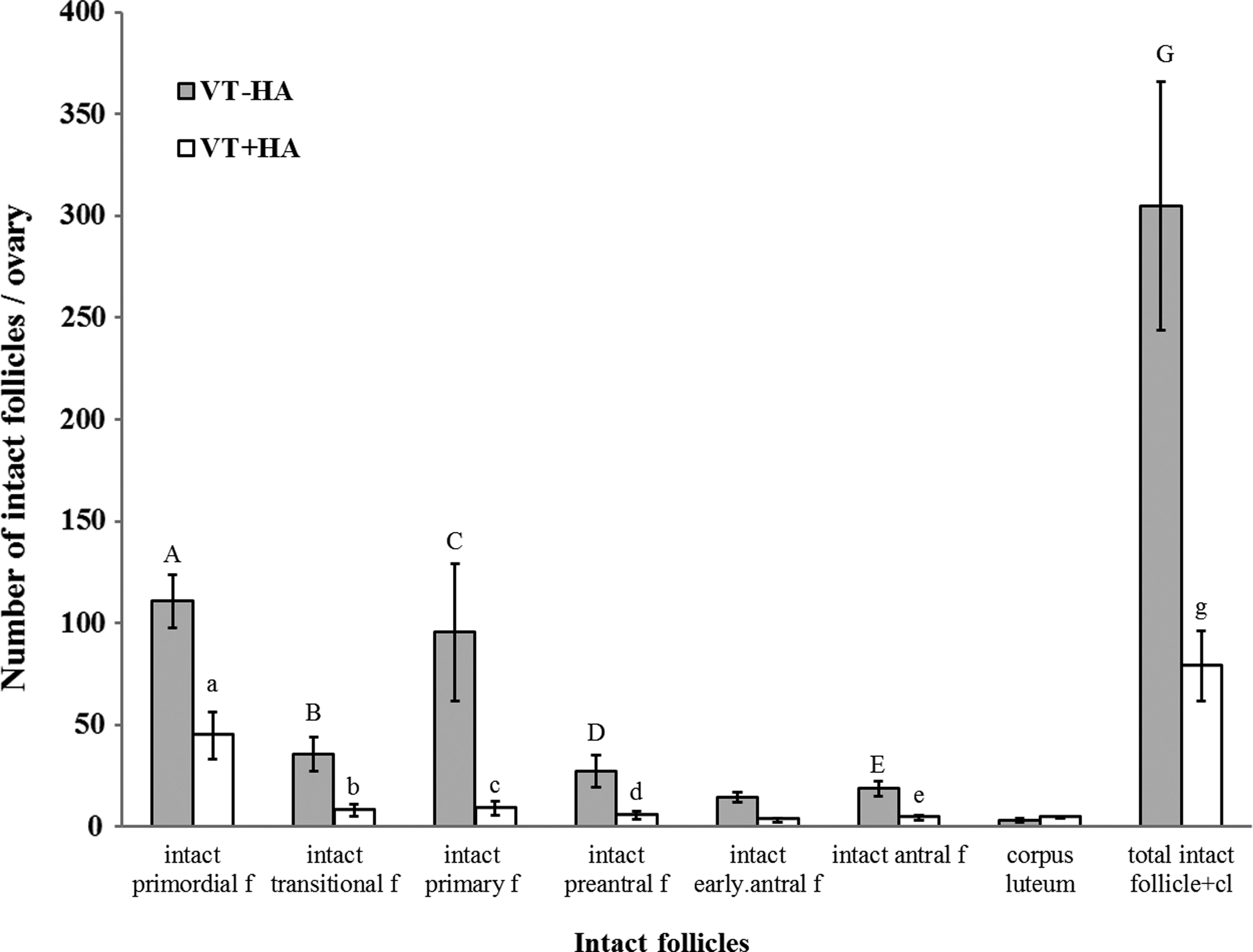
Intact follicles in experimental groups, Data are shown as mean ± SE, n = 5 rats in each group. VT-HA, autotransplanted vitrified ovarian tissue without hyaluronic acid; VT+HA, autotransplanted vitrified ovarian tissue encapsulated with hyaluronic acid. Capital letters vs. the same small letters show significant differences (p < 0.05).
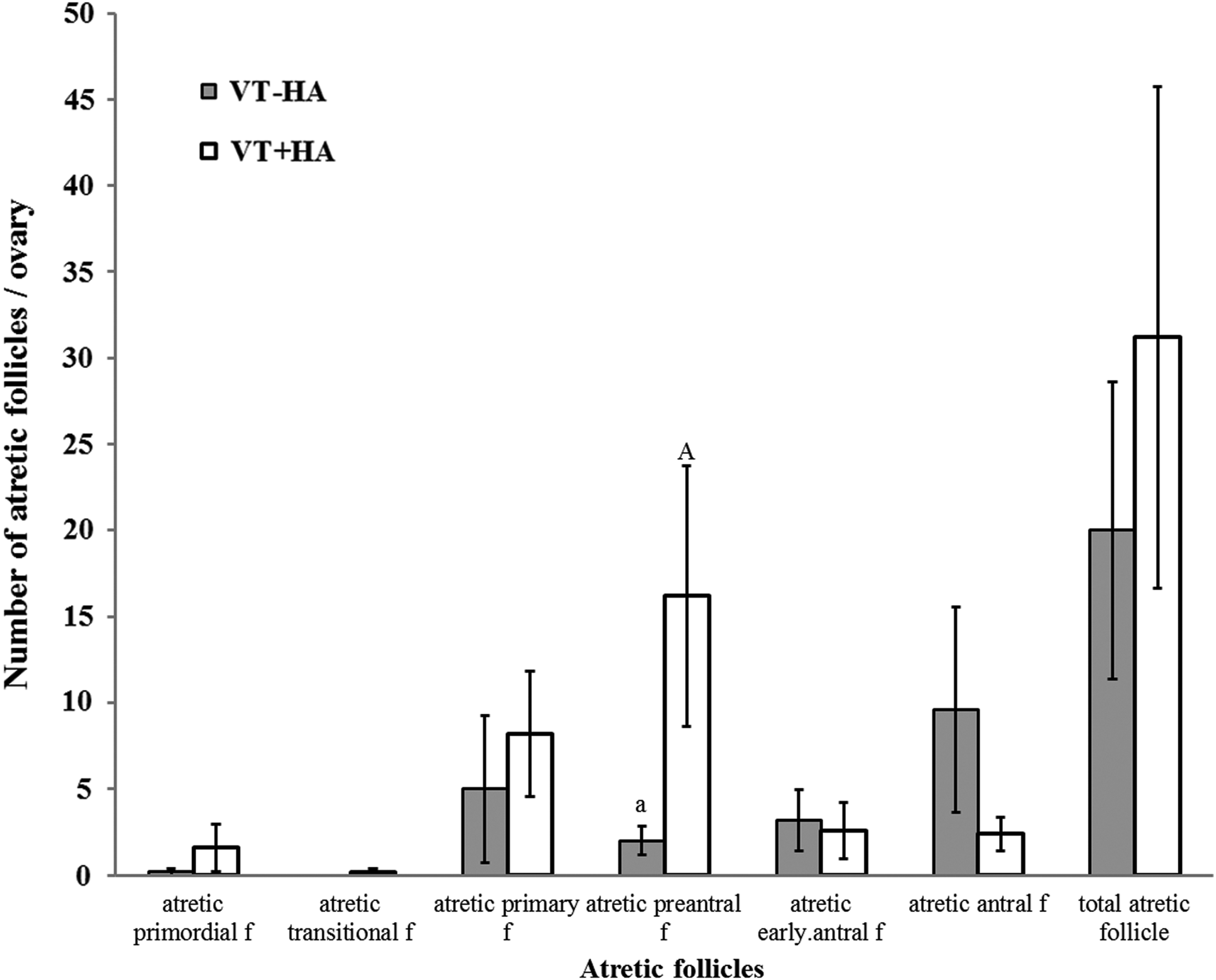
Atretic follicles in experimental groups, Data are shown as mean ± SE, n = 5 rats in each group. VT-HA, autotransplanted vitrified ovarian tissue without hyaluronic acid; VT+HA, autotransplanted vitrified ovarian tissue encapsulated with hyaluronic acid. Capital letter vs. the same small letter shows significant differences (p < 0.05).
Apoptosis incidence in autotransplanted ovary
The percentage of active caspase 3-positive cells was assessed to determine the apoptotic index in grafts (Fig. 5C and 5D). The apoptotic index in the group A (5.08 ± 5.85) was less than group B (9.06 ± 6.73), but no significant difference was observed between them.
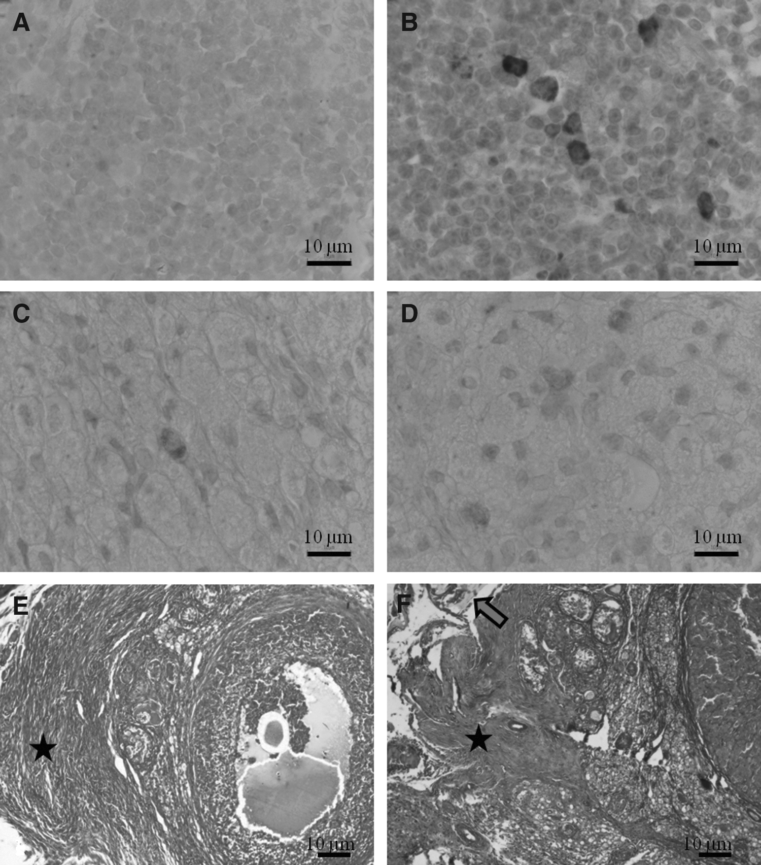
Fibrosis incidence in autotransplanted ovary
Fibrosis was observed in all grafts. Although the area of fibrosis was higher in group A (25.28 ± 17.93) than group B (17.25 ± 4.5), no significant difference was observed between them.
Hormone measurement
The serum level of FSH was measured in experimental, normal, and bilateral ovariectomized rats (Fig. 6). Although serum levels of FSH were higher in group A (14.67 ± 6.76) than group B (8.1928 ± 1.66), no significant difference was observed between this group and normal rats. However, group B showed significantly lower FSH levels than ovariectomized rats (26.62 ± 11.15, p < 0.05).

The serum level of FSH in the experimental groups, data are shown as mean ± SE, n = 5 rats in each group. VT-HA group, vitrified-transplanted without hyaluronic acid; VT + H group, vitrified-transplanted with hyaluronic acid. Capital letter vs. the same small letter shows significant differences (p < 0.05).
Discussion
Successful transplantation of cryopreserved ovarian tissue mainly depends on cryopreservation protocol and transplantation procedure. To prevent ice crystal formation, some researchers preferred to do vitrification rather than slow freezing either for whole or fragmented ovarian tissue.3,27,28 As the literature indicates, the selection of suitable protocol for ovarian tissue vitrification also depends on histological ovarian structure and permeability of different cryoprotectants. Therefore, it is essential to design an optimal vitrification protocol for a given species or animal breed in cryobiology. 3
Numerous studies have evaluated the vitrification protocols and duration of exposure to cryoprotectants for whole or fragmented ovary.13,29–31 Accordingly, Fathi et al. indicated that the combination of EG and DMSO with sucrose is more suitable in comparison with other combinations of cryoprotectant for vitrification of whole rat ovary. 13 Kagawa et al. 32 applied a vitrification protocol for large pieces of ovarian tissue from cattle and humans with an equilibration solution containing 7.5% EG +7.5% DMSO: 25 min and vitrification solution containing 20% EG +20% DMSO +0.5 M sucrose: 15 min. Their results showed this method was successful in restoring ovarian function after autotransplantation of vitrified ovarian tissue in cow. However, Youm et al. 31 suggested a modified protocol based on the Kagawa et al. study 32 with less time for whole mouse ovary with size (∼ 2 × 2 × 2 mm3). 31
In the present investigation, because the dimension of rat ovary is about 4 × 4 × 4 mm3 and its density is higher than mouse ovary, the Kagawa protocol was applied for cryopreservation of whole rat ovary. After transplantation, 100% successful restoration of estrous cycle in both control and experimental groups was achieved. According to this result, we can suggest that the Kagawa's protocol might be useful for rat ovarian cryopreservation too.
On the other hand, transplantation of ovary without vascular anastomosis depends on the growth of new blood vessels for restoration of ovarian function and decrease of ischemic injury. As the literature indicates, quality improvement and apoptosis reduction of ovarian transplantation may be achieved in different ways such as usage of angiogenic factors [fibrin and HA-rich biological glue5,18,19] and considering the optimal transplantation site.33–35 In the present study, a hyaluronic acid hydrogel as a scaffold with probable angiogenic characteristics was used to improve restoration of the estrous cycle and preservation of ovarian reservation after autotransplantation of vitrified rat ovary. The HA hydrogel (Extracel™-HP hydrogel kit) is composed of chemically modified HA containing reactive thiol groups (Glycosil) and chemically modified gelatin containing reactive thiol groups (Gelin-S), which are co-cross-linked with polyethylene glycol diacrylate (Extralink) to form a semisynthetic ECM, which improves vascularization and with a lower occurrence of core necrosis. 36
In rodents, the best method to assess hypothalamic–pituitary–ovarian axis function is through vaginal cytology evaluation. 37 In some experimental studies, analysis of vaginal smear cytology has been used as an indirect and dynamic parameter of ovarian activity re-initiation.6,9,14,38 In our study, restoration of estrous cycle occurred after transplantation in both groups. The mean duration of estrus restoration after transplantation was 14.9 days in autotransplanted vitrified ovarian tissue without HA, and 15 days in autotransplanted vitrified ovarian tissue with HA. Our results were similar to the studies that evaluated rat vaginal smear cytology after transplantation of cryopreserved ovarian tissue.6,39
Despite the angiogenic properties of hyaluronic acid,22,23,36,40 this scaffold has no advantage for acceleration of estrous cycle restoration. Damous et al. found that a hypoxic condition of ovarian tissue prior to the transplantation could be harmful for the graft and delayed re-initiation of estrous cycle. 41 As the histological study indicated, this hypoxic condition might have occurred due to the presence of hyaluronic acid remnants and prevented the acceleration of estrous cycle resumption.
In the absence of the gonads, FSH levels were elevated but in the two transplant groups reached levels of serum FSH found in normal rats. This finding was consistent with daily vaginal cytology data that indicated restoration of ovarian function and control of pituitary gonadotropins. In agreement to our finding, Shikanov et al. also suggested the serum FSH levels decreased after ovarian transplantation. 5
The main purpose of ovarian tissue cryopreservation and transplantation is fertility preservation, 42 so the number of intact follicles is important in subsequent transplantation. Here as opposed to the VT-HA group, the total number of intact different stages of ovarian follicles decreased significantly in autotransplanted vitrified ovarian tissue with HA, and instead many follicle-like structures with luteinized cells and necrotic follicles were observed. Since the vitrification procedure was similar between two groups, the differences could be related to ischemic conditions which might have occurred in the HA group. We also assume that the reduction of ovarian follicles, especially the primordial follicle, could be the main cause of the declining ovarian reserve and deficiency of follicular growth in the HA group.
Moreover, luteinization and necrotic changes were also observed in ovarian stroma after using HA. Some studies have shown that ovarian stromal cells have an important role in follicular survival, development and ovarian function.34,43 In the HA group, damage to stromal cells seems to reduce the number of follicles and surprisingly, the low number of follicles seems to be enough for re-initiation of estrus cycle, but its continuation still needs more investigation.
There were no significant differences between two groups in the incidence of apoptosis. This result is in agreement with the study by Friedman et al., who treated the ovarian graft with HA-rich biological glue. 19 A nonsignificant incidence of higher apoptosis in the HA group could be due to hypoxia that induces apoptosis in the ovarian graft after transplantation. 44
Fibrosis is the formation of excess fibrous connective tissue in an organ or tissue. Birbrair et al. claimed that fibrosis increases during the repair phase of tissue injury. 45 Also, previous studies have shown that fibrosis decreases after heterotopic ovarian transplantation without vascular pedicle increases, and damaged follicles were replaced by the fibrous tissue. 46 47 In contrast, in the present study, damaged follicles were not replaced by fibrous tissue but were instantly replaced by lutein-like cells (prominently in HA group), and no significant difference was observed in the fibrosis index values.
In conclusion, our study demonstrates that hyaluronic acid alone did not show any negative effect on restoration of estrous cycle and for ovarian preservation after autotransplantation. It seems that usage of hyaluronic acid in combination with a growth factor could improve autotransplantation results, and further studies are needed in this area.
Footnotes
Acknowledgments
The authors would like to thank Dr. Reza Salman Yazdi for hormone analysis and Dr. Lakshmi Gopal for editing of manuscript.
Author Disclosure Statement
The research was supported by grants from Royan Institute and Tarbiat Modares University (both located in Tehran, Iran).