
Abstract
The aim of this study was to investigate the effects of different media with or without phenol red or the antioxidant trolox on the successful vitrification of feline ovarian tissue. In a first experiment, ovarian cortical pieces from three cats were vitrified in solutions of Roswell Park Memorial Institute (RPMI)-1640 medium, Minimum Essential Medium, Dulbecco's modified Eagle's medium, or Tissue Culture Medium 199 as basic medium, supplemented or not with 50 μM of trolox, all containing 40% ethylene glycol (EG) and 1 M of sucrose. RPMI-1640 (phenol red–free) without trolox was the only medium that preserved the percentage of morphologically normal preantral follicles similar to control (80%). The main difference between RPMI-1640 and the other media was the absence of phenol red and CaCl2. In a second experiment, ovarian cortical pieces from three cats were vitrified in a solution containing RPMI-1640 as basic medium, 40% EG, 1 M of sucrose, supplemented or not with phenol red or CaCl2 alone, or in combination. It was observed that phenol red supplementation led to follicular degeneration. Finally, to evaluate the interaction between phenol red and the cryoprotectant agent (i.e., EG), ovarian tissue was exposed to RPMI-1640 supplemented with phenol red and EG at different concentrations (10%, 20%, or 40%). There was an inverse relationship between EG concentration and free phenol red in the medium after exposure. It is suggested that vitrification of feline ovarian tissue should be performed in a phenol red–free medium. Medium supplementation with 50 μM of trolox was deleterious for follicular morphology.
Introduction
O
Cryopreservation protocols developed for domestic cats have been applied in wild felines with promising outcomes. 4 Luvoni et al. 5 showed that it is possible to preserve the morphology of feline ovarian follicles at different developmental stages after vitrification of the ovarian cortex. However, the success of this procedure remains limited. For instance, Bosch et al. 6 observed that only 10% of ovarian follicles survived after xenografting of frozen–thawed feline ovarian cortex. When preparing cryopreservation solutions for domestic animals, usually the basic media are Minimum Essential Medium (MEM), Dulbecco's modified Eagle's medium (DMEM), or Tissue Culture Medium 199 (TCM199),7–11 all of which contain the pH indicator phenol red. On the other hand, successful vitrification of human and murine ovarian tissue is performed in phenol red–free media [Roswell Park Memorial Institute (RPMI-1640), 12 phosphate buffered saline (PBS), 13 Dulbecco's PBS 14 ]. Hence, aside from the intrinsic characteristics involving feline gamete sensitivity to chilling, 15 it was hypothesized herein that success of vitrification may also be dependent on the composition of the medium used to prepare the cryopreservation solution.
It has been reported that medium supplementation with antioxidants supports the maintenance of follicular viability and morphology16,17 during cryopreservation of ovarian tissue. In a recent study, the authors showed that 50 μM of trolox not only protected ovarian follicles from freezing damage, but also avoided vacuolization of oocytes and stromal tissue from non-human primates submitted to cryopreservation 11 or ischemic stress. 18 Trolox (6-hydroxy-2,5,7,8-tetramethylchroman-2-carboxylic acid) is hydro- and liposoluble due to a carboxyl group, and acts as a hydroxyl and alkoxyl radical scavenger independently of the solution type (aqueous or lipid). 19 Therefore, medium supplementation was tested with 50 μM of trolox based on the success of the vitrification of feline ovarian tissue.
The present study aimed to investigate the effect of the basic medium with (DMEM, MEM, or TCM199) or without (RPMI 1640) phenol red on the vitrification of ovarian tissue from domestic cats (Felis catus). The possible interaction of this compound with an intracellular cryoprotectant [ethylene glycol (EG)] was also evaluated. Furthermore, medium supplementation with the antioxidant trolox was tested.
Materials and Methods
Chemicals
Unless stated otherwise, chemicals and media used in this study were purchased from Sigma Chemical Co. (St. Louis, MO). For all of the media, the pH was adjusted to 7.4 at room temperature (∼25°C).
Source of ovaries
Nine adult queen cats were subjected to ovariohysterectomy at the Veterinary Hospital of the Federal University of Pará, Brazil. Collected ovaries were allocated to three experiments. For all experiments (Supplementary Fig. S1; Supplementary materials are available online at www.liebertpub.com/bio), immediately after collection, each ovarian pair derived from the same queen was sliced into 2 mm3 pieces and distributed in treatments according to each experiment as follows.
Experiment 1
Each ovarian pair from 3 queens was divided into 9 fragments (2 mm3). One fragment was randomly selected as the fresh control and was fixed in Davidson solution for routine histological evaluation. The remaining eight fragments were exposed to the vitrification solution for 5 min at 20°C. The vitrification solution consisted of 40% EG and 1 M of sucrose in MEM, DMEM, TCM199, or RPMI-1640, either supplemented or not with 50 μM of trolox. After exposure, samples were vitrified.
Experiment 2
Each ovarian pair from 3 queens was divided into 5 fragments (2 mm3). One fragment was randomly selected as the fresh control and fixed in Davidson solution for routine histological evaluation. The remaining 4 fragments were exposed to the vitrification solutions for 5 min at 20°C. Based on experiment 1, vitrification solution consisted of EG +1 M of sucrose in RPMI-1640 medium with or without 0.04 mM of phenol red, 1.8 mM of CaCl2, or both. The choice of adding phenol red or CaCl2 to the medium was based on the main differences in the composition of RPMI-1640 and the other media (Supplementary Table S1) tested herewith in experiment 1.
Experiment 3
Each ovarian pair from 3 queens was divided into 11 fragments (2 mm3). One fragment was randomly selected as the fresh control and fixed in Davidson solution for routine histological evaluation. The remaining 10 fragments were exposed to RPMI-1640 alone or with 1 M of sucrose and EG (0%, 10%, 20%, or 40%) added. Each solution was either supplemented or not with phenol red. After exposure, ovarian fragments were processed for histological analysis, and free phenol red in the exposure medium was measured.
Vitrification
The solid-surface vitrification procedure as well as the protocol for incubating tissue in vitrification solutions for 5 min at 20°C, the dilution regimen to remove the cryoprotectants after warming, and the time and temperature of exposure were chosen based on studies previously described.8,9 In brief, vitrification solutions were prepared just prior to use. Each fragment was initially kept for 5 min at 20°C in 1 mL of the vitrification solution, after which fragments were placed on a cold surface consisting of a hollow cube of aluminum foil partially immersed in liquid nitrogen. The vitrified fragments were transferred into cryovials using nitrogen-cooled forceps, and stored in the liquid phase of a liquid nitrogen tank. Vitrified ovarian fragments were maintained in cryostorage for 1 week. For warming, cryovials were exposed to room temperature (∼25°C) for 1 min, and fragments were separately submitted to cryoprotectant removal. For this step, fragments were immersed in each respective test medium (TM) at 37°C as follows: (1) TM +0.25 M of sucrose (3 min), (2) TM +0.125 M of sucrose (5 min), and (3) TM (7 min). After removal of cryoprotectant, ovarian fragments were fixed for histological analysis.
Histological analysis
After fixation in Davidson solution, ovarian fragments were dehydrated in ethanol, clarified with xylene, and embedded in paraffin wax. Serial sections (5 μm) of ovarian tissue were cut, and every fifth section was mounted on glass slides and stained with hematoxylin and eosin. All sections were examined using a light microscope (Leica) at a magnification of 200×. Preantral follicles were defined as follicles with an oocyte surrounded by either one flattened and/or cuboidal layer or several layers of only cuboidal granulosa cells. To avoid counting a follicle more than once, preantral follicles were counted only in the sections where their oocyte nucleus was observed. Follicular quality was evaluated based on the morphological integrity of the oocyte, the granulosa cells, and the basement membrane. 7 In brief, preantral follicles are classified as (1) histologically/morphologically normal when they contain an intact oocyte and intact granulosa cells; (2) degenerated grade 1 when their oocyte nucleus has become pyknotic; and (3) degenerated grade 2 when the oocyte is shrunken and its nucleus pyknotic, and when granulosa cells may have detached from the basement membrane and have become enlarged in volume.
Determination of free phenol red in the medium and its cytotoxicity
For a standard curve, serial 2-fold dilutions of phenol red in RPMI-1640 in final concentrations of 0.8, 0.4, 0.16, 0.08, and 0.04 mg/mL were prepared. RPMI-1640 alone was used as blank. Phenol red adsorption was detected using a UV/Vis spectrophotometer (TU-1800SPC, China) at 560 nm wave length, as reported in a previous study.
20
Samples exposed to vitrification solutions as stated in experiment 3 were fixed for histological analysis to determine cell quality via morphology. After exposure, tissues were submitted to cryoprotectant removal as described above, and the exposure medium was evaluated for free phenol red in the solution. For this, exposure solutions were separately centrifuged (15,000 g for 10 min), and supernatants were submitted to phenol red measurement using the UV/Vis spectrophotometer at 560 nm wavelength. The amounts of free phenol red in the medium were calculated according to the equation:
where Co and Ce are the phenol red concentrations (μg/mL) contained in the original tissue exposure medium and in the supernatants, respectively, and Ceg is the concentration of EG in the exposure medium (mg/mL).
Statistical analysis
Data are presented as mean ± standard deviation (SD), and comparisons were performed using one-way analysis of variance (ANOVA) and Tukey's post hoc test using Prism v6.04. Differences were considered significant when P < 0.05.
Results
Experiment 1: medium composition affects feline follicular survival after vitrification
A total of 2,768 preantral follicles were evaluated. The rates of morphologically normal preantral follicles were similar to the control (80% ± 12%) only when ovarian tissue was vitrified in RPMI-1640 without trolox (50% ± 38%). The highest degeneration rates were observed in feline ovarian tissue vitrified in a medium composed of DMEM, where only 5.6% ± 5.6% of the follicles appeared morphologically normal. Trolox did not improve the rates of morphologically normal preantral follicles, and even significantly decreased the percentages (25% ± 19%) of normal follicles vitrified in a RPMI-1640-based medium (Fig. 1).

(
Experiment 2: phenol red and not calcium impairs follicular survival to vitrification
A total of 988 preantral follicles were evaluated. The rates of morphologically normal preantral follicles were similar to the control (67% ± 8%) only when ovarian tissue was vitrified in RPMI-1640 without (42% ± 26%), or supplemented with calcium chloride (52% ± 12%). Phenol red led to a significant increase in the degeneration rates of the preantral follicles from vitrified ovarian tissue (17% ± 5%), regardless of whether it was combined with calcium chloride (10% ± 9%; Fig. 2).
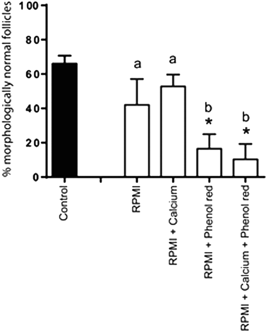
Percentages (mean ± SD) of morphologically normal follicles in control and in vitrified ovarian fragments. Effect of RPMI-1640 supplementation with 1.8 mM of calcium, 0.04 mM of phenol red, or both. Vitrification was performed in the presence of 40% EG and 1 M of sucrose. *Differs from control; a,bdifferences when comparing treatments (P < 0.05).
Experiment 3: damage driven by exposure to phenol red is intensified in the presence of 20% and 40% EG
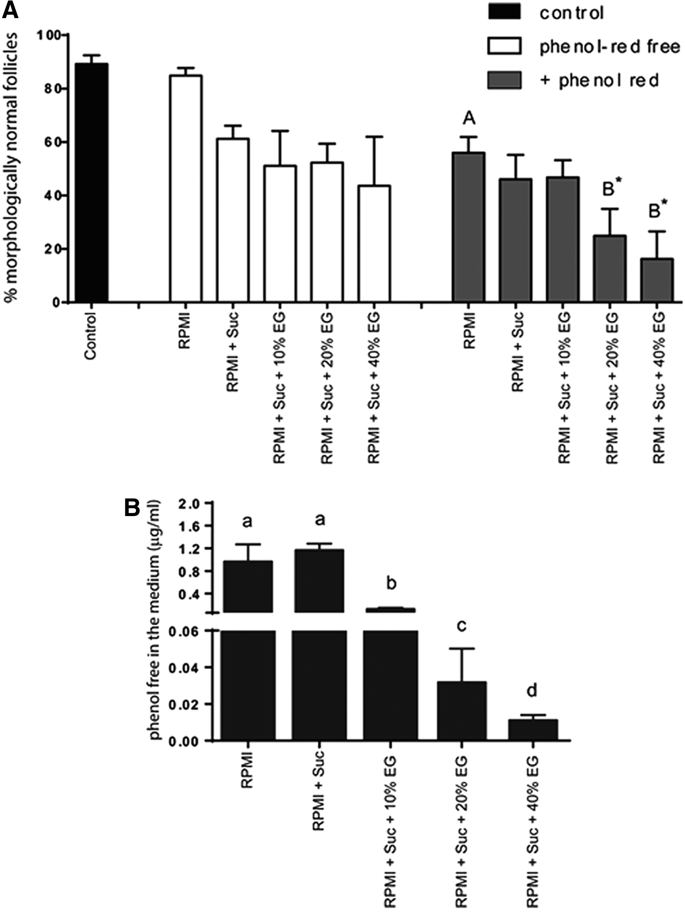
A total of 2,842 preantral follicles were evaluated immediately after exposure to different solutions with or without phenol red in their composition. The rates of morphologically normal preantral follicles were similar to the control (89% ± 4%) in all treatments. However, when ovarian tissue was exposed to a vitrification solution where RPMI-1640 was supplemented with phenol red and contained 20% (25% ± 10%) or 40% EG (16% ± 10%), a significant decrease in the percentage of normal follicles was detected (Fig. 3A).
(
To verify experimentally the speculation that the toxic effect of phenol red was increased in interaction with EG, the concentration of phenol red free in the medium after ovarian tissue exposure was evaluated. It was observed that an EG concentration dependent effect occurred, where increases in EG concentration from 10% to 20% and from 20% to 40% resulted in a significant decrease in the levels of phenol red free in the medium (Fig. 3B).
Discussion
The present study showed the detrimental effect of phenol red on the morphology of vitrified feline preantral follicles. Four commercial media used for the preparation of cryopreservation solutions (DMEM, MEM, TCM, and RPMI-1640) were tested. Among these, RPMI-1640 is commonly used without phenol red or calcium chloride supplementation. Also, based on previous studies in non-human primates,11,12 50 μM of trolox was added to the vitrification solution as a strategy to counteract follicular loss due to oxidative stress. Inconsistent with observations in non-human primates, 50 μM of trolox in the medium led to a significant increase in follicular loss. It has been shown that the antioxidant trolox may act as a pro-oxidant, depending on the environment, but such a mechanism is still not completely clarified. 21 It is likely that for feline ovarian tissue, this concentration is already giving rise to the U-shaped biphasic effect, or this effect is simply caused by the vitrification process, different from what was observed in frozen–thawed non-human primate ovarian tissue. 11 Degenerated preantral follicles presented ooplasm vacuolization as reported before in cryopreserved ovarian tissue from other species,11,13,22,23 and might be related to inappropriate dehydration of the tissue, perhaps together with osmotic stress. 24 In the present study, degeneration was not only related to osmotic stress caused by vitrification solution, but also by the medium used as the basis.
The main difference between RPMI-1640 and the other tested media was the absence of calcium chloride and phenol red. Therefore, extra experiments were performed adding these compounds to RPMI-1640, followed by ovarian tissue vitrification. The phenol red but not calcium was responsible for the follicular degeneration. The use of calcium-free cryopreservation solutions containing EG have been indicated for the preservation of oocytes, mostly mature ones or those harvested from antral follicles, since a rise in calcium may lead to zona pellucida hardening. 25 However, this is not a challenge for immature oocytes from preantral follicles, which are not yet provided with cortical granules. Although RPMI-1640 has no calcium chloride in its composition, calcium nitrate is present.
Although phenol red in cell culture media is well accepted, many commercial vitrification media used in clinics are phenol red–free, since this pH indicator is unnecessary in such a process. For instance, successful recent studies with human and mouse cells used phenol red–free media.13,26,27 However, these authors did not evaluate or did not consider discussing the absence of phenol red in the basis medium. Zhu et al. 20 reported phenol red as the causative agent of toxicity in HeLa cells culture in a serum-free medium. The present study also performed all vitrification processes in a serum-free system, and a decrease in cell survival in medium supplemented with phenol red was observed. In the study by Zhu et al., 20 the internalization of phenol red in HeLa cells was related to its interaction with carbon nanoparticles present in the culture medium. The present study evaluated the relationship of phenol red and EG at different concentrations in the exposure medium to evaluate such an interaction. Interestingly, there was an inverse relationship of free phenol red with the concentration of EG in the exposure medium. For instance, with the increase of EG concentration from 10% to 40%, there was a decrease of free phenol red in the exposure medium. Although the scientific nature behind this phenomenon is unclear, it is suggested that the intracellular cryoprotectant EG interacted with phenol red facilitating its permeation into the tissue, similar to the findings with carbon nanoparticles, 20 hence explaining the decrease in free phenol red in the exposure medium and the increase in ovarian follicles degeneration.
In conclusion, feline ovarian tissue can be best vitrified in a phenol red–free solution, and the use of 50 μM of trolox does not improve follicular survival. Vitrification is still under improvement, and the susceptibility of feline ovarian follicles to degeneration must be considered. For instance, the use of high concentrations of cryoprotectants plays an important role in follicular loss due to osmotic shock. Therefore, the combination of cryoprotectants with intracellular solutions, for example HypoThermosol, 28 might appear as an alternative to protect the ovarian follicles against stress and cryoprotectants toxicity.
Footnotes
Acknowledgments
This research was supported by CNPq, Brazil. D.C.B. was supported by a grant from CAPES.
Author Disclosure Statement
No conflicting financial interests exist.
References
Supplementary Material
Please find the following supplemental material available below.
For Open Access articles published under a Creative Commons License, all supplemental material carries the same license as the article it is associated with.
For non-Open Access articles published, all supplemental material carries a non-exclusive license, and permission requests for re-use of supplemental material or any part of supplemental material shall be sent directly to the copyright owner as specified in the copyright notice associated with the article.