
Abstract
Routine techniques for the isolation of human peripheral blood mononuclear cells (PBMCs) include density centrifugation with Ficoll-Paque and isolation by cell preparation tubes (CPTs) and SepMate tubes with Lymphoprep. In a series of experiments, these three PBMC isolation techniques were compared for cell recovery and viability, PBMC population composition, and cell functionality, aiming to provide a starting basis for the selection of the most appropriate method of PBMC isolation for a specific downstream application. PBMCs were freshly isolated from venous blood of healthy male donors, applying the different techniques in parallel. Cell recovery and viability were assessed using a hemacytometer and trypan blue. Immunophenotyping was performed by flow cytometry. Cell functionality was assessed in stimulated (100 ng/mL staphylococcal enterotoxin B [SEB]) and unstimulated 24 hours PBMC cultures, with cytokine production and lactate dehydrogenase (LDH) release as readout measures. PBMC isolation by SepMate and CPT resulted in a 70% higher recovery than Ficoll isolation. CPT-isolated populations contained more erythrocyte contamination. Cell viability, assessed by trypan blue exclusion, was 100% for all three isolation techniques. SepMate and CPT isolation gave higher SEB-induced cytokine responses in cell cultures, for IFNγ and for secondary cytokines. IL-6 and IL-8 release in unstimulated cultures was higher for CPT-isolated PBMCs compared to Ficoll- and SepMate-isolated PBMCs. LDH release did not differ between cell isolation techniques. In addition to criteria such as cost and application practicalities, these data may support selection of a specific PBMC isolation technique for downstream analysis.
Introduction
H
Multiple studies have compared different PBMC isolation techniques.4–8 In these earlier studies, PBMCs were isolated using multiple techniques and compared in specific downstream analyses. In addition, storage time of unprocessed blood before isolation, cryopreservation, and processing by different laboratories was evaluated. In our study, we evaluated three routine PBMC isolation techniques as follows: the classic Ficoll approach, isolation by cell preparation tubes (CPTs) by Becton Dickinson, and isolation by SepMate tubes with Lymphoprep by STEMCELL Technologies with freshly collected blood. Notably, to our knowledge, the SepMate isolation method was not compared to the CPT isolation method in prior studies. We focused on cell recovery and viability and population composition. In addition, we evaluated the functionality of isolated PBMCs by stimulation with superantigen staphylococcal enterotoxin B (SEB). SEB cross-links T-cells with antigen-presenting cells and stimulates a part of the donor's T-cell population.9,10 This cross-linking bypasses ZAP-70 signaling of T-cells to drive robust IFNγ secretion, which in turn stimulates additional inflammatory cytokines from other cell types, including monocytes. 11 We report these data describing cell recovery, viability, and functionality to provide a starting basis when selecting the most appropriate method of PBMC isolation for a specific downstream application.
Materials and Methods
PBMC isolation
Venous blood was collected from healthy volunteers into sodium-heparin tubes (Becton Dickinson) or CPTs containing sodium heparin (Becton Dickinson) after obtaining written consent in accordance with the Declaration of Helsinki and guidelines for Good Clinical Practice.
Parallel PBMC isolations were performed by three different techniques, within 1 hour of blood collection. CPT containing sodium heparin (Becton Dickinson), Ficoll-Paque Premium (density of 1.077 g/mL; GE Healthcare), and Lymphoprep using SepMate tubes (STEMCELL Technologies) were used according to the manufacturer's instructions. CPT uses Ficoll as the density medium; however, the tube contains a gel barrier made of thixotropic polyester. Although Lymphoprep and Ficoll are both composed of polysaccharides and diatrizoate, there are slight differences between the two density media. Ficoll-Paque contains edetate calcium disodium as a chelating agent, whereas Lymphoprep does not contain a chelating agent.
Three different technicians performed the isolations. The three isolation protocols are summarized in Figure 1. In the Ficoll-Paque technique, heparinized blood is diluted twofold with phosphate-buffered saline (PBS) and layered on top of Ficoll-Paque in a 50 mL conical polypropylene tube (Becton Dickinson). After centrifugation (400 g, 40 minutes, no brake), PBMCs are collected using a plastic Pasteur pipette and stored in a 50 mL polypropylene tube (Becton Dickinson). For the SepMate technique, heparinized blood is diluted twofold with PBS and layered on top of Lymphoprep. The main differences with the Ficoll technique are that SepMate tubes have an insert making layering easier and the centrifugation protocol (1200 g, 10 minutes, brake on). PBMCs are collected by pouring supernatant into a 50 mL polypropylene tube. The procedure for PBMC isolation using CPT function differs significantly from the Ficoll and SepMate approach. In CPTs, the density gradient is present underneath a gel barrier. Blood is drawn directly into the tube without any further dilution and centrifuged directly at 1800 g for 30 minutes with the brake on, which limits sample handling. PBMCs are collected by pouring supernatant into a polypropylene tube. Collected PBMCs were washed twice with PBS without calcium and magnesium (500 g, 5 minutes, room temperature; Gibco, Thermo Scientific). Cells were counted using a hemacytometer with trypan blue (Lonza) to determine cell viability.
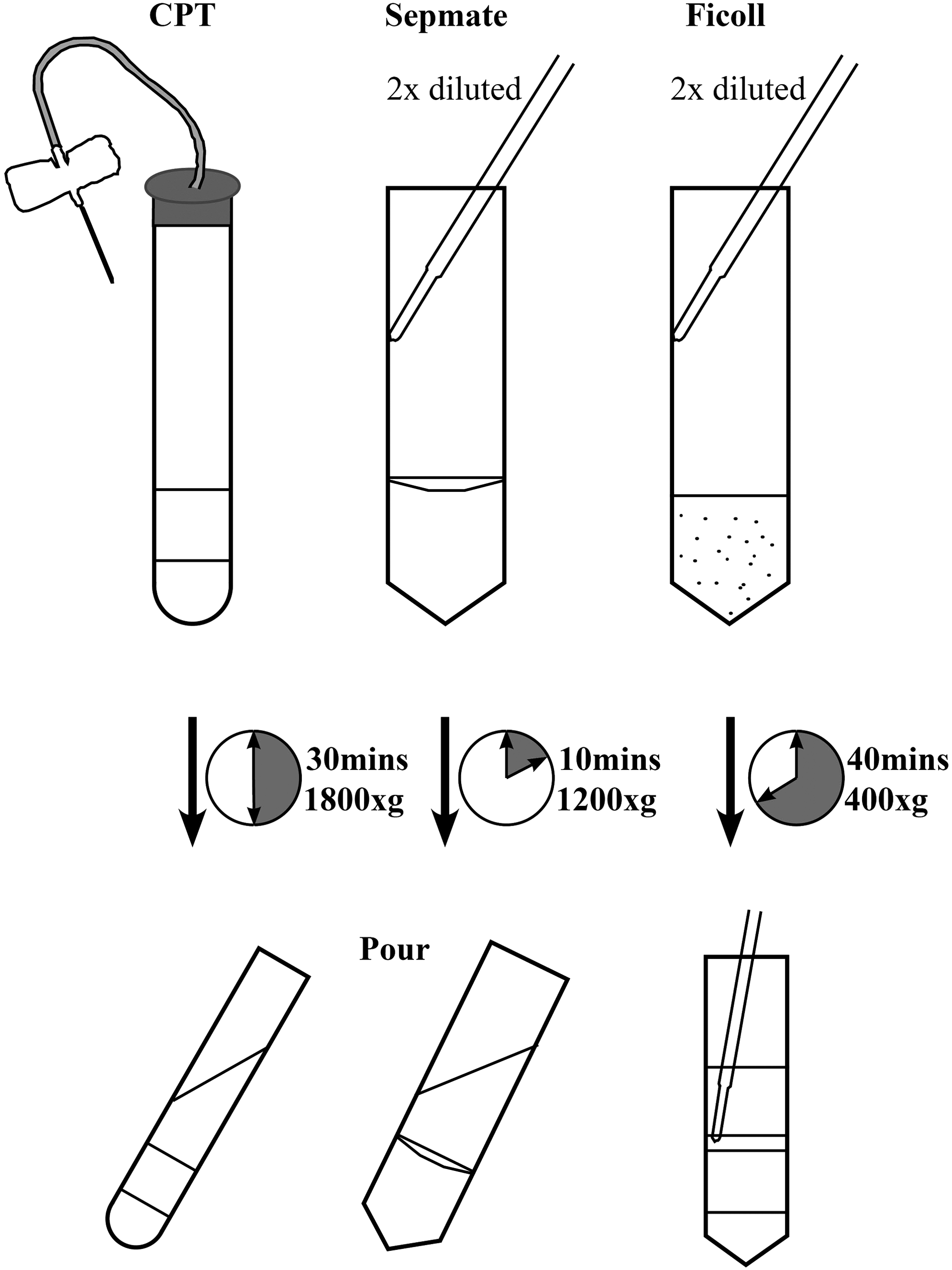
Schematic presentation of the three isolation techniques. Blood is diluted twofold in the SepMate and Ficoll techniques and used undiluted with CPT. Centrifugation time and speed differ for all three techniques. PBMC collection for Ficoll requires pipetting, whereas for the other techniques, PBMCs can be simply poured off. CPT, cell preparation tube; PBMC, peripheral blood mononuclear cell.
Immunophenotyping of PBMC populations
Immunophenotyping was performed by flow cytometry using CD3-APC (clone OKT3; eBioscience), CD20-APC (clone 2H7; eBioscience), CD56-APC (clone CMSSB; eBioscience), and CD14-PE-Cy7 (clone 61D3; eBioscience) antibodies. Samples were measured with a FACS Canto II system (BD Biosciences) and analyzed with FlowJo software v10.
PBMC culture and stimulation
Duplicate PBMC cultures of 5 × 10⁵ cells per tube in RPMI1640 media (Gibco, Thermo Scientific) were set up in polystyrene 5 mL round-bottom tubes (Becton Dickinson). Cells were stimulated with SEB from Staphylococcus aureus (Sigma-Aldrich) at a final concentration of 100 ng/mL. Unstimulated control cultures were included for comparison. Cytokine release was measured in the culture supernatant using the V-PLEX Human Proinflammatory Kit (IL-β, IL-6, IL-8, and TNFα; Meso Scale Discovery). IFNγ was measured by ELISA (Mabtech). Lactate dehydrogenase (LDH) release was measured in the supernatant using the Pierce LDH Cytotoxicity Assay Kit (Thermo Fisher Scientific) and expressed as the percentage of the total LDH present in lysates of unstimulated cells.
Results
Recovery and viability
PBMCs were isolated from three volunteers in parallel using the three methods described earlier. This experiment was performed by an experienced technician (Fig. 2A). The lowest cell recovery was observed for the classic Ficoll approach, amounting an average 6 × 10⁵ cells/mL of whole blood. Cell recovery was higher for the SepMate tubes and CPT (8 × 10⁵ and 13 × 105 cells/mL of whole blood, respectively). In another experiment, cell recovery was assessed for PBMC fractions, isolated from blood of a single donor by two technicians (Fig. 2B). Again, for both technicians, the lowest recovery was observed for the Ficoll approach and higher recoveries for the SepMate tubes and CPT. Overall, cell recovery was higher for the PBMC isolations performed by technician 2 (61%, 36%, and 42% higher compared to technician 1 for CPT, Ficoll, and SepMate, respectively), demonstrating operator-dependent differences. After isolation, cell viability was assessed by trypan blue exclusion. For all three techniques, viability was 100% directly following isolation (data not shown).

PBMC recovery per milliliter whole blood.
Population composition
Although not specifically quantified in this study, microscopic examination during counting revealed more erythrocyte contamination for CPT-isolated populations compared to Ficoll- and SepMate-isolated populations. To assess the composition of the isolated PBMC populations, monocytes (CD14), T-cells (CD3), B-cells (CD20), and NK-cells (CD56) were measured by flow cytometry (Fig. 3). The composition of the PBMC population varied between subjects. The major population was T-cells (65%–70%) followed by B-cells (5%–22%), NK-cells (5%–14%), and monocytes (4%–12%). Only minor differences were observed between isolation techniques, demonstrating that there is no preferential loss of any specific cell subset.
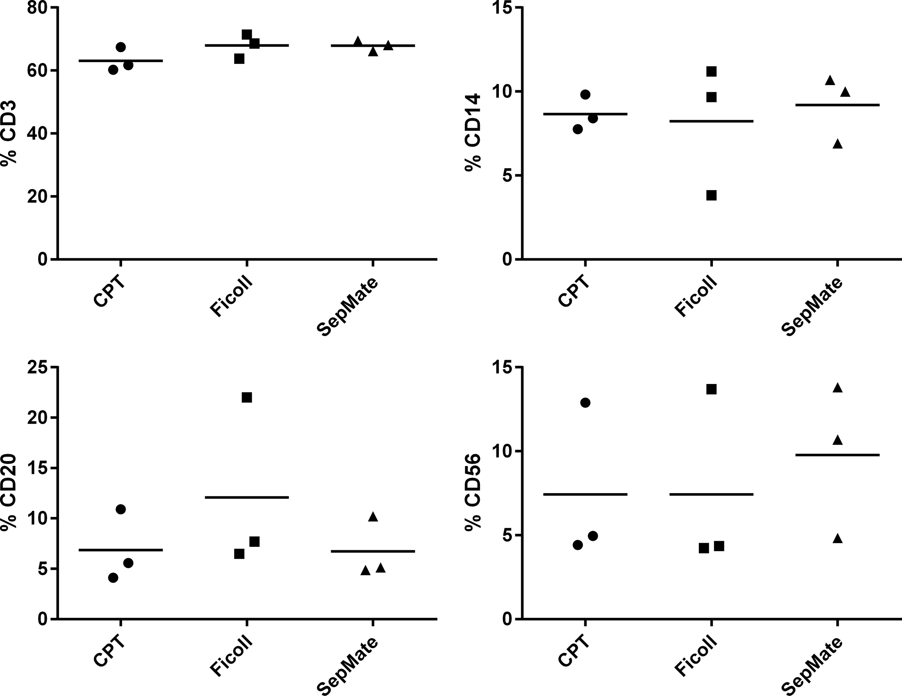
PBMC composition by flow cytometry. PBMCs were isolated and stained with anti-CD3 (T-cells), -CD14 (monocytes), -CD20 (B-cells), and -CD56 (NK-cells). Percentage positive cells are shown per donor, the mean is indicated by the bar.
Cell functionality
To assess the functionality of PBMCs after isolation, the response of PBMCs to a superantigen stimulation (100 ng/mL SEB, 24 hours) was quantified by the release of IFNγ, IL-1β, IL-6, IL-8, and TNFα (Fig. 4A). Stimulation with SEB resulted in IFNγ release, which varied between PBMC donors. Cultures prepared from Ficoll-isolated PBMCs secreted lower levels of IFNγ than cultures prepared from CPT- or SepMate-isolated PBMCs (average 22 ng/mL vs. 62 and 65 ng/mL for Ficoll, CPT, and SepMate, respectively). In addition, the release of IL-1β, IL-6, IL-8, and TNFα, as a secondary response to SEB stimulation, was lower for Ficoll-isolated PBMCs compared to CPT- and SepMate-isolated PBMCs. When comparing the three subjects of IFNγ responses, we observed the same rankings when PBMCs were isolated using CPT and SepMate (e.g., the same subject was the highest responder in both cases), but this differed when cultures were prepared by Ficoll. Evaluation of spontaneous cytokine release in unstimulated cultures also exhibited some differences. IL-6 and IL-8 release in unstimulated cultures was highest for CPT-isolated PBMCs compared to SepMate- and Ficoll-isolated PBMCs, whereas other cytokines measured were absent or very low.
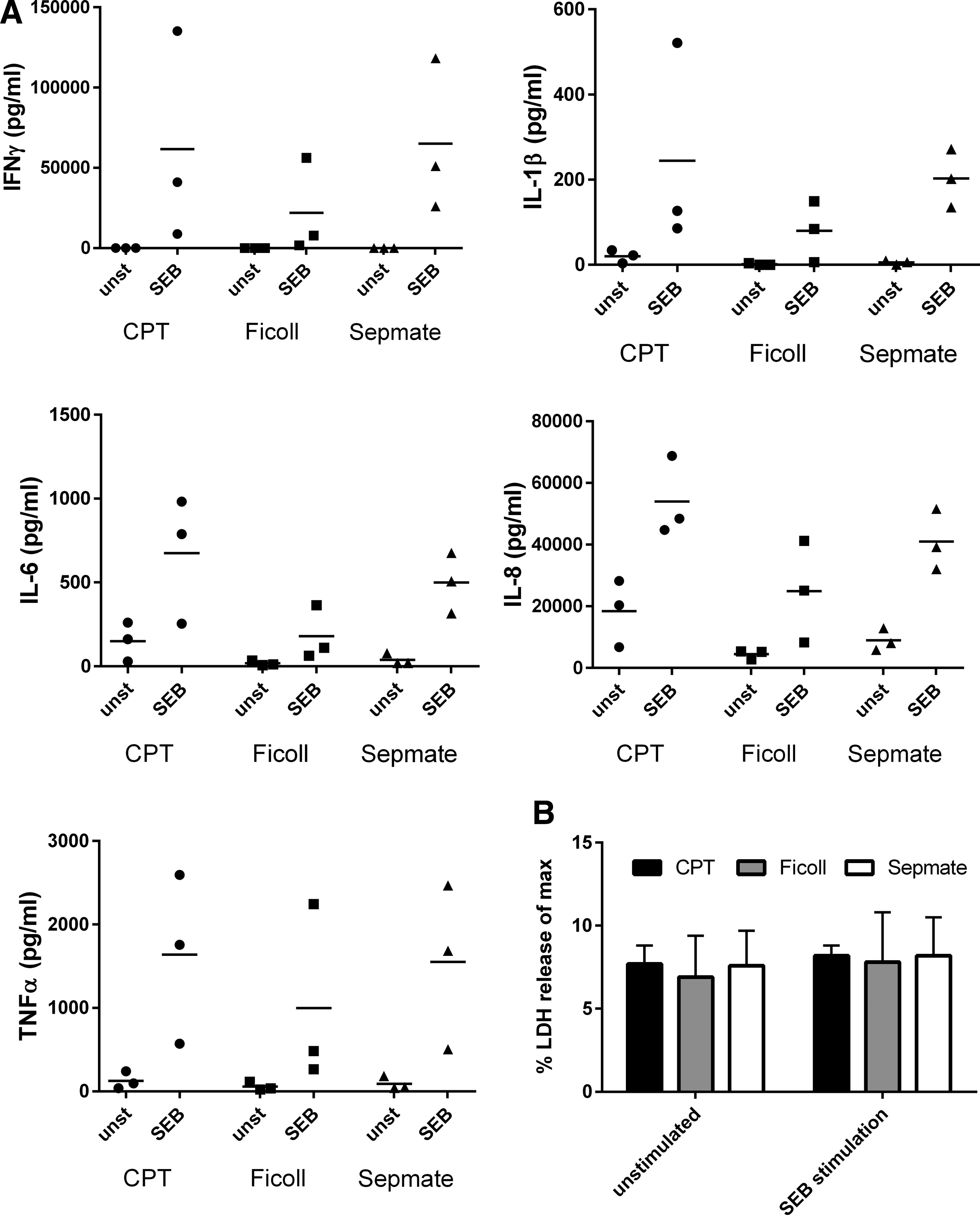
Cytokine and LDH release in PBMC cultures. PBMCs were isolated from three donors and cultured for 24 hours in the absence or presence of 100 ng/mL SEB.
The LDH release was assessed for all cultures, as a quantitative measure for cytotoxicity or cell stress (Fig. 4B). Overall, no significant differences were observed between PBMC isolation techniques or between the stimulated and unstimulated conditions. LDH release (expressed as percentage of LDH levels in cell lysates from unstimulated cells) was ∼7%, indicating good viability of all PBMC cultures.
Discussion
Three routine PBMC isolation techniques were evaluated, focusing on cell recovery and viability, population composition, and cell functionality. The techniques comprised the classic Ficoll approach, isolation by CPTs, and isolation by SepMate tubes with Lymphoprep. A summary of the main findings is provided in Table 1.
CPT, cell preparation tube; PBMC, peripheral blood mononuclear cell; SEB, staphylococcal enterotoxin B.
Cell recovery was lowest for the Ficoll approach. PBMC isolation by CPT and SepMate tubes resulted in ∼70% higher cell counts. It was readily observed that more erythrocyte contamination was found for CPT-isolated populations compared to Ficoll- and SepMate-isolated populations as observed by eye when counting cells by hemacytometer. It should be stressed that although the applied PBMC isolation technique is an important determinant for cell recovery, the experience of the technician performing the isolations is equally important: differences up to 60% in cell recovery were observed between a well-trained but inexperienced technician and a technician experienced in cell isolations. Cell viability, assessed by trypan blue exclusion, was optimal for all three isolation techniques. The composition of the PBMC population varied between subjects, as described before, 12 but did not differ between PBMC populations isolated by different techniques. The recovery of specific minority populations of cells (e.g., dendritic cells) may be dependent on the applied isolation technique, but this was not investigated in the current study.
To assess the effect of the applied isolation technique on the functionality of isolated cells, PBMCs were subjected to a superantigen challenge. SEB is known to drive a robust IFNγ secretion by T-cells, which in turn induces secretion of other cytokines. 11 SEB-induced cytokine release differed between isolation techniques. Cultures prepared from Ficoll-isolated PBMCs secreted lower levels of IFNγ and showed differences between subjects compared to cultures prepared from CPT- or SepMate-isolated PBMCs. In addition, the release of IL-1β, IL-6, IL-8, and TNFα was lower for Ficoll-isolated PBMCs compared to CPT- and SepMate-isolated PBMCs. Evaluation of spontaneous cytokine release in unstimulated cultures also displayed differences. Spontaneous IL-6 and IL-8 release was higher for CPT-isolated PBMCs compared to Ficoll- and SepMate-isolated PBMCs, with Ficoll-isolated PBMCs showing the lowest spontaneous cytokine release. IL-8 release due to sample handling is a common phenomenon and may relate to, for example, exposure to increased extracellular calcium levels. 13 Furthermore, it should be noted that there are differences in the composition of the density media, and the presence of a gel barrier in the CPT may potentiate cell activation. Notably, higher spontaneous IL-8 release corresponded to higher specific cytokine release in response to SEB stimulation. This may reflect compounding signals from isolation and activation with antigen, and care should be taken when interpreting such data. For other cytokines tested, spontaneous release was absent or very low, with no major differences between isolation techniques. LDH release, as a quantitative measure for cytotoxicity or cell stress, did not differ between cell isolation techniques or between the stimulated and unstimulated conditions.
PBMC isolation by CPTs and SepMate tubes is straightforward and involves limited sample handling. The classic Ficoll isolation is attractive from a cost perspective, but in practice, this technique is relatively laborious. We have demonstrated that isolation techniques based on SepMate and CPT result in a higher recovery than Ficoll isolation, although for CPT-isolated populations significant erythrocyte contamination may be observed. In addition, PBMCs isolated by SepMate and CPTs give higher cytokine responses in cell cultures, both for stimulated and unstimulated conditions, which demonstrate functionality of isolated cells, but which may also relate to cell activation due to sample handling.
Footnotes
Author Disclosure Statement
No conflicting financial interests exist.