
Abstract
Tears covering the ocular surface are important biofluids containing thousands of molecules, including proteins, lipids, metabolites, nucleic acids, and electrolytes. Tears are valuable resources for biomarker research of ocular and even systemic diseases. For application in biomarker studies, tear samples should ideally be stored using a simple, lowcost, and efficient method along with the patient's medical records. For this purpose, we developed a novel Schirmer's strip-based dry method that allows for storage of tear samples in vacuum bags at room temperature. Using this method, tear protein patterns can also be preserved. Liquid chromatography–mass spectrometry/mass spectrometry analysis of proteins recovered by the dry method and traditional wet method showed no significant difference. Some tissue/organ-enriched proteins were identified in tear samples, thus tears might be a good window for monitoring changes of these tissues or organs. This dry method facilitates sample transportation and enables the storage of tear samples on a large scale, increasing the availability of samples for studying disease biomarkers in tears.
Introduction
T
Tears show promise as biofluids for biomarker studies and should be preserved along with a patient's medical record. This is a critical step in validation, which facilitates biomarker research and its translation from the bench to the bed. The primary methods for collecting tears are using the Schirmer's strip and glass capillary tube, followed by flash-freezing at −80°C. 11 Cryopreservation of tears cannot absolutely prevent the degradation of proteins, as the samples contain various enzymes and hydrolases. In addition, use of the required cold chain process during sample transportation is challenging and costly.
Here, we dried the Schirmer's strip soaked with tears and stored the strip in a vacuum bag. Importantly, the proteins were dry, preventing their degradation and enabling preservation at room temperature.
Materials and Methods
Ethical statement
The consent procedure and study protocol were approved by the Institutional Review Board of the Institute of Basic Medical Sciences, Chinese Academy of Medical Sciences (Project No. 007-2014). Written informed consent was obtained from each subject.
Tear collection and preservation
Tear samples were collected from seven healthy volunteers by Schirmer's type I tear test without using local anesthesia. No volunteers had a recent history of ocular disease or contact lens usage. The Schirmer's strips were inserted for 5 minutes in the lower eyelid in a standard manner in both eyes for the same subject. Schirmer's strips from both eyes of the volunteer were cut longitudinally into two halves immediately after sampling. Half of the sample was added to a 2-mL Eppendorf tube snap-frozen at −80°C for 2 weeks (Fig. 1A). The other half of the sample was stored by the dry method for 2 weeks (Fig. 1B). The strip soaked with tears was dried using a hair dryer (Philips HP8200) for 2–3 minutes, and then the strip was placed in an aseptic plastic bag. The bag was then sealed using a kitchen vacuum sealer and stored at room temperature.
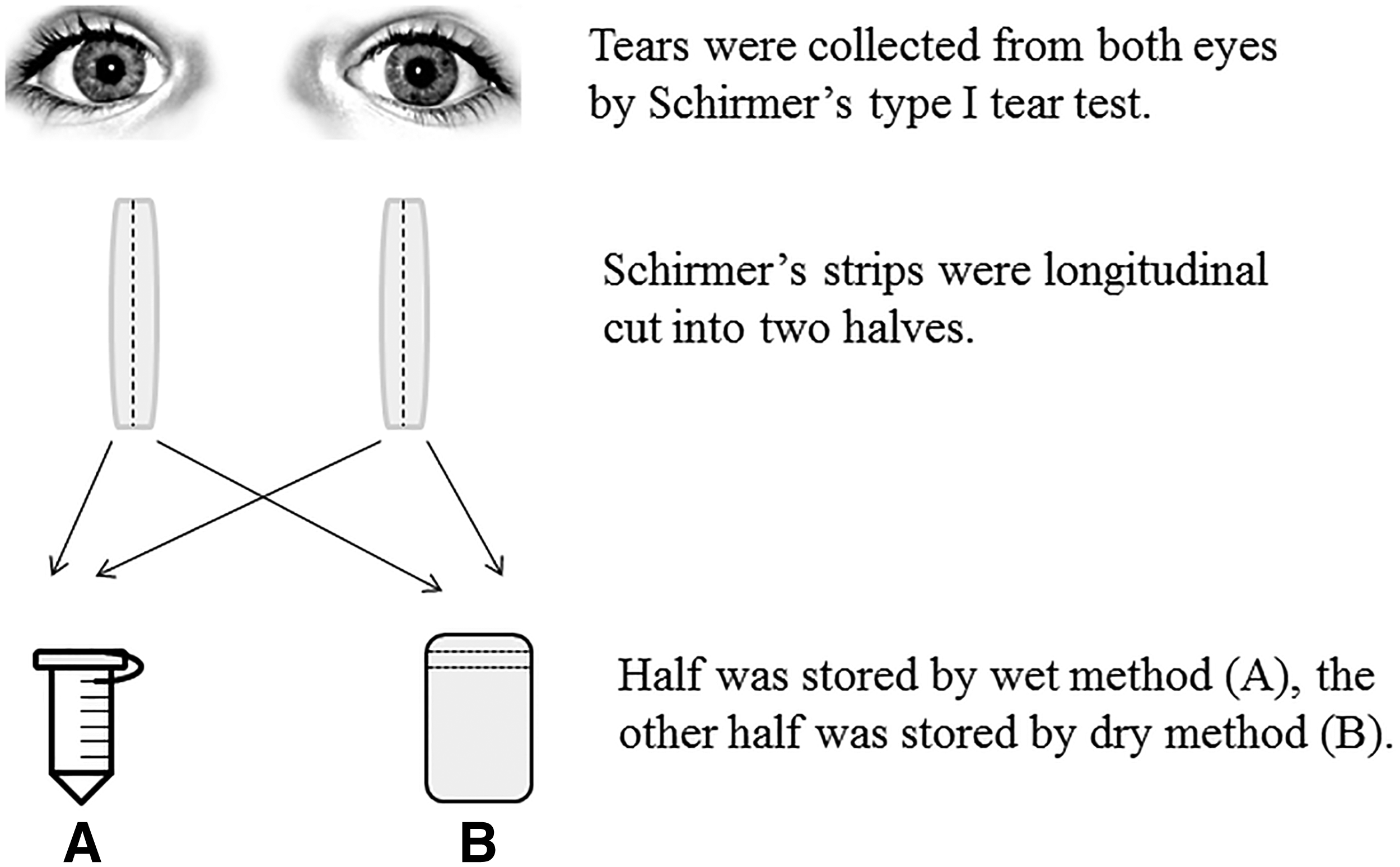
Workflow of the collection and preservation of tear samples.
Protein extraction
The strip was cut into small pieces and transferred into a 0.6 mL tube. Next, 200 μL elution buffer (100 mM NH3HCO3, 50 mM NaCl) was added and gently shaken for 2 hours at room temperature. The tube was punctured at the bottom with a cannula, placed in a larger tube (1.5 mL), and centrifuged at 12,000 g for 5 minutes. 12 The filtrate in the outer tube was collected and quantified by the Bradford method before sodium dodecyl sulfate polyacrylamide gel electrophoresis (SDS-PAGE) analysis.
Tryptic digestion
Tear proteins were digested by filter-aided sample preparation methods. 13 In brief, a 200 μg protein sample was loaded on the 10 kD filter unit (Pall), and 200 μL UA (8 M urea in 0.1 M Tris-HCl, pH 8.5) was added to the filter unit and centrifuged at 12,000 g for 40 minutes. Then, 200 μL ABC (0.05 M NH4HCO3 in water) was added and centrifuged. Dithiothreitol solution (4.5 mM dithiothreitol in ABC) was added to the filter unit and incubated for 1 hour at 37°C. The filter units were centrifuged at 12,000 g for 30 minutes. Iodoacetamide solution (10 mM iodoacetamide in ABC) was added to filter unit and incubated in the dark for 30 minutes at room temperature. The filter units were centrifuged at 12,000 g for 30 minutes. Then the concentrate was dissolved in 50 mM NH4HCO3. Proteins were digested with trypsin (4 μg) for 14 hours at 37°C. The digested peptides were desalted using Oasis HLB cartridges (Waters). The resulting peptides were desalted and dried by a SpeedVac (Thermo Fisher Scientific, Waltham, MA). The reproducibility of digestion was estimated, and the details are included in the Supplementary Data (Supplementary Data are available online at www.liebertpub.com/bio).
Liquid chromatography–mass spectrometry/mass spectrometry analysis
The digested peptides were dissolved in 0.1% formic acid and loaded on a trap column (75 μm × 2 cm, 3 μm, C18, 100 Å). The eluent was transferred to a reversed-phase analytical column (50 μm × 150 mm, 2 μm, C18, 100 Å) by a Thermo EASY-nLC 1200 high-performance liquid chromatography system. Peptides were analyzed using a Fusion Lumos mass spectrometer (Thermo Fisher Scientific). The Fusion Lumos was operated on data-dependent acquisition mode. Survey mass spectrometry (MS) scans were acquired in the Orbitrap using a 350–1550 m/z range with the resolution set to 120,000. The most intense ions per survey scan (top speed mode) were selected for collision-induced dissociation fragmentation, and the resulting fragments were analyzed in Orbitrap. Dynamic exclusion was employed with a 30 seconds window. Three technical replicate analyses were performed for each sample.
Data analysis
The mass spectrometry/mass spectrometry (MS/MS) spectra were processed using Mascot software, using the human proteome database (UniProtKB/Swiss-Prot release January 10, 2014). The FASTA file contained 20120 protein sequences. Search parameters were set as follows: 10 ppm precursor mass tolerance, 0.02 Da fragment mass tolerance, two missed cleavage sites allowed in the trypsin digestion, cysteine carbamidomethylation as fixed modification, and oxidation (M) as variable modifications. Protein identifications were accepted if they could be established at >91.0% probability 14 to achieve a false-discovery rate of <1.0%, and if they contained at least one identified peptide.
Results
SDS-PAGE analysis of proteins recovered from tears stored by wet and dry methods
To estimate the effectiveness of storing tear proteins in a dry state at room temperature, proteins were recovered from strips that had been stored for 2 weeks by the wet method and dry method, and then separated by SDS-PAGE and stained with silver. As shown in Figure 2, the same tear sample stored by the wet and dry methods exhibited similar legible patterns, suggesting that these two methods have similar effectiveness for protein preservation.

SDS-PAGE analysis of tear proteins recovered from wet method and dry method. In each lane, 5 μg tear proteins was loaded, stained with silver. M, marker; 1, 2, 3, 4, 5, 6, 7, different volunteers; W, wet method (strips soaked with tears were snap-frozen at −80°C); D, dry method (strips soaked with tears were dried and stored in a vacuum bag at room temperature). SDS-PAGE, sodium dodecyl sulfate polyacrylamide gel electrophoresis.
Liquid chromatography–MS/MS identification of proteins recovered from wet method and dry method
To further evaluate the preservation effectiveness of the dry method, the exact species of proteins in the tear samples stored by the wet and dry methods, from four subjects, were identified by liquid chromatography (LC)–MS/MS (Supplementary Table S1). After label-free quantification, identification of proteins for three LC–MS/MS technical replicates prepared by the wet method revealed 316 shared proteins (456, 434, and 417 in each replicate), with an overlap rate of 72.5% (overlap rate = shared proteins/mean proteins × 100%) (Fig. 3A). Simultaneously, 290 shared proteins were identified (397, 394, and 395 in each replicate) within the replicates prepared by the dry method, with an overlap rate of 73.4% (Fig. 3B). A total of 240 proteins were identified as common by these two methods, with an overlap rate of 79.2% (Fig. 3C), which was similar to that of the LC–MS/MS technical replicates (Supplementary Figs. S1, S2 and S3). After preservation, some proteins showed differences between the wet method and dry method; thus, in each study, the same collection and preservation method should be used.
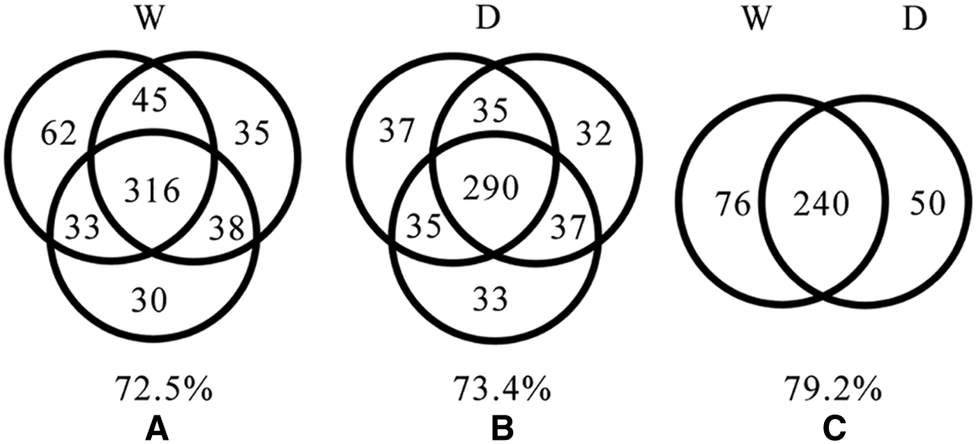
LC–MS/MS identification of tear samples from volunteer 1 preserved by two methods.
To further compare these two methods, a spectral counting method was used to estimate each identified protein's abundance. 15 We drew a correlation curve of identified protein's abundance between the dry and the wet methods. The correlation coefficient (R2) of protein abundance was 0.9987, 0.9974, 0.9969, and 0.9993, respectively, for volunteer 1, 2, 3, and 4 (Fig. 4 and Supplementary Figs. S4, S5 and S6). The results showed good correlation of protein abundance between these two methods. The proteins that were only preserved in the wet or dry method were low abundance proteins.

Correlation curve of identified protein's abundance between dry method and wet method for volunteer 1. Spectral counting method was used to estimate each identified protein's abundance.
Discussion
Recently, Uhlén et al. reported a tissue-based map of the human proteome, describing the expression and distribution of human proteins across 44 different tissues and organs, both at the mRNA (32 tissues) and protein levels. 16 In our study, we identified 514 tear proteins, and then we compared each of them with the tissue-enriched proteome. In all, 365 proteins that are highly enriched in different tissues and organs were also identified in tears, and 132 proteins corresponding to 132 protein-encoding genes that are highly expressed in different tissues and organs were also detected in tears (Supplementary Table S2). This is an observation that tissue/organ-enriched proteins are present in tears. There is no known mechanism as far as we know. At this point, we can only propose the possibility that if those organs had functional and/or structural changes, proteins enriched in those organs may be released in a different quality and quantity into the blood. These changes may somehow reach the tears and be reflected in proteins in tears. Therefore, tears might be a good window for monitoring changes in these tissues or organs. These proteins are not specific to those organs. They may also be made by the tear gland locally.
According to the qualitative and quantitative results of LC–MS/MS, proteins only preserved in the wet or dry method were low abundance proteins. This is very likely caused by the proteomics strategy adopted in this study. LC–MS/MS with data-dependent acquisition (DDA) was used, and it is based on signal intensity used for the precursor-ion selection, which results in an incomplete sampling of the peptide mixture generated to represent the proteome. The analytical reproducibility of peptide identification obtained using DDA-based methods is about 75% overlap between technical replicates. 17 We suggest that most of the proteins were preserved in both methods. Even if there are differences, it would not be a problem when the same method is used in one study, as we suggested.
In addition to proteins, other biomolecules in tears were preserved on the strip by the dry method, including lipids, metabolites, nucleic acids, and electrolytes. Since all tears were soaked on the strip, only water and some volatile matter were lost during the drying procedure. In this study, we analyzed the proteins only, considering the time consumption and experimental techniques. Seven volunteers were included. We focused on estimating the stability of this new dry method. This procedure included tear collection using Schirmer's strip, protein preservation on dry and vacuum station, protein elution from the strip, and the protein identification using SDS-PAGE and LC–MS/MS. Except for the preservation part, the other three parts have proven to be reproducible, respectively.1,12,13,17 To prove that this dry method is stable, we studied reproducibility of all four parts as a whole. Seven samples involves seven technical replicates, which may not be large enough for a biomarker study, but when considering that every step had been proved reproducible, it should be enough to prove the stability of the whole method.
We first reported the dry method for preserving tear samples. The most significant difference between the dry and wet methods is with the preservation procedure. Using the wet method (the primary method), after tear collection, the Schirmer's strip is flash-frozen at −80°C. The disadvantage is that cryopreservation of tear samples cannot absolutely prevent the degradation of proteins, as the samples contain various enzymes and hydrolases. In addition, use of the required cold chain during sample transportation is challenging and costly. According to the dry method, after tear collection, the Schirmer's strip is dried and stored in a vacuum bag. The advantage is that the proteins were dry, preventing their degradation and enabling preservation at room temperature. Therefore, a higher degree of dryness and vacuum should keep tear samples at room temperature for a longer period.
Thus, the dry method is applicable for establishing libraries of stored tear samples for long periods, and can simplify studies of disease biomarkers in tears.
Footnotes
Supporting Information
Acknowledgments
This work was supported by the National Key Research and Development Program of China (grant number 2016YFC1306300), the National Basic Research Program of China (grant number 2013CB530850), and the Fundamental Research Funds for the Central Universities (2015KJJCB21), Beijing Natural Science Foundation (7173264), and 2016 pumch Science Fund for Junior Faculty (pumch20162.27).
Author Disclosure Statement
No conflicting financial interests exist.
References
Supplementary Material
Please find the following supplemental material available below.
For Open Access articles published under a Creative Commons License, all supplemental material carries the same license as the article it is associated with.
For non-Open Access articles published, all supplemental material carries a non-exclusive license, and permission requests for re-use of supplemental material or any part of supplemental material shall be sent directly to the copyright owner as specified in the copyright notice associated with the article.