
Abstract
The practical requirements of islet transplantation necessitate that a large quantity of pancreatic islets be cryopreserved for a long period of time in a simple and convenient manner. We cryopreserved rat islets (size range 101–150 μm in mean diameter) by vitrification with either a Cryotop® device or a ø = 57-μm nylon mesh device in units of 10 islets, or by conventional freezing with a Bicell® vessel in units of 50 islets. Postwarm/thaw survival rates of the islets were 68.1% ± 5.9%, 64.1% ± 3.5%, and 47.7 ± 1.2% following Cryotop vitrification, nylon mesh vitrification, and Bicell freezing, respectively (p < 0.05). Glucose-stimulated insulin secretion in the two vitrification groups (stimulus index [SI] = 3.1–3.9) was superior to that in the freezing group (SI = 0.8). Additional experiments involved scaling-up the cryopreservation process using the nylon mesh device in units of 10, 50, or 100 islets. Increased numbers of islets per device had no adverse effects on cryosurvival (58.6%–68.5%) or insulin secretion potential (SI = 2.8–4.2). As the nylon mesh device does not require the handling of individual islets with glass pipettes, pre- and postvitrification islet treatment is less complicated. Therefore, nylon mesh can serve as a simple cryodevice for the vitrification of large quantities of rat pancreatic islets.
Introduction
T
Conventional cell freezing protocols have been used for islet cryopreservation in rats and humans.6–8 While a freezing protocol is advantageous because a large quantity of isolated islets can be handled with a relatively simple procedure, the inevitable formation of ice crystals may lead to impaired insulin secretion function of freeze–thawed islets.9–11 This dysfunction in post-thaw islets results in the requirement of transplanting more than double the IEQ, compared with fresh islets, to achieve euglycemia.7,11 However, a vitrification protocol, which can avoid ice crystal formation by use of an extremely high cooling–warming velocity, is superior to conventional freezing in terms of glucose-stimulated insulin secretion and the expression of β-cell function-associated genes. 12 Different devices such as Cryotop®, 12 open-pulled straw, 13 and hollow fiber 14 have been used for the vitrification of isolated rodent islets. However, vitrification protocols have a technical limit as large quantities of islets cannot be loaded onto or into each device (recommended quantity 10–12, possible upper limit 20–30).
Matsumoto et al. 15 reported that as many as 65 bovine immature cumulus–oocyte complexes (COCs) can be vitrified–warmed using nylon mesh as a cryodevice, with subsequent work by Abe et al., 16 successfully producing a calf from these COCs. Because of the similar size characteristics between COCs and pancreatic islets, our present study was conducted to investigate the capacity of a nylon mesh device for vitrification of large quantities of rat pancreatic islets.
Materials and Methods
Experimental design
Rat pancreatic islets (n = 10 or 50 per group) were subjected to one of three cryopreservation protocols (Cryotop vitrification, nylon mesh vitrification, or Bicell® freezing) in a single replicate, and their postwarm/post-thaw survival rates were assessed by fluorescein diacetate (FDA)/propidium iodide (PI) double staining. Glucose-stimulated insulin secretion (stimulus index: SI; 20 mM/3 mM) was measured with an Enzyme-linked Immunosorbent Assay (ELISA) Kit for functional analysis. Nylon mesh was selected as the cryodevice for the larger number of islets (10, 50, or 100 islets per single operation). The survival and SI values of the vitrified–warmed islets were measured to determine whether the nylon mesh enabled the scale-up of islet numbers in a single operation.
Chemicals and animals
Unless otherwise indicated, chemicals used in the present study were purchased from Sigma-Aldrich Corp. (St. Louis, MO). Specific pathogen-free Brown Norway rats were purchased from Japan SLC, Inc., (Shizuoka, Japan). Rats were housed in an environmentally controlled room with a 12-h dark/12-h light cycle at a temperature of 23 ± 3°C, with free access to a laboratory diet (NMF; Oriental Yeast Co., Ltd., Tokyo, Japan) and tap water. All experimental animal procedures were reviewed and approved by the Animal Care and Use Committee of the Shinshu University (Nagano, Japan). Animals were treated humanely and the standards conformed to those of current ethical animal research practices.
Isolation of rat islets
Pancreatic islets were isolated from male rats at 8–12 weeks old. Briefly, the bile duct of the rats was cannulated with a fine plastic tube, and the pancreas was distended with ∼8 mL of Liberase™ TL (Thermolysin Low) solution (1 WU/mL Liberase in cold Hanks' balanced salt solution [HBSS]). The pancreas was excised, minced, and incubated at 37°C for 30 minutes and then the digested tissues were purified on a discontinuous Histopaque gradient that was layered with Histopaque1119, Histopaque1077, and 2% fetal bovine serum (FBS; HyClone™, GE Healthcare Life Sciences, Logan, UT) in HBSS. Islets with a size of 101–150 μm in mean longest and widest diameter were handpicked using capillary pipettes under a stereomicroscope and cultured for 24 hours in 2 mL of RPMI-1640 (Product No. 11835-030; Life Technologies, Inc., Rockville, MD) supplemented with 10% FBS and antibiotics (100 U/mL penicillin and 100 μg/mL streptomycin) at 37°C in a humidified atmosphere of 5% CO2 in air until use for cryopreservation.
Cryopreservation
The Cryotop vitrification protocol was conducted as described previously. 12 Briefly, islets were equilibrated with 7.5% ethylene glycol (EG; Wako Pure Chemical Industries Ltd., Osaka, Japan) and 7.5% dimethyl sulfoxide (Me2SO; Wako Pure Chemical Industries, Ltd.) in RPMI-1640 supplemented with 20% FBS for 3 minutes at ambient temperature (25 ± 2°C) and then transferred into a vitrification solution (VS) comprising 15% EG, 15% Me2SO, and 0.5 M sucrose as cryoprotective additives (CPAs) in RPMI-1640 containing 20% FBS for 60 seconds at ambient temperature. Within this 60-second period, 10 islets were loaded onto the polypropylene strip of a Cryotop device (Kitazato Corp., Shizuoka, Japan) with a minimal volume of the VS and then quickly plunged into liquid nitrogen (LN2).
After storage for at least 1 week, islet warming was performed by immersing the polypropylene strip of Cryotop into RPMI-1640 containing 20% FBS and 1 M sucrose at 38.5°C for 1 minute. Following warming, islets were transferred to RPMI-1640 containing 20% FBS and sucrose in a stepwise manner (0.5, 0.25, and 0 M sucrose for 3, 5, and 5 minutes, respectively).
The Nylon mesh vitrification protocol was conducted as described previously, 16 with some modifications to enable the original protocol developed for bovine COCs to be adapted to rat islets. Briefly, a 10-mm triangle sheet was cut from commercially available nylon mesh (pore size ø = 57 μm; Sansyo Co., Ltd., Tokyo, Japan) and processed to form a triangular pyramid by folding in quarters. Islets were equilibrated with 7.5% EG and 7.5% Me2SO in RPMI-1640 containing 20% FBS for 3 minutes at ambient temperature and then placed onto the center triangle of the sterilized nylon mesh device using a capillary pipette. Immediately thereafter the equilibration solution was removed by placing the device on a sterilized filter paper (Kimwipes®; Nippon Paper Crecia Co., Ltd., Tokyo, Japan), and the islets on the device were exposed to VS (15% EG, 15% Me2SO, and 0.5 M sucrose in RPMI-1640 containing 20% FBS) for 60 seconds at ambient temperature using sterilized tweezers. Within this 60-second period, the device was placed on new sterilized Kimwipes and then quickly plunged into LN2.
After storage in LN2-filled 50-mL conical tubes at least for 1 week, islet warming was performed by immersing the device into RPMI-1640 containing 20% FBS and 1 M sucrose at 38.5°C for 1 minute. Following warming, islets on the device were transferred to RPMI-1640 containing 20% FBS and sucrose in a stepwise manner (0.5, 0.25, and 0 M sucrose for 3, 5, and 5 minutes, respectively). Islets were released from the device during the last 5-minute exposure to the sucrose-free solution using a capillary pipette. A schematic diagram of the nylon mesh vitrification protocol is shown in Figure 1.

Outline of islet vitrification with nylon mesh device.
A conventional freezing protocol was conducted using Bicell bio freezing vessels according to the manufacturer's instruction manual (Nihon Freezer Co., Ltd., Tokyo, Japan). Briefly, islets were exposed to 15% Me2SO in RPMI-1640 containing 10% FBS and antibiotics for 15 minutes at ambient temperature and then cooled to 4°C for 15 minutes. Fifty islets were placed in a precooled cryotube containing 500 μL of the abovementioned CPA solution, and the cryotubes were packed into a Bicell vessel. Subsequently, the Bicell vessel was placed in a −80°C deep freezer overnight (estimated cooling rate, −0.5°C/min during the first 3 hours) and then the cryotubes were transferred into LN2.
After storage for at least 1 week, the cryopreserved islets were thawed by gently warming the cryotubes in a 37°C water bath for 1 minute. Following thawing, islets were transferred to RPMI-1640 containing 10% FBS, antibiotics, and sucrose in a stepwise manner (1.0, 0.5, 0.25, and 0 M sucrose for 2, 3, 5, and 5 minutes, respectively).
Survival assay
Islet survival was assessed by double staining with membrane the exclusion dyes FDA and PI. An aliquot of 10 islets in each group was stained with 25 μg/mL FDA and 25 μg/mL PI for 30 seconds in the dark. After three washes in phosphate-buffered saline, micrographs depicting FDA (green) and PI (red) fluorescence were taken using an epifluorescence microscope (IX73; Olympus Corp., Tokyo, Japan). The fluorescent area was quantified using ImageJ software (https://imagej.nih.gov/ij/). Survival rate was calculated as: 100 × FDA-positive area divided by total of FDA-positive + PI-positive area.
Insulin secretion
Islet functionality was assessed by static glucose-stimulated insulin secretion assay. Briefly, post-thaw/postwarm islets (10 each per group) were washed thrice with RPMI-1640 containing 10% FBS and 3 mM glucose and incubated for 1 hour at 37°C in a humidified atmosphere of 5% CO2 in air. The islets were then stimulated by transfer into RPMI-1640 containing 10% FBS and 20 mM glucose and incubated for 1 hour as described above. At the end of incubation, supernatants were collected and stored at −80°C until an insulin assay was conducted. The basal and stimulated insulin levels (ng/islet/h) were determined using an ELISA Kit for rat insulin (MIoBS, Inc., Kanagawa, Japan), and the SI was defined as the stimulated insulin level in response to 20 mM glucose divided by the basal level in response to 3 mM glucose.
Statistical analysis
Percentage data were subjected to arcsine transformation before statistical analysis. Differences between groups were assessed by one-way analysis of variance (ANOVA). When ANOVA was significant, differences among values were analyzed by Tukey's Honest Significant Difference test for multiple comparisons. Data were considered statistically significant at p < 0.05.
Results
Comparison of islet revivability among different cryopreservation protocols
All islets cryopreserved were recovered after thawing/warming (100% recovery). Survival of frozen–thawed islets (Bicell group) was significantly lower compared with vitrified–warmed islets (both Cryotop and Nylon mesh groups), as shown in Figure 2. While the insulin secretion level in response to 3 mM glucose was higher in frozen–thawed versus vitrified–warmed islets, the insulin secretion levels stimulated by 20 mM glucose were comparable among the three groups, as shown in Table 1. Therefore, the SI value for the Bicell group (0.8) was significantly lower or tended to be lower than values for the Cryotop and Nylon mesh groups (3.9 and 3.1, respectively). For reference, noncryopreserved fresh islets were isolated from the same donor rat colony, with a survival rate of 92.1% ± 1.5% and an SI value of 7.3 ± 0.8 (four replicates) obtained.
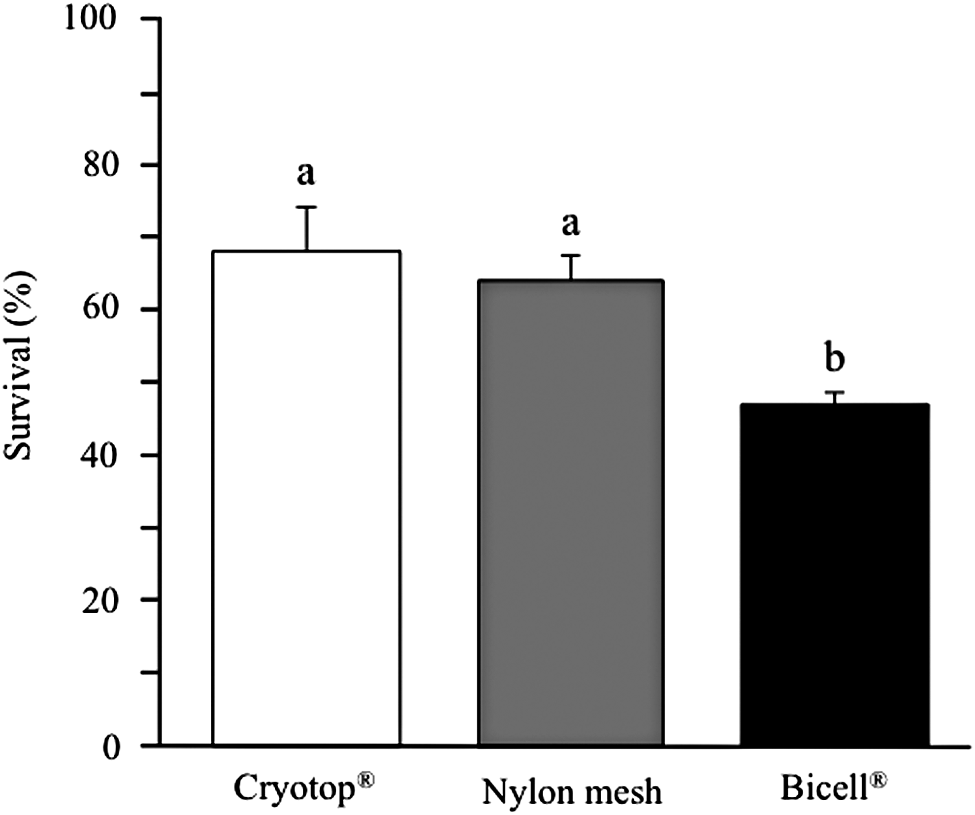
Survival of islets cryopreserved by Cryotop® vitrification, nylon mesh vitrification, or Bicell® freezing. Data are expressed as the mean ± SEM of six replicates in each group. a,bDifferent superscripts represent significantly different groups (p < 0.05). SEM, standard error of the mean.
Data are expressed as the mean ± SEM of six replicates in each group.
Different superscripts represent significantly different groups (p < 0.05).
SEM, standard error of the mean.
Sample size effect on islet revivability following nylon mesh vitrification
Both the exposure of islets to VS before cooling and the stepwise CPA dilution of postwarm islets were achieved without using glass capillaries to maintain a tight time schedule for each step (Fig. 1). Almost all islets were recovered after nylon mesh vitrification/warming in a unit of 10 islets (100%, 160/160), 50 islets (99.3%, 397/400), and 100 islets (98.9%, 791/800). Small debris located in the periphery of postwarm islets after CPA dilution (Fig. 3) disappeared within a few hours of recovery culture. When the number of islets loaded onto a nylon mesh device increased from 10 to 50 or 100, the survival rate of vitrified–warmed islets did not change significantly, as shown in Figure 4. In addition, the number of samples per device did not affect glucose-stimulated insulin secretion in the postwarm islets, which was within the normal range (SI 2.8–4.2), as shown in Table 2.

Morphology of rat islets vitrified–warmed using a nylon mesh device versus frozen–thawed using Bicell vessel.
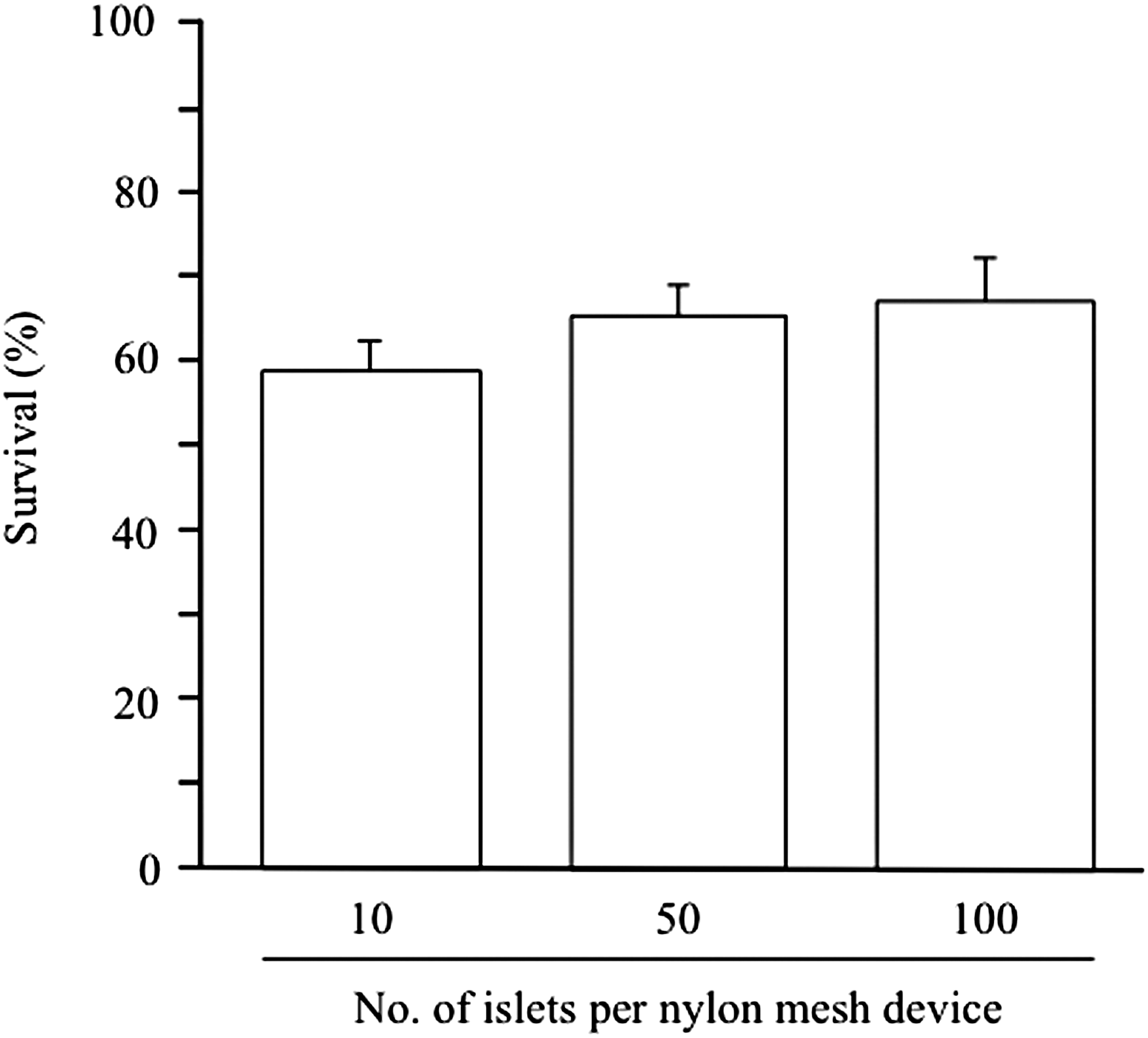
Effect of sample size per nylon mesh device on the survival of vitrified–warmed rat islets. Data are expressed as the mean ± SEM of eight replicates in each group. No significant differences were detected among three groups.
Data are expressed as the mean ± SEM of eight replicates in each group.
No significant differences were detected among three groups.
Discussion
Using nylon mesh devices for the vitrification of rat pancreatic islets overcomes the technical scale limit faced by some cryodevices during islet cryopreservation.12–14 We successfully used the same pre- and postvitrification CPA treatments reported previously for Cryotop vitrification 12 during nylon mesh vitrification, as assessed by cryosurvival and glucose-stimulated insulin secretion (Fig. 2; Table 1). The Cryotop vitrification protocol is recommended over the Bicell freezing protocol, 12 despite the injurious aspect of highly concentrated CPAs. However, the Cryotop vitrification protocol may be unsuitable for large-scale operation (>30 islets per device) because necessary treatments—such as CPA equilibration, VS exposure, VS minimization, and CPA dilution—are difficult to achieve within the required time frame. Dysfunctional islets were recovered from conventional Bicell freezing, despite the capacity for cryopreservation of large quantities of pancreatic islets, as reported previously.12–14
Practically acceptable levels of cryosurvival and SI were obtained after scaling up to 100 islets per nylon mesh device (Fig. 4; Table 2). This suggests that the nylon mesh device offers promising advantages, including easy handling of large quantities of islets and minimization of VS volume for ultrarapid cooling/warming (Fig. 1). Effectiveness of islets cryopreserved for the longer period of time remained to be examined from a viewpoint of practical importance. The solid surface vitrification (SSV) protocol, which was originally developed in bovine COCs, 17 allows cryopreservation of large quantities of islets (100 islets per 40-μL microdrop). 13 However, islet handling with glass capillaries may be time-consuming and labor intensive. The hollow fiber vitrification (HFV) protocol has been used for mouse, porcine, and bovine embryos18–20 and for mouse islets. 14 The HFV protocol simplifies the processes of CPA addition/dilution because of the microdialyzable characteristics of the hollow fiber device and has been applied to a maximum of 40 mouse embryos 18 or 25–35 islets 14 in a single device.
As in previous reports, we used membrane integrity-based measurement following FDA/PI double staining to quantify the morphological survival of islet preparations.21–23 However, a methodological limitation of this assay (two-dimensional analysis of three-dimensional structure) has been raised.7,24 Therefore, glucose-stimulated insulin secretion analysis was also performed to evaluate the function of cryopreserved islets, and the SI values (20 mM/3 mM glucose) of rat islets vitrified with nylon mesh device ranged from 2.8 to 4.2 (Tables 1 and 2). Sasamoto et al. 13 reported that the SI value of vitrified rat islets in the original SSV protocol was only 1.1 and that modification in the VS composition (EDT324) increased the SI value up to 6.4. In addition, Nagaya et al. 14 failed to demonstrate the SSV suitability with the EDT324 solution for mouse islets, but reported a SI value (28 mM/2.8 mM glucose) of 3.5 when the HFV protocol was applied.
There are no distinct SI criteria to distinguish functional and dysfunctional islets, because the glucose concentration used for stimulation varies from 15 to 33 mM. Nevertheless, islets with a SI value of larger than 3 have been empirically regarded as those maintaining functional insulin secretion and are recommended for transplantation. 25 The islet insulin secretion potential measured in vitro is closely related to the in vivo response for glycemic control.7,13 Increasing the islet number per nylon mesh device (from 10 to 100) resulted in a mild increase in cryosurvival (58.6%–68.5%) and a decrease in SI (4.2 to 2.8), requiring IEQ-based further investigation for the presence and significance of a negative correlation.
In conclusion, nylon mesh can serve as cryodevice for vitrification of large quantities of rat pancreatic islets. Such nylon mesh vitrification is practically advantageous because islets can be handled for CPA addition/dilution without glass capillary micropipetting.
Footnotes
Acknowledgments
This work was supported, in part, by a Grant-in-Aid for Scientific Research from Japan Society for the promotion of Science (JSPS; 16K07985 to S.H.) and a grant of Leading Advanced Projects for Medical Innovation from Japan Agency for Medical Research and Development (LEAP/AMED; to M.H.).
Author Disclosure Statement
No conflicting financial interests exist.