
Abstract
Introduction:
Cryopreservation provides an efficient way to preserve cells for a broad range of medical applications, including cell therapy. In clinical practice, cells are frozen in solutions containing dimethyl sulfoxide (DMSO) cryoprotectant agents (CPAs) to reduce their damage during the cooling process. In the current cell preservation methods, polysaccharides such as dextran, a nonpenetrating CPA, are used. However, the cell viability decreases when the solution concentration in polysaccharides increases.
Materials and Methods:
To overcome this limitation, we have developed a dextran-based hydrogel (PSH) as a new CPA. Three molecular weight PSHs (PSH40, PSH70, and PSH500) were synthesized. The physicochemical characteristics of PSHs were studied. Then, their biocompatibility properties were studied in vitro in BALB/c 3T3 cells according to ISO standard 10993-5/12. Crystallization temperature (Tc), that is, ice-crystal formation, was determined using the thermocouple method. Finally, PSHs were used as CPAs in a slow freezing procedure of BALB/c 3T3 cells with Voluven® (Fresenius Kabi, Sèvres, France), and were compared with the DMSO procedure.
Results:
Our results showed that PSHs were biocompatible and did not modify the osmolality of the Voluven cryopreservation solution. PSHs decreased the Tc when compared with the DMSO procedure. Furthermore, without adding DMSO, PSH500 cryopreserved the viability of BALB/c 3T3 cells, and the result was similar to that of the control conditions.
Conclusion:
PSH500 could represent an alternative to DMSO. It could be used as a new medical device while avoiding DMSO side effects on patients.
Introduction
C
To reduce damage from freezing, CPAs are required. They lower the solution freezing point, decrease the effect of the cryopreservation medium itself, and reduce ice-crystal formation, thus limiting cell damage. 8 Two types of CPAs can be used, the intra- and extracellular CPAs. 9 The most clinically used intracellular CPA is dimethyl sulfoxide (DMSO).4,10 However, several issues have been identified including cell toxicity when it is used at high concentrations.11,12 When the cell solution injection containing DMSO is directly grafted in a patient, adverse effects may occur, ranging from headaches, nausea to cardiac and/or respiratory arrest.13–16 Among nonpenetrating CPAs, polysaccharides are used to increase extracellular osmotic pressure, regulate cell dehydration during cooling, limit extracellular crystal formation by increasing the viscosity, and prevent rapid rehydration during warming. 17 Dextran, a clinical-grade biocompatible polysaccharide, was added to commercial hypothermic or freezing solutions such as Perfadex® or CryoSolve™.17–19 However, the cell viability decreases for dextran concentrations higher than 400 nM in solutions.
Hydrogels are 3D networks obtained after chemical or physical crosslinking. Among them, hydrophilic polymers can swell and retain a large amount of water, release drugs, and act as cell carriers or scaffolds for tissue engineering.20–27 Hydrogel properties depend on their chemical structure conferred by the molecular weight (MW) of the polymer used, as well as the choice of the crosslinking agent. 28 Thanks to their properties, hydrogels are widely used in different applications, including cell encapsulation, drug-controlled release, and plaster in skin wound repair.
In previous studies, we have developed a dextran-based hydrogel (PSH) for cell and vascular tissue engineering.25,27 We have shown that PSHs were biocompatible, biodegradable in vitro and in vivo, and could act as a drug delivery platform.22,25,27 Based on these results and to overcome the limitation of polysaccharides in the freezing solutions, we have developed new PSHs suitable for cell and tissue cryopreservation.
As the final purpose was to develop a new clinical device, we decided to use only clinical- or pharmaceutical-grade dextran in the synthesis. In this study, we synthesized three PSHs based on 40 kDa (PSH40), 70 kDa (PSH70), and 500 kDa (PSH500) MW dextran. We studied their physicochemical characteristics and biocompatibility properties according to ISO standards 10993-5 and 10993-12.29,30 We then determined the crystallization temperature, that is, the ice-crystal formation of PSHs using the thermocouple method, with Voluven® (Fresenius Kabi, Sèvres, France), a clinical freezing solution containing 6% hydroxyethyl starch in a sodium chloride solution (0.9%) and 0% proteins. Finally, PSHs were used as CPAs both in the absence and in the presence of DMSO in the slow freezing procedure of BALB/c 3T3 cells.
Materials
Cell culture
Cell culture plastics and cryotubes were obtained from Corning (Fontenay-Sous-bois, France). Dulbecco's modified Eagle's medium (DMEM), minimum essential medium alpha, phosphate-buffered saline (PBS), Hank's balanced salt solution, fetal calf serum (FCS), antibiotic-antimycotic solution, and trypsin-EDTA were purchased from Gibco by Life Technologies (Carlsbad, CA). Trypan blue was purchased from Sigma (St. Louis, MO). DMSO was purchased from WAK-Chemie (Steinbach, Germany). Saline solution (NaCl 0.9%) was obtained from Laboratoire Aguettant (Lyon, France). Voluven was purchased from Fresenius (Kabi, France SA).
Dextran-based hydrogel synthesis
Pharmaceutical-grade dextrans of different MWs (40, 70, and 500 kDa) were obtained from Pharmacosmos (Holbaek, Denmark). The other chemical products were obtained from Sigma.
Methods
PSH powder synthesis and characterization
PSH production
The PSH was prepared as previously described.24,25 First, polysaccharides were dissolved in water. The chemical crosslinking of polysaccharides was carried out in sodium hydroxide using the crosslinking agent, trisodium trimetaphosphate (STMP), at 25% (w/w, Sigma). The polysaccharides mixed with STMP were incubated at 50°C for 1 hour. The resulting PSH was washed thoroughly with PBS (pH 7.4) and crushed with a grinder (Grindomix, Retsch, France). It was dehydrated using ethanol/water baths and dried in an oven to constant weight. After a sieving step (AS 200, Retsch), the PSH (250–500 μm granulometry) was sterilized under UV light and kept at room temperature.
For this study, three PSHs were synthesized from dextrans with clinical-grade MWs of 40, 70, and 500 kDa, and named, respectively, PSH40, PSH70, and PSH500.
PSH phosphate content
As previously described, 50 mg of PSH powders (n = 9) was heated in 10% nitric solution at 105°C.24,25 Solutions of ammonium metavanadate and ammonium heptamolybdate were then added to the dissolved PSH. Results were read at 405 nm absorbance with a spectrophotometer (Infinite M200 Pro, Tecan) and compared with the phosphate calibration curve to determine the phosphate concentration.
PSH swelling ratio determination
For the swelling ratio (SR), 10 mg of PSH40, PSH70, or PSH500 (n = 9) was swollen in 1 mL of Voluven with 0%, 2%, 5%, and 10% DMSO, or in saline solution at room temperature for 1 hour to obtain a final concentration of 10 mg/mL. The excess solution was then removed, and the SR was calculated using the following formula:
PSH osmolality
PSH solution's osmolality (n = 3) was measured using a Micro-Osmometer Type 6® (Löser Messentechnik, Berlin) according to the manufacturer's protocol. PSHs were swelled in Voluven or Voluven/DMSO (10 mg/mL), and the supernatant's osmolality was measured after 1 hour at room temperature.
Particle size distribution in swollen PSH
The size distribution of PSH was determined by the light scattering technique using laser diffraction in a Mastersizer 3000 (Malvern). PSH (5 mg/mL, n = 3) was swollen in saline solution (NaCl 0.9%) or in Voluven, and added to the dispersion unit of the Mastersizer® as described by the manufacturer.
Crystallization temperature determination
PSH powders were swollen in Voluven (10, 20, or 40 mg/mL concentration) without or with 2%, 5%, or 10% of DMSO (n = 5). Temperature probes of a thermocouple (Multi-Channel Thermometer, Consort T851; Bioblock Scientific, Illkirch, France) were introduced in each sample. The tubes containing the probes were then placed in an automatic freezer (Freezal, Air Liquide, Courbevoie, France) set at a cooling rate of 2°C/min from 10°C to −40°C. The temperature decrease was automatically monitored until it reached −40°C. This reaction was exothermic, and the crystallization temperature (Tc) was the coldest temperature measured before the supercooling peak.
In vitro PSH evaluation
Cell culture
BALB/c 3T3 clone A31 cells (ATCC) were plated in a 75°cm2 culture flask at 5 × 10³ cells/cm2, cultured in DMEM supplemented with 10% FCS (v/v) and 0.1% antibiotic-antimycotic (v/v) (complete medium [CM]) at 37°C, 5% CO2, and changed every 2 days.
Biocompatibility of PSH determined by MTT assay
For this assay, 5 × 10³ cells/cm2 BALB/c 3T3 clone A31 cells (ATTC, CCL-163™) were seeded in a 48-well plate in CM until 80% cell confluency. PSH extracts were prepared according to ISO 10993-5 and 10993-12 standards. 29 PSHs (200 mg/mL, n = 6) were shortly incubated in CM for 24 hours in a humidified atmosphere containing 5% CO2 at 37°C. The supernatants were then recovered (pure extract or 100%) or diluted in complete culture medium (12.5% final concentration), according to ISO 10993-5 and 10993-12 standards, and added to the BALB/c 3T3 cell well plate. After 24 hours in culture, supernatants were removed, and the cells were washed twice using PBS. The evaluation of the cell viability was then carried out using thiazolyl blue tetrazolium bromide assay (MTT; Sigma) as recommended by the manufacturer. CM and CM containing 10% DMSO were used as a positive or negative control. The cell viability was calculated as the percentage ratio of cell viability versus the positive control condition (CM).
Cell cryopreservation in PSH
BALB/c 3T3 cells, clone A31 (ATTC, CCL-163) were cryopreserved in Voluven containing PSH40, PSH70, and PSH500 in 0%, 2%, 5%, or 10% DMSO. A solution containing 90% FCS and 10% DMSO was used as the control (CFCS). For that purpose, PSH powder was swollen in cryopreservation solutions (12 mg/mL final concentration), and 300,000 BALB/c 3T3 cells were added. For all samples, a slow freezing procedure was used (Freezal, Air Liquide, Courbevoie, France) according to the hospital biobank with a first cooling rate at 2°C/min from 10°C to −5.2°C where an automatic seeding was performed. The second cooling rate was 2°C/min to −40°C. The final rate was 10°C/min to −120°C. At the end of the freezing procedure, the samples were stored in liquid nitrogen.
Cell apoptosis evaluation by Annexin V determination
After 7 days of storage, sample vials were heated in a water bath at 37°C, and the solutions were filtered (40 μm cell strainer; Falcon®; Fontenay-Sous-bois) to separate the cells from the PSH. The BALB/c 3T3 cell viability was then assessed using APC Annexin V Apoptosis flow cytometry Detection Kit with 7-AAD, according to the manufacturer's instructions (Ozyme, Biolegend, Montigny-Le-Bretonneux, France). In brief, after cell centrifugation (1200g, 5 minutes), the cells were incubated with APC Annexin V and 7-AAD in Annexin V Binding Buffer. After 15 minutes of incubation, the cell apoptosis was determined using a LSRII® cytometer (Becton Dickinson, Rungis Complexe, France). For each sample, >10,000 events were analyzed. The analyses were performed using the BD FACSDiva™ (Becton Dickinson) software. Results were expressed as the ratio of the CFCS.
Statistical analysis
The statistical analysis was carried out using one-way analysis of variance, followed by Tukey's and Dunnett's multiple comparison tests. Statistical tests were performed using GraphPad Software (version 6, La Jolla, CA). Differences were considered as being significant when p < 0.05.
Results
PSH synthesis and characterization
The chemical synthesis was carried out using pharmaceutical-grade compounds. Three types of dextran-based hydrogels were obtained, PSH40, PSH70, and PSH500, with a yield of PSH of 80% ± 20% (n = 6 for each composition).
Phosphate content
A significant difference (p < 0.05) was observed between PSH500 and PSH40 (102.8 ± 6.00 μM and 116.6 ± 3.95 μM, respectively), while no significant difference was observed for PSH70 (Table 1).
Dextran-Based Hydrogel Characterizations
PSH40, PSH70, and PSH500 were synthesized from clinical-grade molecular weight dextran of 40, 70, and 500 kDa, respectively, as previously described.24,25 Resulting PSHs were crushed indoor to obtain a powder. PSH phosphate content and PSH swelling in saline solution or in clinical solution, that is, Voluven containing 0%, 2%, 5%, and 10% DMSO, were determined. Results are expressed as mean ± SD (n = 9). Statistical analysis was performed using ANOVA, followed by Tukey's multiple comparison test. *Comparison of phosphate content between PSH40 and PSH500 (p < 0.05); #Comparison of PSHs swelling between Voluven with or without DMSO to saline solution (#p < 0.05; ##p < 0.01; ###p < 0.001).
ANOVA, analysis of variance; DMSO, dimethyl sulfoxide.
PSH swelling ratio and osmolality determination
The swelling of the PSH powder was studied in two different solutions, that is, saline solution and Voluven. As shown in Table 1, no significant difference was found between the PSHs in saline solution. When PSHs were swollen in Voluven, the SR significantly decreased in PSH40 and PSH70 (p < 0.01). No significant difference was observed for PSH500. After adding 2%, 5%, and 10% DMSO in Voluven, the SR was at steady state regardless of the PSH MW. As shown in Table 2, the PSHs did not modify the osmolality. The osmolality of cryopreservation solutions increased significantly (p < 0.05) after adding DMSO and depending on DMSO concentration. Similar results were observed when DMSO was added to the PSH cryopreservation solutions.
Osmolality of Voluven Solution Containing PSH40, PSH70, PSH500 and Increasing Dimethyl Sulfoxide Concentration
Each value represents the mean ± SD (n = 3).
Particle size distribution in swollen PSH powder
Figure 1 shows the particle size distribution of PSH40 (Fig. 1a), PSH70 (Fig. 1b), and PSH500 (Fig. 1c) swollen in saline solution or in Voluven. The lower average size of the particles was between 310 μm (PSH70 and PSH500) and 352 μm (PSH40) in saline solution or in Voluven. In saline solution, 17% of the particles had an average size of 859 μm (PSH40) and 756 μm (PSH70). In Voluven, 19% of the particles had an average size of 756 μm (PSH40) and 666 μm (PSH70). In PSH500, regardless of the solution, 12% of the particles had an average size of 860 μm. For all PSHs, the maximal average size was between 2200 and 3000 μm.

Size distribution of dextran-based hydrogel analyzed using the Malvern Matersizer Microplus laser diffractometer technique. PSH40
Crystallization temperature determination
Table 3 shows the Tc determinations according to the PSHs and their concentrations using the thermocouple method during the slow freezing procedure. Without DMSO, PSHs at a concentration of 20 mg/mL were able to significantly decrease the Tc (−11.14°C ± 1.46°C PSH40, p < 0.01; −7.8°C ± 3.11°C PSH70; −10.26°C ± 2.17°C PSH500, p < 0.05) when compared with the Voluven (−2.47°C ± 0.43°C). A significant decrease of Tc was observed at a PSH concentration of 40 mg/mL. Furthermore, PSH500 in Voluven decreased the Tc without any DMSO addition. To obtain a similar effect on Tc without PSH, the addition of 2% DMSO was needed in the Voluven solution.
Crystallization Temperature (°C) of Voluven Solution Containing PSH40, PSH70, PSH500 and Increasing Dimethyl Sulfoxide Concentration
Each value represents the mean ± SD of five experiments performed once. The results were compared with the control condition; that is, Voluven without PSH and without DMSO (bold values). Statistical analyses were carried out using ANOVA, followed by Dunnett's multiple comparison test (*p < 0.05; **p < 0.01; ***p < 0.001).
Tc, crystallization temperature.
In vitro PSH evaluation
PSH cell biocompatibility
The viability of BALB/C 3T3 cells after 24 hours of culture in the presence of PSH extracts was assessed by measuring the metabolic activity of cells (Fig. 2). As shown in Figure 2, all samples exhibited cell viability between 85% and 100% when compared with CM (control group).
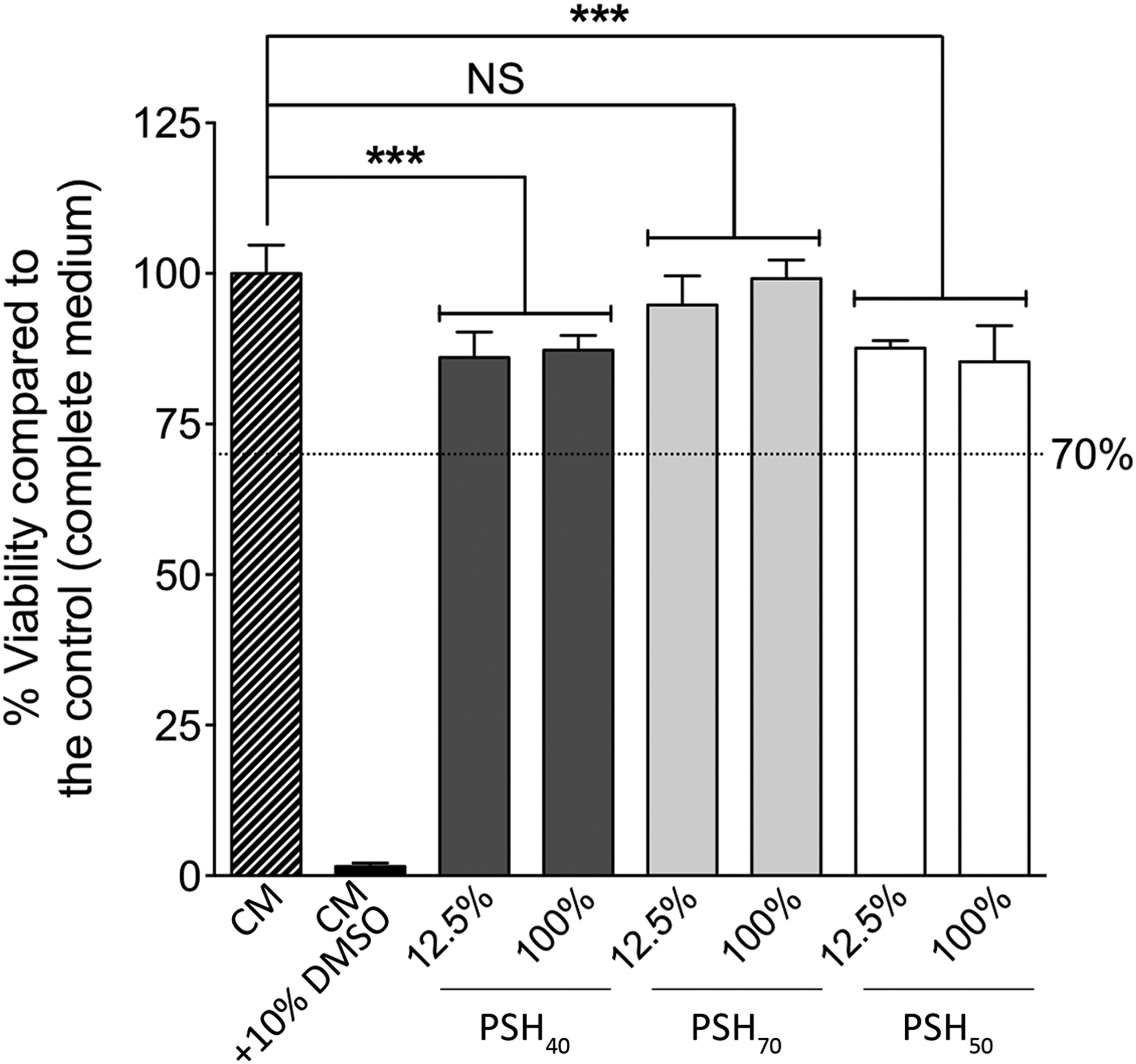
Dextran-based hydrogel BALB/c 3T3 cell biocompatibility. Cytotoxic effects of PSH40 (dark gray bar), PSH70 (clear gray bar), or PSH500 (white bar) of PSH-diluted extracts (12.5%) or PSH pure extracts (100%) were evaluated on BALB/c 3T3, clone A31 cells (ATTC, CCL-163) by MTT assay. Complete culture medium (CM) and CM containing 10% DMSO were used as a positive or negative control, respectively. Results were expressed in percentage of cell viability as compared with the positive control (CM) (n = 6/condition). Statistical analyses were performed using ANOVA, followed by Tukey's multiple comparison test (***p < 0.001, NS). According to the ISO 10993-5 and 10993-12, 29 samples were considered as being biocompatible >70% of viability (dotted line). NS, nonsignificant.
Cell preservation after cryopreservation in PSH
PSH500 used as a CPA without DMSO (Fig. 3a) protected from apoptosis (2.1-fold, (p < 0.001) as compared with Voluven alone. Interestingly, PSH500 preserved cells from apoptosis regardless of DMSO concentration as compared with CFCS (1.20-fold; 0% DMSO [Fig. 3a], 1.19; 2% DMSO [Fig. 3b], 1.26; 5% DMSO [Fig. 3c] and 1.1; 10% DMSO [Fig. 3d]).
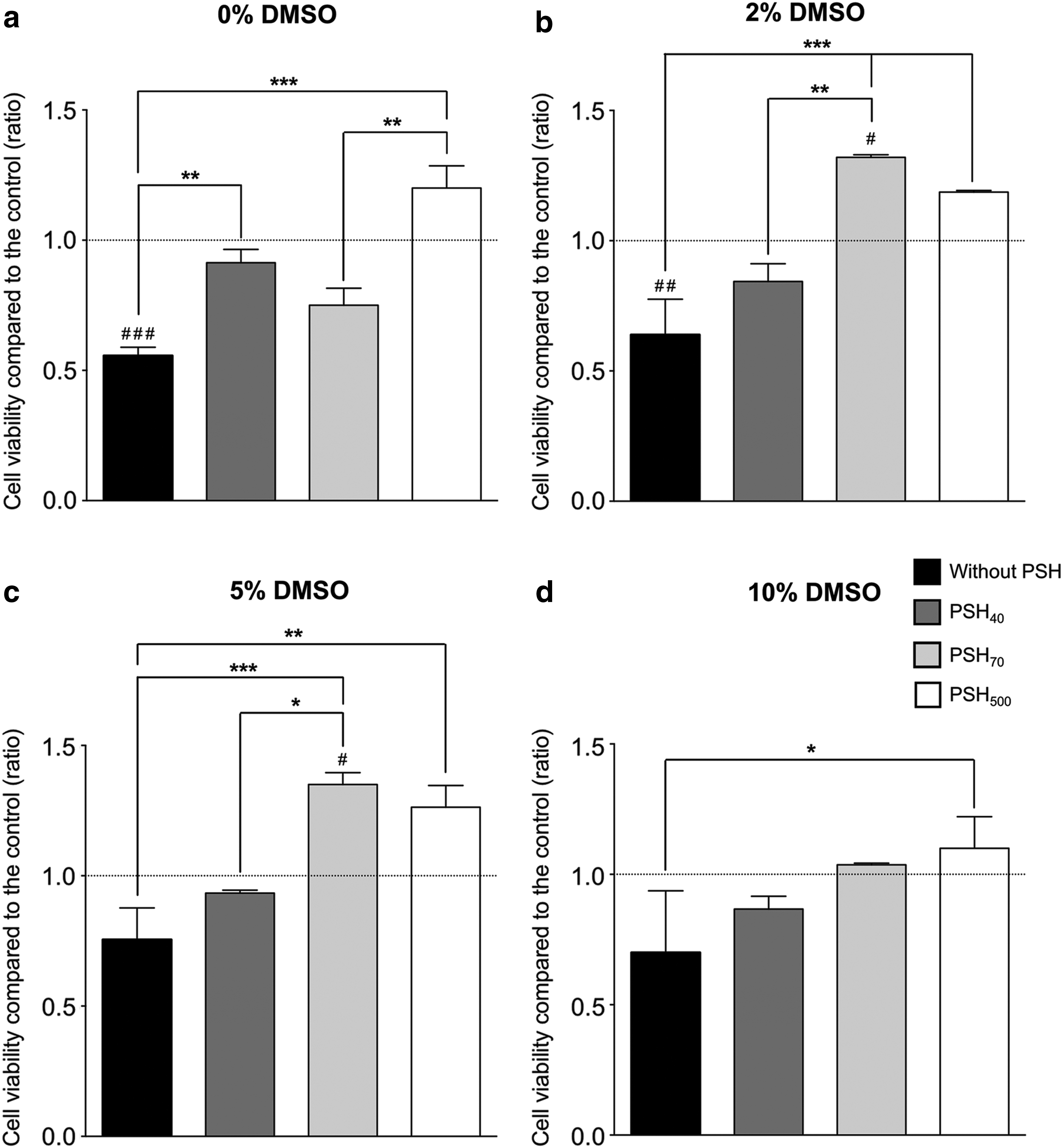
BALB/c 3T3 cell viability after cryopreservation in dextran-based hydrogel. After cryopreservation of BALB/c 3T3 cells in Voluven with PSH40 (dark gray bar), PSH70 (clear gray bar), or PSH500 (white bar) at 40 mg/mL, or without PSH (black bar) and
When PSH40 and PSH70 were used as CPAs without DMSO, cells were protected from apoptosis as compared with Voluven alone (1.6-fold [p < 0.01] and 1.3-fold, respectively). However, without DMSO, PSH40 and PSH70 exhibited lower protection than CFCS.
Adding 2% or 5% DMSO to PSH70 (Fig. 3b, c) increased 1.4-fold the capacities of CPA to preserve cells. No significant difference was observed with PSH40.
Adding 10% of DMSO to PSHs did not modify the cell effects as compared with CFCS.
Discussion
Currently, one of the main challenges of cell therapy and tissue transplantation lies in the quality of sample cryopreservation, which has a significant impact on successful grafting. The end-result of the cryopreservation depends on the CPA, the choice of a freezing protocol, and the selection of the cryopreservation solution. A large number of cryopreservation solutions used in cell therapy have been developed by cell transplant centers or by manufacturers.
Polysaccharides in solution exhibit cryopreservation properties. 31 However, their efficiency depends on their concentration in the solution. The higher the concentration, the lower the cell viability. 4 To overcome this limitation, we have used polysaccharides in a hydrogel form.
During the freezing process, the cells are usually embedded in the gel throughout the gel formation, by either chemical crosslinking or freezing processes.32–34 One limitation was represented by the production of uniform, reproducible, and stable capsules or a cellularized hydrogel without cell membrane damage. 32 As reported in the literature, if cell separation from gel was needed, a gel enzymatic digestion step was performed before use, and could decrease the cell viability.35–38 Thanks to our process, cells could be easily retrieved by adding medium to the sample, dispersing them from the gel, as they were not embedded in a hydrogel capsule but surrounded by a swelling powder. Since the swelling gels exhibited a low gel particle size distribution (between 310 and 352 μm), a single filtration using a cell strainer (<200 μm) can be used to separate the cells from the gel, avoiding a centrifugation step and therefore decreasing cell damage. Furthermore, the limitation presented by the production of a uniform, reproducible, and stable capsule or a cellularized hydrogel without cell membrane damage during the process was overcome.32,33,37,38
In this study, we chose a slow freezing procedure for the cell therapy cryopreservation according to the Saint Louis Hospital Biobank procedures. This clinical standard protocol allowed the cryopreservation of a large volume of cells ranging from 20 to 150 mL requested for cell therapy, and was not time-consuming for the operator.
The osmolality of the solution is a key point in freezing because it could increase during ice-crystal formation and damage the cell by fast dehydration. 8 The osmolality can be influenced by both the gel type and the freezing medium. To be able to set the pharmaceutical composition of the freezing solution, we used Voluven, a clinical cryopreservation solution.39–41 Voluven meets the requirements of the U.S. Food and Drug Administration, and is compliant with European regulations. It does not contain any DMSO or CPA, and exhibits an osmolality similar to blood serum.40,42 In this study, we have shown that in Voluven, the osmolality of PSHs remains in the physiological range. As already observed, the osmolality of the solution containing PSHs increases if DMSO is added. Adding PSH40, PSH70, or PSH500 did not influence the osmolality of the solution. Furthermore, thanks to their swelling properties, PSHs may act like a sponge, allowing a slow rehydration and preventing hyperosmolality post-thawing, which is harmful to the cell.44,45 As observed, all PSHs were able to swell in the cryopreservation solution. Hydrogel physical properties depend on their components and the concentration of the crosslinking agent during synthesis.28,46,47 Because the phosphate contents were similar in all PSHs, the differences observed in the swelling properties were related to the MWs of the PSHs. As observed with other gels, this could be explained by the spatial rearrangement linked to the configuration and the length of the polymer chains.48,49
CPAs and polymers decreased the Tc, that is, the temperature of the first ice-crystal formation.50–52 Evaluating the effect of PSHs on Tc values could help determine the optimal cryopreservation solution. Adding DMSO to PSH40 and PSH70 was needed to reach the Tc of the Voluven/DMSO solution. DMSO is the most clinically used CPA despite its side effects.41,43,53 Reducing the use of DMSO is still challenging, and several polymer solutions such as polyvinylpyrrolidone have been developed for that purpose. 54 Interestingly, our results have shown that PSH500 alone (40 mg/mL) decreases Tc similarly to the Voluven/2%–10% DMSO solutions.
Given that CPAs have been classified as medical devices, we performed biocompatibility studies according to EN ISO 10993-5 and 10993-12 standards.29,30 As observed, all PSHs were biocompatible, and the viability was >70% with pure extracts (87.27% ± 2.44%, 99.16% ± 3.06%, and 85.31% ± 6.00% for PSH40, PSH70, and PSH500, respectively).
Based on these results, we compared the effects of PSHs (40 mg/mL) on BALB/c 3T3 cell viability after cryopreservation under standard conditions. Remarkably, PSH500 without any DMSO addition cryopreserved the BALB/C 3T3 cell viability similarly to the standard procedure. Furthermore, the cryopreservation solution (PSH500-Voluven) did not contain any FCS or human albumin, preventing the risks associated with human or animal by-products.
Numerous works have shown that polymers' efficiency in cryopreservation was related to the MW. An increase in MW increased the cryoprotective effect. 55 Wang et al. showed that dextran 40 kDa incorporated into an alginate/gelatin hydrogel could have an effect on the cryopreservation of adipose stem cells by reducing the DMSO concentration. This effect goes through the increase in viscosity, and the reduction of the ice nucleation and crystal formation rate. 56 Our findings were consistent with this, because the addition of at least 2% DMSO was required when freezing in the presence of PSH40 or PSH70. An increase in the dextran MW avoided DMSO addition, preventing cell membrane damage. 57
Conclusion
In conclusion, PSH500 seems to be an alternative to DMSO for cell preservation and could be used as a new medical device. In addition, PSH500 gel is reliable, and could be easily produced on a large scale at low cost. No new equipment or specific personnel training is required for cell cryopreservation. Further studies are in progress to better understand the cryoprotective effects of PSHs and their potential use in the long-term storage of cells.
Footnotes
Acknowledgments
The authors thank the Agence de Biomédecine and the SATT Ile-de-France for their financial support, as well as Dr. Anne Baudot and Pr Gerard Louis, Inserm 1148, Paris-Diderot University, for allowing them to use the thermocouple instruments. They are grateful to Graciela Pavon-Djavid and Aicha Chouiakh for the discussions and the reading of this article.
Author Disclosure Statement
No conflicting financial interests exist.