




Abstract
Leukocytes function as central effectors in innate immunity (such as phagocytosis) as well as adaptive immunity (e.g., antigen-dependent T cell activation), and serve as an important resource in the fields of translational medicine, precision medicine, and cell therapy. Isolation of leukocytes from whole blood is necessary for high-quality RNA and downstream research. This process is susceptible to the variability of many factors, such as blood collection, isolation reagents, and extraction methods. In this study, three methods were applied for leukocytes separation, followed by RNA extraction and quality testing to evaluate the methods. Results showed that leukocytes were purified using lymphocyte separation medium (LSM), optimized LSM method, or red blood cell lysis buffer (RBC lysis), and RNA quality met the basic requirements for downstream studies. Although considering the simplicity of the procedure and RNA quality from donated samples, the RBC lysis method should be recommended to biobanks for further research.
Introduction
O
Leukocytes are found throughout the body, and are derived from multipotent cells in the bone marrow known as hematopoietic stem cells. All leukocytes contain nuclei, which distinguish them from the nuclei-free cells of blood (red blood cells and platelets). Past studies in multiple systems have revealed functions of leukocytes in cell movement such as migration,1–3 rolling,4–6 and immune system functions such as immune activation7–10 and downstream effector response.11–14 Extracellular vesicles include apoptotic bodies, exosomes, and microvesicles, which play important roles in disease as well as the therapeutic candidate of tumors. Circulating vesicles released from leukocytes contain potentially valuable biological information for biomarker discovery in primary and secondary prevention of clinical disease.1,15,16 Also, baseline and post-treatment changes in leukocytes, including lymphocytes, eosinophils, neutrophils, neutrophil to lymphocyte ratio, and monocytes counts, are promising to become routinely available blood markers in therapy.17–19 Thus, leukocytes are of great value for clinical research.
Preservation of leukocytes is an important process in biobanking activity and biospecimen preservation. RNA quality tests are important to evaluate the quality of leukocytes. In this study, we developed four methods to purify leukocytes and performed quality qualification, in which direct centrifugation, lymphocyte separation medium (LSM), optimized LSM, and red blood cell lysis buffer (RBC lysis) were used. The results showed that high-quality RNA could be obtained with the LSM, optimized LSM, and RBC lysis methods, with the goal to develop the optimal method for leukocytes isolation and preservation using evidence-based practices in this biobanking activity. We found that the RBC lysis method was the best method in terms of practical applications.
Materials and Methods
Informed consent
Signed consent was provided by all the donors for research use of their blood samples.
Blood sampling
Whole blood was collected into EDTA anticoagulation tubes. All tubes were rotated vertically and gently after blood collection for 2 minutes. Four different methods were used for leukocytes purification. The cell pellet was first resuspended in 1 mL of TRIzol. Figure 1 shows the flowchart.

Schematic of sampling and processing protocol. Whole blood was collected in EDTA anticoagulation tubes. Four methods were used to separate leukocytes. RNA quality was tested after freezer storage.
Method 1: direct centrifugation
Whole blood was fractionated by centrifuging at 900 g for 5 minutes at room temperature. Using a transfer pipette, the plasma layer was aspirated down to approximately within 2 mm from the buffy coat layer, while not disturbing the leukocytes layer. Then the buffy coat layer was aspirated (usually contains leukocytes and a low proportion of plasma and red blood cells), followed by aliquoting the leukocytes and placement into collection tubes. Five conditions were tested for long-term storage. (A) Transfer the leukocytes directly to −80°C freezer. (B) Place the leukocytes tube into liquid nitrogen and transfer to a −80°C freezer. (C) Wash the leukocytes with phosphate buffered solution, place the leukocytes tube into liquid nitrogen, and transfer to a −80°C freezer. (D) Wash the leukocytes with phosphate buffered solution, suspend in TRIzol reagent, and transfer to a −80°C freezer. (E) Suspend in RNAlater reagent, rotated gently at 4°C overnight, wash the leukocytes with phosphate buffered solution for three times, and transfer to a −80°C freezer.
Method 2: LSM (Sigma number 10771)
First bring the medium to room temperature and add 5 mL medium to the bottom of the tube. Carefully add 5 mL of whole blood onto the medium, then centrifuge 400 g for 30 minutes at room temperature. After centrifugation, carefully aspirate the upper layer and transfer the opaque interface. Wash the cells by adding 10 mL of phosphate buffered solution and mix gently. Centrifuge the mixture at 250 g for 10 minutes. Resuspend the cell pellet with 5 mL of phosphate buffered solution and centrifuge at 250 g for 10 minutes. Repeat washing of the cells and transfer to a −80°C freezer.
Method 3: optimized LSM (Sigma number 10771)
Centrifuge at 800 g for 20 minutes at 4°C using 4 mL of whole blood. Aspirate the plasma layer and add 4 mL of phosphate buffered solution and mix gently. Add 4 mL medium to the bottom of tube. Carefully add 4 mL of blood-phosphate buffered solution mixture onto the medium, then centrifuge at 400 g for 30 minutes at room temperature. After centrifugation, carefully aspirate the upper layer and transfer the opaque interface. Wash the cells by adding 6 mL of phosphate buffered solution and mix gently. Centrifuge at 300 g for 5 minutes. Resuspend the cell pellet with 5 mL of phosphate buffered solution and centrifuge at 300 g for 5 minutes. Repeat washing of the cells and resuspend cell pellet in 1 mL of TRIzol and transfer to a −80°C freezer.
Method 4: RBC lysis
Centrifuge at 800 g for 20 minutes using whole blood. Carefully aspirate 300 μL of leukocytes and add 1 mL RBC lysis buffer. Mix gently for 10 minutes. Centrifuge at 400 g for 5 minutes. Aspirate the supernatant and add 1 mL of RBC lysis buffer. Mix gently and centrifuge at 400 g for 5 minutes. Aspirate the supernatant and add 1 mL of phosphate buffered solution, mix gently, and centrifuge at 400 g for 5 minutes. Repeat once and transfer to −80°C freezer.
RNA extraction
RNA preparation using TRIzol
In brief, add 0.2 mL of chloroform to 1 mL of TRIzol and shake the tube by hand for 15 seconds. Centrifuge at 12,000 g for 15 minutes at 4°C. Place the aqueous phase into a new tube and add 0.5 mL of 100% isopropanol. Place at −80°C for 30 minutes and centrifuge at 12,000 g for 15 minutes. Remove the supernatant and leave the RNA pellet. Wash the pellet with 1 mL of 75% ethanol and centrifuge at 7500 g for 5 minutes at 4°C. Remove the supernatant, air dry the pellet for 5 minutes, and resuspend the RNA pellet into RNase-free water.
RNA quality analysis
The concentration of RNA was determined by Nano-Drop (Thermo Scientific) and integrity of 100–200 ng RNA was determined by an Agilent 2100 bioanalyzer (Agilent Technology) and accompanied software.
All operations were implemented according to the manufacturer's instructions.
Results
To determine the best conditions for leukocytes isolation and storage, we developed four methods for leukocytes preparation followed by RNA extraction for quality testing. The ratio of 28S/18S and RNA integrity were measured to evaluate the quality.
First, method 1 of direct centrifugation was used for leukocytes isolation. Five conditions were implemented to isolate leukocytes. Total RNA was prepared for integrity testing to estimate the effect of leukocytes purification. Low RNA integrity number (RIN) values were observed under these conditions (Fig. 2), which indicated that RNA quality did not meet the need for downstream research.
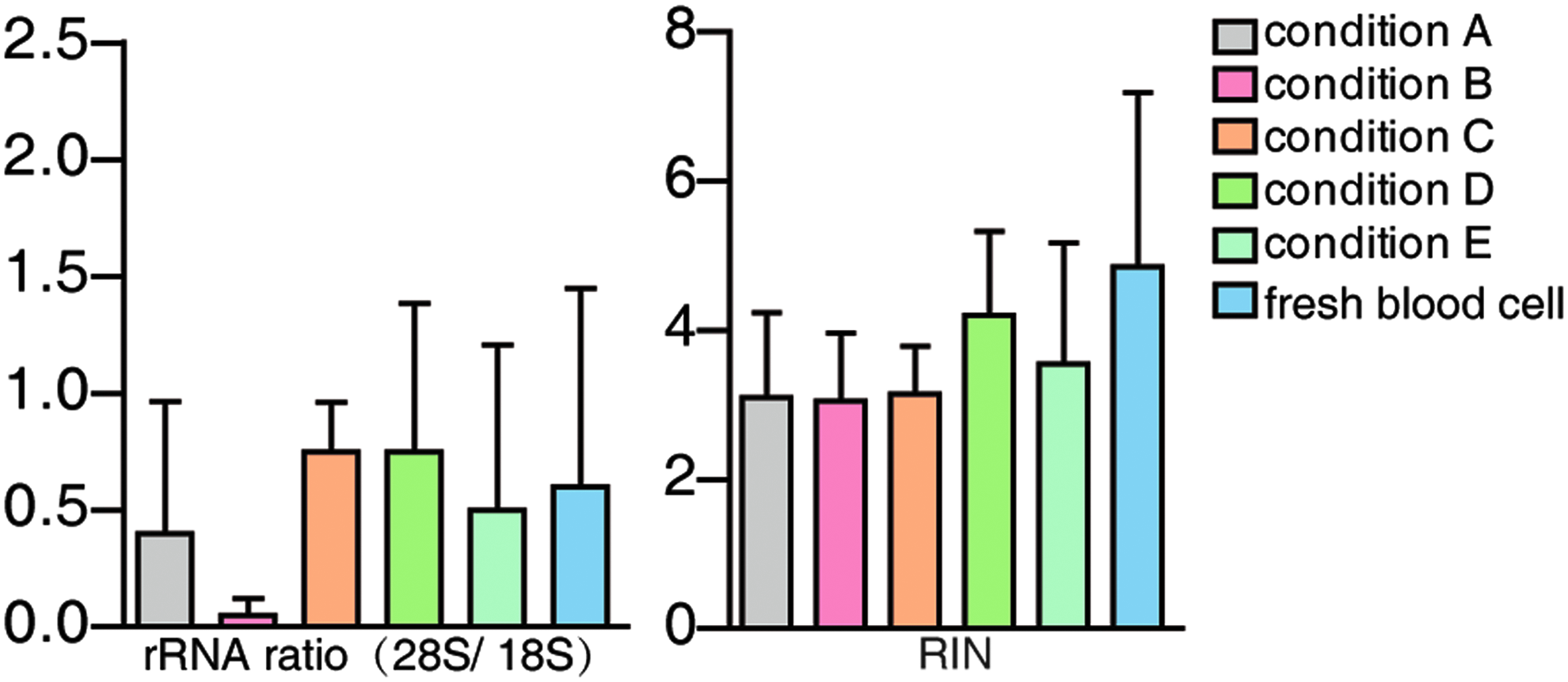
Assessment of RNA quality by direct centrifugation method. Left panel: rRNA ratio test, 28S/18S. Right panel: RNA integrity test, RIN. All values are shown as the mean ± SD. RIN, RNA integrity number; SD, standard deviation.
To better purify leukocytes, three methods were developed. Method 2 was more effective among the leukocytes separation methods. Compared with method 1, the mean value of RIN was >9, which indicated that relatively purified leukocytes were obtained (Fig. 3). Although leukocytes were more purified, method 2 was more time consuming. To obtain plasma and optimize the procedure for clinical research, we developed method 3, in which the time of centrifugation and the volume of wash buffer were decreased. Similar results were observed (Fig. 4). The mean value of RIN was >8.9, which was a higher value than the LSM method. Moreover, we developed a new method (RBC lysis) to purify leukocytes as described in method 4 (Fig. 5). The mean value of RIN was >8.2, and this method produced similar results to optimized LSM method. Moreover, compared with methods 2 and 3, this method consumed less time. Finally, in the clinical validation of our method 4, we chose 18 random persons, of which the average age was >65 years. In our previous study, it was difficult to purify high-quality leukocytes from older people. The results showed that high-quality RNA was obtained, which indicated this method could be used for leukocytes separation (Fig. 6).
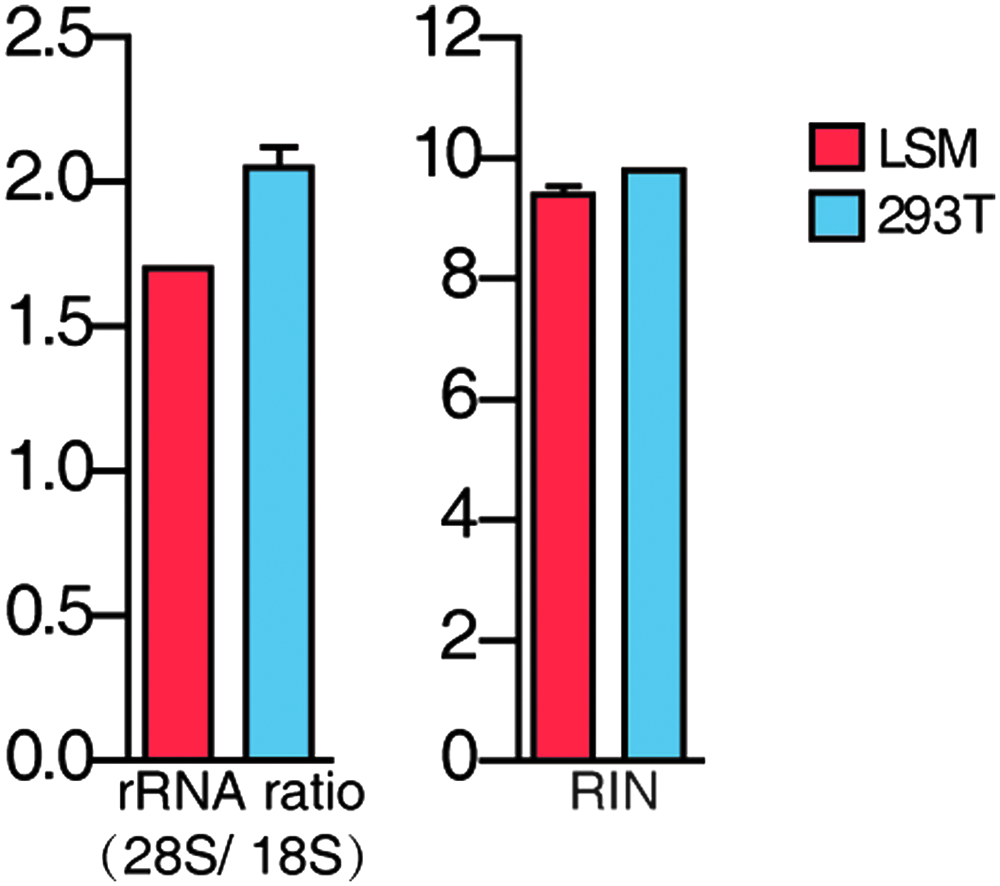
Assessment of RNA quality by LSM method. Left panel: rRNA ratio test, 28S/18S. Right panel: RNA integrity test, RIN. All values are shown as the mean ± SD. RNA extraction from 293T cells as positive control. LSM, lymphocyte separation medium.
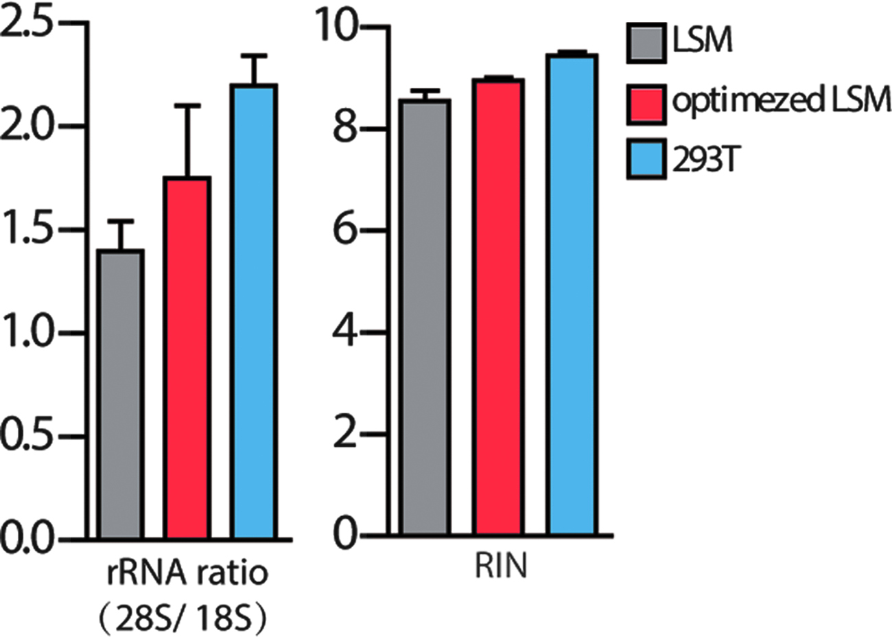
Assessment of RNA quality by optimized LSM method. Left panel: rRNA ratio test, 28S/18S. Right panel: RNA integrity test, RIN. All values are shown as the mean ± SD. 293T cells as positive control. RNA extraction from 293T cells as positive control.
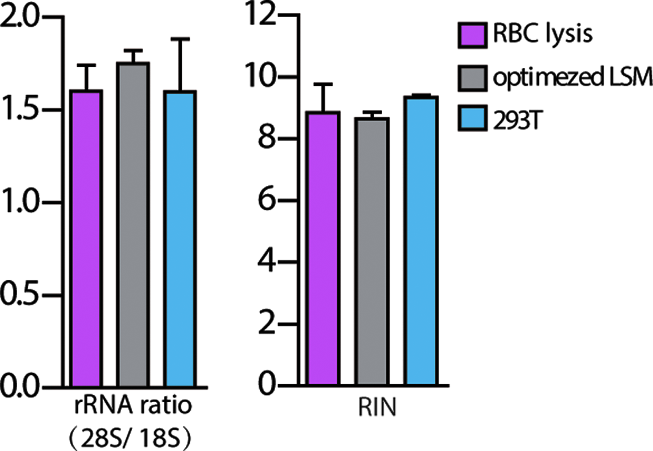
Assessment of RNA quality by RBC lysis method. Left panel: rRNA ratio test, 28S/18S. Right panel: RNA integrity test, RIN. All values are shown as the mean ± SD. RNA extraction from 293T cells as positive control. 293T cells as positive control. RBC lysis, red blood cell lysis buffer.

Validation of RBC lysis method. Validation with clinical samples. Left panel: rRNA ratio test, 28S/18S. Right panel: RNA integrity test, RIN. All values are shown as the mean ± SD.
Discussion
All fractions of whole blood, such as plasma, platelet, leukocytes, and red blood cells, can be used for downstream research. Previous studies have indicated that RNA could be stabilized by EDTA. But several investigations have cast doubt on this finding.20–22 Besides the functions of cell migration, intracellular signaling and intercellular communication, leukocytes are important for immune response, 23 vaccine development,24,25 and clinical trials.26,27 As valuable resources, high-quality clinical samples are important for clinical research. So, preservation of high-quality of leukocytes is a significant challenge in biobanking.
In this study, we developed four methods for leukocytes purification, followed by RNA extraction and quality tests to evaluate the purification procedures. Method 1 was the simplest among the four, and five conditions were used for isolation and preservation of leukocytes. The results indicated poor-quality RNA could not meet the need for downstream applications. It is possible that platelets, red blood cells, and leukocytes were centrifuged down together. RNA was contaminated by degraded RNA in the platelet and red blood cells at high centrifugal speed. 28 Next, to remove platelet and red blood cells contamination, low centrifugal speed and commercial reagents were applied. According to the instructions of LSM, the time-cost steps were developed. The mean value of RIN demonstrated that the RNA was sufficient for most experiments. However, leukocytes could be prepared by this method and plasma should also be isolated for other applications. To meet this requirement, the optimized LSM method was developed. Plasma was extracted by centrifugation before leukocytes purification. Plasma could be preserved for further research and the RNA extracted was of high quality. However, although high-quality leukocytes could be obtained by LSM and optimized LSM, these two methods consumed a lot of time. Finally, we developed a new method for leukocytes preparation using RBC lysis. First, plasma was separated for downstream research, and the mean value of RIN by this method was >8, which could be applicable for downstream research. To further confirm this method, we randomly chose 18 older persons for leukocytes purification, for which it is difficult to obtain high-quality leukocytes.
To our surprise, the mean value of rRNA ratio was <2, except for the 293T cells, which served as positive controls. The manipulation was carried out according to the instruction, and the methods used for leukocytes isolation need to be further optimized.
According to the standard operating procedures from the U.S. National Cancer Institute's Biospecimen Research Database, direct centrifugation was the most popular method used by researchers. However, as the results indicated, leukocytes could be contaminated by platelet and red blood cells resulting in poor RNA quality. The LSM method was also applied by other laboratories, but more time consuming was the main problem. This method could not meet the need for large-scale samples application. The RBC lysis method we developed was less time consuming and simple when compared with previous procedures. In addition, the RBC lysis method made leukocytes isolation from large-scale samples much easier.
Also, disadvantages of these methods existed. RNA was the only standard for quality control, but more parameters should be included such as quality of DNA and protein. In our following research, RNA, DNA, and protein analyses will be implemented for quality control to evaluate the final effect of leukocytes purification.
Conclusion
In summary, this study indicated that three methods could be used for leukocytes purification and preservation. Compared with direct centrifugation, LSM, and optimized LSM, RBC lysis is the simplest method and could be applied for large-scale collections of samples. The RNA quality may vary between different samples from different sources. This method allows the proper leukocytes isolation and preservation in biobanking activities.
Footnotes
Acknowledgments
We express our sincere thanks to our colleagues in Shanghai Clinical Research Center. This work was supported by The National Key Research and Development Program of China (program number 2016YF1201804), the Shanghai Science and Technology Innovation Action plan (program number 16DZ0500600), and the Special Development Fund of Shanghai Zhangjiang National Innovation Demonstration Zone (program number 201701-XH-A2023–004).
Author Disclosure Statement
No conflicting financial interests exit.