
Abstract
Background:
The ability to preserve living cells or stem cells is critical for their use in cell therapy, especially for regenerative, reproductive, and transfusion medicine. This article addresses the low survival rates of cells and their loss of function during traditional freezing and banking (cells in a liquid medium with cryoprotectants).
Aim:
In this article, we developed multiple emulsions (water-in-oil-in-water type) for the effective encapsulation and cryopreservation of cells. In multiple emulsions, the oil drops, acting as a protective membrane, contain even smaller water droplets with encapsulated living cells, dispersed in the continuous water phase.
Materials and Methods:
The multiple emulsions with HEK293 cells encapsulated in the internal alginate droplets were successfully prepared in a Couette-Taylor flow biocontactor. The cryoprotectants (sucrose/dimethyl sulfoxide-DMSO) were located within the internal or external or both water phases of the emulsions. Encapsulated and non-encapsulated cells were frozen to −80°C (cooling rate: −1°C/min) and then transferred to liquid nitrogen (−196°C) for 24 hours. The standard rapid warming procedure was applied to thaw samples. Cell proliferation and viability were measured by using the AlamarBlue™ assay after recovery of cells.
Results:
The results showed that the viability of cells encapsulated in the internal droplets of multiple emulsions, and then cryopreserved, was significantly higher, up to 27.9%, than that observed for cells conventionally cryopreserved (non-encapsulated cells in water). Moreover, the effective cell-loaded multiple emulsions-based banking method allowed DMSO—toxic cryoprotectant—to be eliminated from the cryopreservation system.
Conclusion:
The proposed approach of the cryoprotection of cells encapsulated in multiple emulsions could minimize cell damage, degradation, and their loss during freezing
Introduction
In view of the increasing role of human cell-based therapies in modern regenerative, transfusion, and reproductive medicine,1–3 the development of an effective cell banking method that can preserve the function and life of cells, before clinical applications, has become a critical issue. 4 Traditional cryopreservation, which is the most common and economical way to preserve cells, involves freezing cells suspended in a liquid medium with cryoprotective agents (e.g., dimethyl sulfoxide-DMSO), storage in liquid nitrogen, and thawing. However, this form of preservation of biological material is subject to a high risk of irreversible damage to the cells by: (i) formation of ice crystals in the extracellular space and mechanical damage of the cell membrane as a result of crystal growth, (ii) dehydration of the cells by osmotic water transport from the cell to the external environment (exposure of cells to a harmful level of hypertonic salt concentration), and (iii) formation of ice crystals in the intercellular space due to the super-cooling of cells (destruction of cells from the inside).5,6
Currently, the quality and quantity of cells obtained after thawing remains unsatisfactory, despite the availability of various freezing techniques, alternative vitrification and lyophilization, 3 modern laboratory equipment for the preparation, freezing, and storage of cellular material, 7 and the encapsulation of cells before banking in a protective solid or gel capsules 8 and targeted cryopreservation. 9
Coupling microencapsulation with different cryopreservation protocols, such as slow cooling 10 or 3-step slow cooling with controlled ice nucleation, is one of the strategies developed 11 to increase the availability of cells for cell-based therapies. However, the most promising, and most recent proposals, concern carriers in the form of structured core-shell hydrogel microcapsules 12 (formed via double emulsions as templates), including carriers with liquid cores 13 to increase cell viability and specific functions. One novel approach proposes low cryoprotectant vitrification, by combining microencapsulation of cells in core-shell hydrogel constructs, with nanoparticles–mediated nanowarming for successful stem cells cryopreservation. 14
Another approach proposes low cryoprotectant vitrification, by combining double emulsion-templated core-shell hydrogel microcapsules, with nanoparticles–mediated nanowarming for successful stem cells cryopreservation. 14 When it comes to the encapsulation of cells in structured carriers with liquid cores, multiple emulsions may be one such carrier that is suitable for addressing the problem of the low survival rate of cells and their loss of function. 15
Multiple emulsions (Fig. 1) represent hierarchically dispersed liquid systems in which small droplets of an internal phase of one liquid are incorporated within larger drops of another liquid, the membrane phase. Both the internal and membrane phases are dispersed in a continuous external phase. Multiple emulsions may be of the “water-in-oil-in-water” (W/O/W-internal/membrane/external phases) type, in which a W/O emulsion is dispersed as drops in an aqueous phase or the reverse “oil-in-water-in-oil” (O/W/O) type, where an O/W emulsion is dispersed in an oil phase. In this case, the term “multiple emulsions” is interchangeable with the term “double emulsions.”
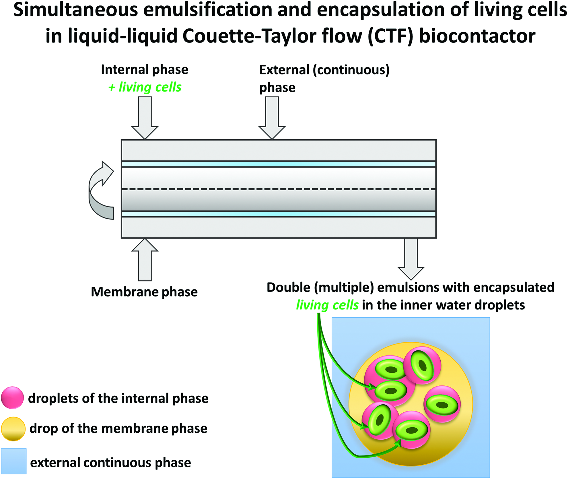
One-step preparation method of double (multiple) emulsion in a Couette-Taylor flow biocontactor and the structure of double emulsion. Color images are available online.
There are also emulsions denoted as W1/O/W2, and O1/W/O2 when the compositions of the internal and external phases are different. Multiple emulsions may also represent higher order dispersed systems, if they consist of more than three phases, for example, W/O/W/O. Multiple emulsions contain more interfaces than simple emulsions; thus, they have a large interfacial area, which significantly influences heat and mass transfer process rates. The importance of multiple emulsions lies in their internal structures, enabling the co-existence of different types of phases, which are able to encapsulate ingredients that are soluble in water and/or in oil. The presence of a membrane phase, between the internal and external phases, protects the encapsulated ingredients from the external environment, and it permits the sustained and controlled release of the encapsulated ingredients. Moreover, the release rates are well determined by the drop sizes and the physicochemical properties of the liquid phases of emulsions. In general, the stability of multiple emulsions is low; therefore, they require the presence of stabilizers and hydrophobic and hydrophilic surfactants.
For practical use, the most promising, to date, have been double emulsions. Multiple emulsions are created by one- or two-step methods. The most common are two-step mechanical agitation, or homogenization. In the first step, simple emulsions are formed, which are then re-emulsified in an oil or water phase, respectively, to form multiple emulsions, in the second step. One-step methods consist of a simple emulsion phase inversion, using microfluidic systems,12,16 membrane or microchannel emulsification, 16 and rotor-stator agitation, including a Couette-Taylor flow (CTF).15,17,18
The multiple emulsions in a liquid and solid form as nano-/microparticles have been successfully used: (i) in controlled drug release therapies,19,20 (ii) in cellular therapies: carriers of living cells/stem cells for tissue engineering, regenerative medicine, and transplantation or the protection of probiotics/cells before therapeutic application2,15,21; and (iii) as a programmable microenvironment for cellular studies.
22
This article proposes an approach that could minimize cell damage, degradation, and loss during freezing
Materials and Methods
Cell line
The cell line used was a human embryonic kidney 293 (HEK293) mito-TEV-EGFP-FLAG transfected with a plasmid, containing a GFP tag (Institute of Biochemistry and Biophysics-PAS, Poland).
Cell culture
The cells were grown in culture medium (DMEM with glucose (HyClone), 10% FBS (Gibco), 1% Penicillin/Streptomycin (Life Technologies)) in an incubator (37°C, 5% CO2), on 10-cm cell culture dishes for 80%–90% confluence, and then they were passaged by using 0.25% trypsin with 0.1% EDTA (HyClone). The handling of cells was carried out according to the legal provisions and rules of the University of Warsaw.
Preparation of W1/O/W2 emulsions
The multiple (double) emulsions with encapsulated cells were prepared by a one-step method in a CTF biocontactor, Figure 1. The liquid streams of the internal phase with living cells (W1), and the membrane (O), and the external (W2) phases, were continuously introduced and mixed into the annular gap between the coaxial cylinders of the biocontactor (rotating inner cylinder and stationary external cylinder). During the emulsification process, the cells encapsulated in the inner phase (W1) formed fine droplets incorporated within large drops of the membrane phase (O), which, in turn, were dispersed in the external continuous phase (W2). Samples of multiple emulsions were collected and prepared for cryopreservation, once the steady state of the hydrodynamic parameters in the biocontactor was achieved. A detailed description of the one-step emulsification method and a CTF can be found in our previous papers.15,17,18,23 The internal phase (W1) of multiple emulsion contained 1 × 10⁶cells/mL HEK293 cells+freezing medium (DMEM high glucose (Sigma)+FBS 20%(v/v), (Gibco))+highly purified sodium alginate (0.5 w/v%-low concentration to prevent strong cross-linked hydrogel formation), (Sigma)+with or without cryoprotectant: sucrose (23 w/v%), (Sigma). The membrane phase (O) of the emulsion consisted of light-liquid paraffin (Sigma)+(5 w/v%) Span 83 (Sigma), and the external phase (W2) was formed by Mili-Q water+(0.25 w/v%) Tween 80 (Sigma).
The location of cryoprotectants (sucrose/DMSO) in the multiple emulsions is presented in Table 1. The preparation conditions in the CTF-biocontactor included: the rotational frequency of the inner cylinder 350–900 rpm, and the volumetric flow rates of the internal/membrane/external phases: 10–60/10–60/40–200 mL/min, in a ratio of 1:1:2. The gap width of the biocontactor was 0.0025 m, whereas the length of the cylinders was 0.40 m. Before emulsification, the CTF-biocontactor was sterilized with ethanol. The process of the preparation of multiple emulsions with encapsulated cells in the biocontactor was carried out in a laminar air flow chamber (laminar class II biological safety cabinet, TopSafe-Biohazard).
Location of Cryoprotectants in Multiple (Double) Emulsions EI-EVI with Encapsulated Cells and the Composition of Non-Encapsulated Cells (Free Cells in Freezing Medium)
The drops sizes of double emulsions and the encapsulation efficiency (EE) of HEK293-cells.
Characterization of emulsion structure
The observation of multiple emulsion structures was carried out with a fluorescence microscope (Zeiss AxioImager.M2) connected with a digital camera. The size and drop size distributions of emulsions were measured by using image analysis software Image-Pro Plus 4.5 (Media Cybernetics). At least 1500 drops of the membrane phase and 3000 droplets of the internal phase were analyzed for each emulsion sample.
The stability of emulsions
The stability of multiple emulsions during cryopreservation was evaluated by examining the morphology of the internal and membrane phase drops over time, using a fluorescence microscope-Zeiss AxioImager.M2.
The encapsulation efficiency of HEK293-cells
The encapsulation efficiency (EE) of cells was calculated as the ratio of the number of living cells encapsulated in the drops of the internal phase of the emulsion to the total cell number observed in the microscopic image (Zeiss AxioImager.M2) of the emulsion. The microscopic observation of the location of living cells in the emulsion was conducted with green filter visualization of induced green fluorescent protein (GFP) synthesis, whereas multiple emulsion structure by transmitted light. To induce the transcription of the fused GFP, tetracycline (0.1 mg/mL) was added to the grown medium 24 hours before encapsulation. For each sample of the emulsion, at least 300–500 drops were analyzed.
Cryopreservation
The cryogenic vials (Nunc®), each with the samples (cells in emulsions EI-EVI and free cells, Table 1) of 1.0 mL, were kept in a freezing box (Mr. Frosty) filled with isopropanol (Chempur), (cooling rate: −1°C/min) at −80°C in an ultra-low temperature freezer (New Brunswick). After 24 hours, vials were transferred to liquid nitrogen (cryostorage Dewar-LS6000 LabSystem, Taylor-Wharton) for the next 24 hours. Then, samples were thawed at 37°C in a water bath for 15 minutes (the standard rapid warming method).
Recovery of cells
The recovery (release) of cells was conducted with regards to cells encapsulated in multiple emulsions before freezing and then after thawing. Samples of 1 mL of the investigated cell-loaded emulsions EI-EVI, all containing the culture medium (4 mL), were incubated at 23°C for 10 minutes. In the case of thawed emulsions, the cryogenic vials were rinsed with 1 mL of NaCl (0.9%v/v) to recover residual cells. Then samples with the culture medium were centrifuged for 5 minutes at 23°C, 400 rpm (Eppendorf 5810R). During centrifugation, the complex structure of emulsions was destroyed.
After centrifugation, the supernatant was removed, leaving ∼1 mL of a concentrated solution of (i) free cells in liquid medium or (ii) encapsulated cells: cells enclosed in cross-linking alginate constituting the internal phase of the emulsions EI-EVI. In the case of the cell-loaded emulsion, before freezing, the cells were recovered from the alginate by adding 20 μL of 0.5 M EDTA (chelating reagent) solution at pH 8.0 to sequester Ca2+ ions, mixed for 3 minutes at 37°C (Infors-htEcotron), and finally diluted by adding the 25 mL of growth medium to avoid damaging the cells by EDTA. Then, the solution with cells was centrifuged (5 minutes, 23°C, 400 rpm), supernatant was removed, and cells were resuspended in the culture medium.
The viability of cells
The viability of cells was measured after recovering the cells from emulsions EI-EVI and for free cells cryopreserved in a liquid medium (non-encapsulated cells) before and after freezing
Cell proliferation and viability assay
The assay was performed by using the REDOX indicator reagent AlamarBlue™ (Invitrogen), basically according to the manufacturer's instructions. Exponentially growing cells at a density of 1000 cells/well were seeded in 96-well plates, and an aliquot of AlamarBlue (1:10 v/v) was added to the wells after 20 hours. The relative fluorescence units (RFU) was measured (Ex. 560 nm/Em. 590 nm) after 5 and 24 hours of reagent addition, using the Multimode detector (DTX880-Beckman coulter). In the case of the cryopreservation study, the subsequent measurements were taken every 24 hours for 3 days.
All materials, glassware, and apparatus used were sterile. Each measurement was performed in triplicate and repeated three times. All results are presented as a (mean ± SE).
Results and Discussion
The preparation of multiple emulsion with encapsulated cells
The multiple (double) emulsions with encapsulated cells were prepared by one-step emulsification in the CTF biocontactor, which offers uniform shear rates, high mass transfer coefficients, and gentle mixing conditions that are especially important for engineering cells15,17,18,23,24 (Figure 1). At the first stage of research, the mixing intensity in the CTF-bioreactor was determined to avoid damage to shear-sensitive cells. As the results showed, cell viability of 93.2% ± 3% may be achieved with a mixing intensity that corresponds to the rotational frequency of the inner cylinder in the CTF biocontactor at 540 rpm, with the flow rates of the liquid phases: internal/membrane/external = 30/30/60 mL/min, and the gap size of 2.5 mm. The next stage of the study was to create the emulsion-based carriers with encapsulated living cells for cryopreservation experiments and storage in liquid nitrogen (−196°C). The best possible multiple emulsions EI-EVI, considering mean drop size, the stability and (EE), were selected for cryopreservation (Table 1).
The concept of the cryoprotection of cells encapsulated in multiple emulsions
The proposed concept involves the encapsulation of cells in aqueous internal droplets of W1/O/W2 emulsions before freezing (Table 1 and Figure 2). The internal droplets are suspended in oil drops forming the “flexible” membranes, which isolate the encapsulated cells from the external aqueous environment. In this way, it is expected that the cells will be protected from the negative influence of ice crystals forming in the aqueous phases of the emulsions, and from the possible toxic concentration of cryoprotectants.

The concept of the cryoprotection of cells encapsulated in multiple emulsions versus conventional cryopreservation. Color images are available online.
The ability of oil membranes to protect the cells is due to the much lower freezing point of oil than aqueous phases (the temperature at which ice crystals form and destroy the cells). In addition, the resulting state in the form of internal drops of the emulsions lowers the freezing point of this phase, which should also minimize the destruction of the encapsulated cells. 25 Also, the oil phase of multiple emulsions during freezing remains in a liquid form (flexible) for a long time. In connection with these characteristics of the proposed multiple emulsion-based carriers, a greater viability of encapsulated cells can be expected than that required for free cells (non-encapsulated cells), as shown in Table 1.
An evaluation of cell-loaded multiple emulsions for cells' cryopreservation and banking
To evaluate the multiple emulsions as carriers for the cryopreservation of cells, the effect of cryoprotectant type, concentration, and location in the emulsion on the post-thaw cell viability and proliferation was examined in comparison to non-encapsulated cells. Six multiple emulsions (EI-EVI) were tested, representing two groups (EI-EIII, and EIV-EVI, Table 1), each characterized by the same average drop size and EE of cells.
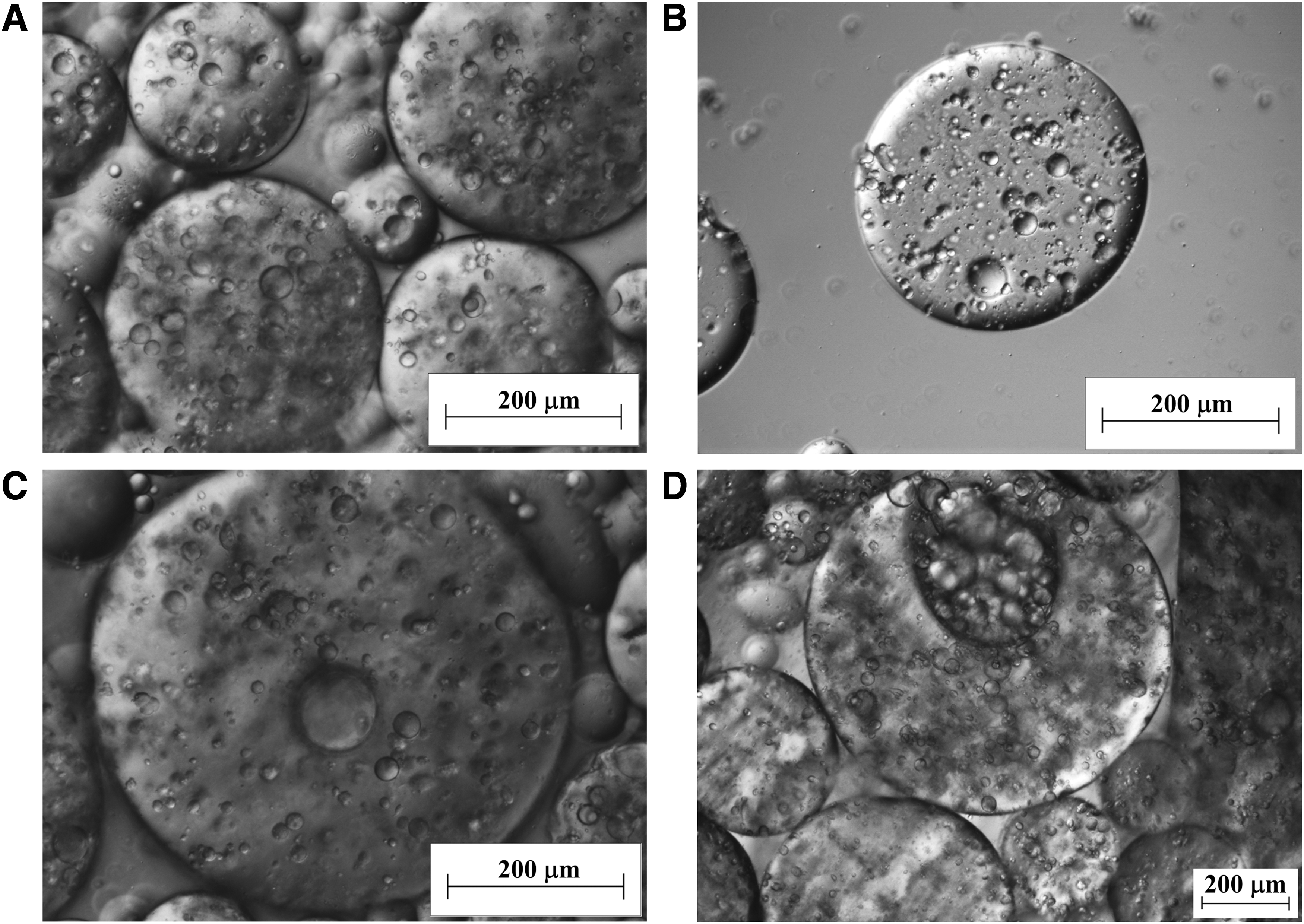
The results showed that cryoprotectant location was the most important, as a factor affecting the average drop sizes of emulsions and thus cell EE and stability of emulsions. The non-penetrating and penetrating cryoprotectants (sucrose/DMSO) were located within the internal, or external, or both aqueous phases of six groups of multiple emulsions that EI-EVI created in the CTF-biocontactor (Table 1). The microscopic analysis of the emulsions prepared with sucrose localized in both aqueous phases (EV), before and after freezing

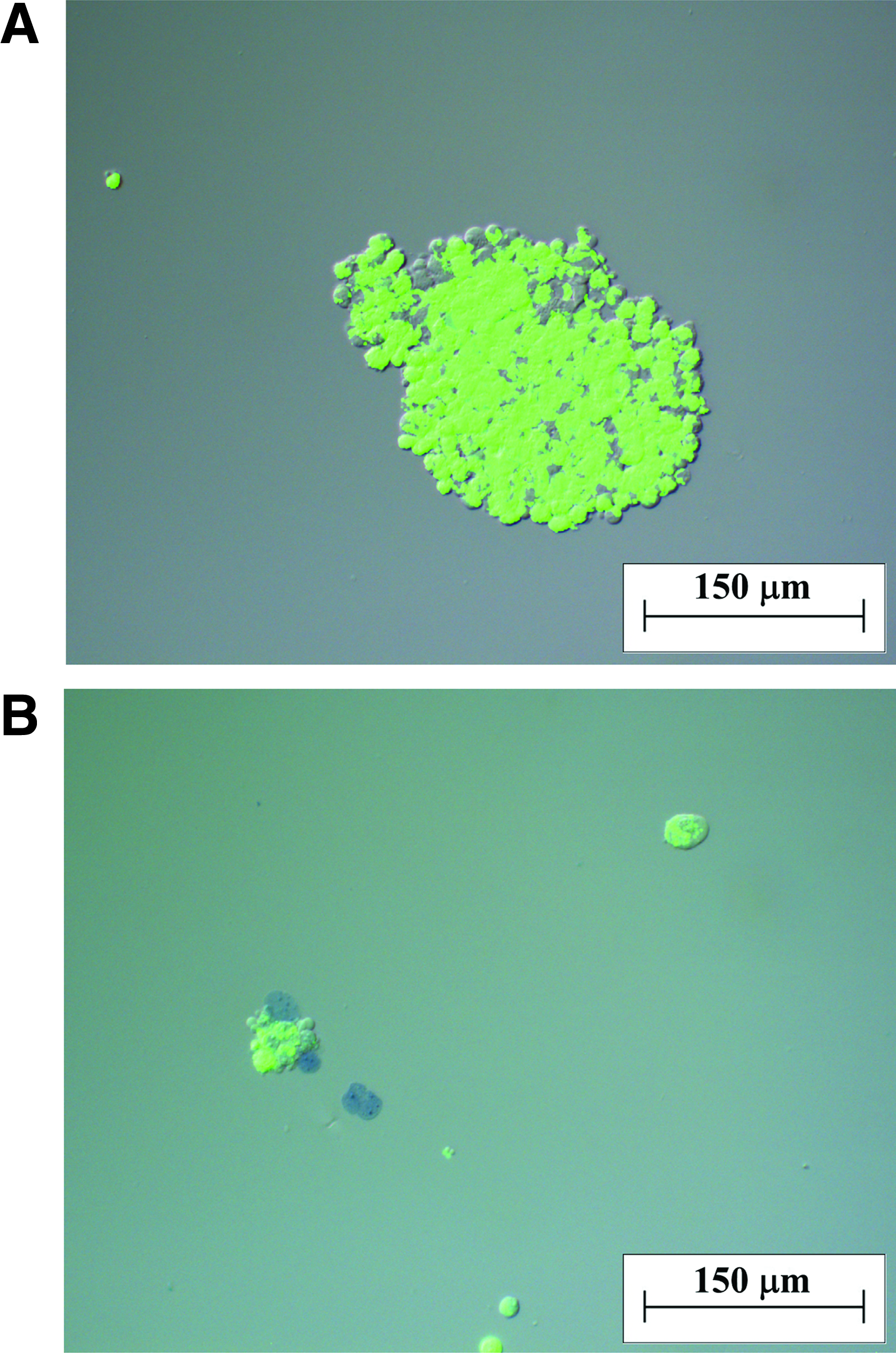
The microscopic images of multiple emulsion EV with encapsulated cells:
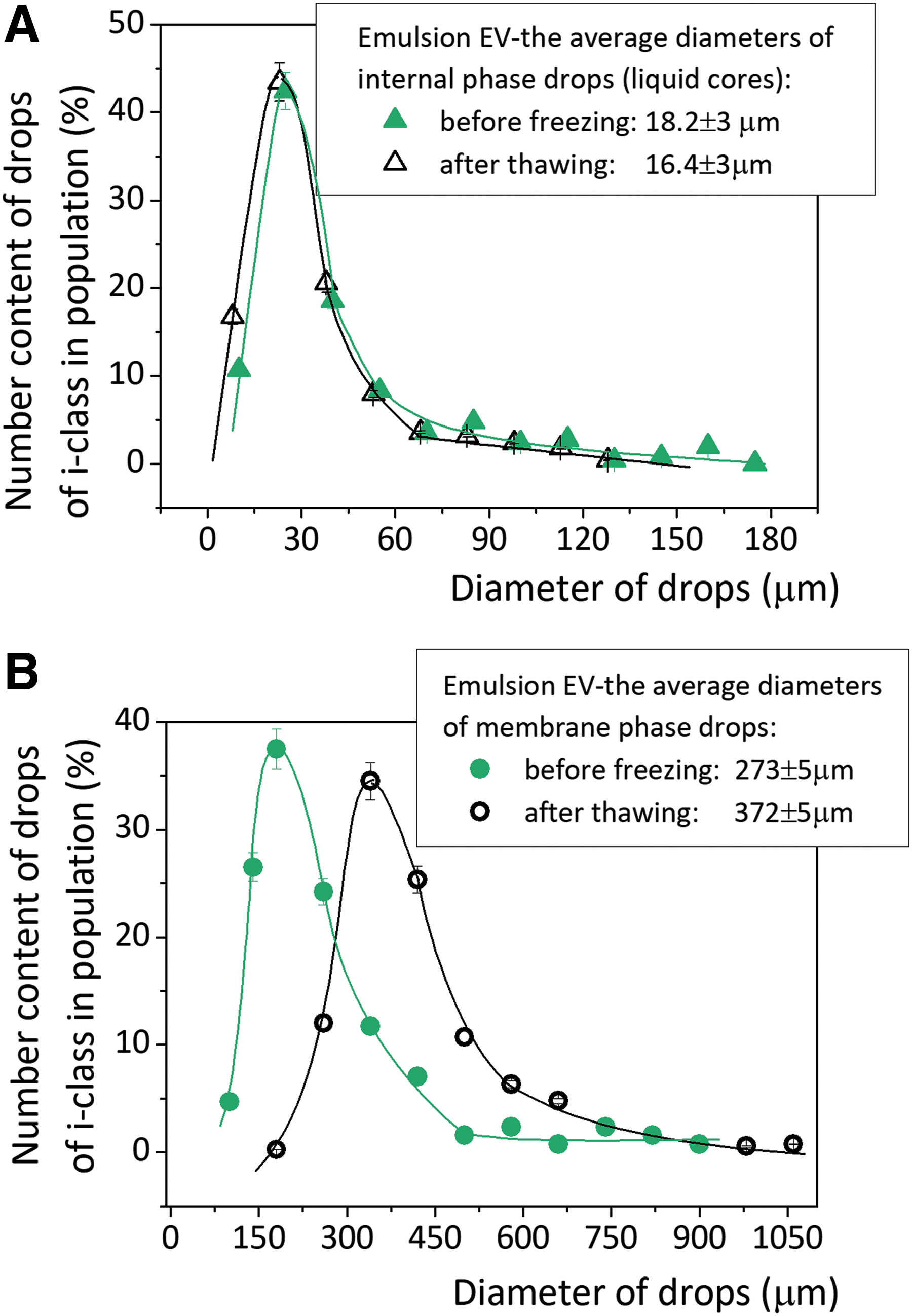
The drops size distributions of:
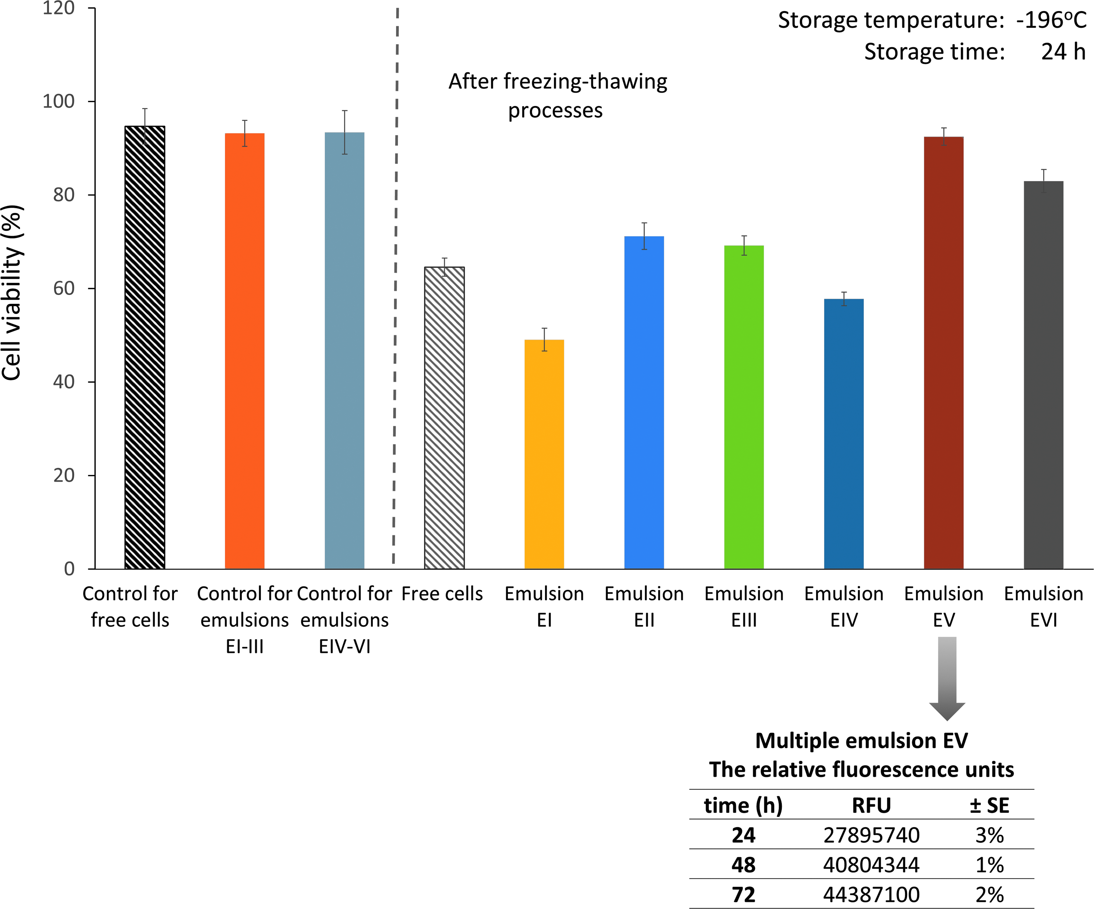
The comparison of the viability of cells cryopreserved in multiple emulsions (EI-EVI) and cells non-encapsulated (free cells), and the proliferation ability of emulsion EV. The control samples: cells non-encapsulated (control free cells), cells encapsulated in multiple emulsion EI-EIII without cryoprotectant in the internal phase droplets (control EI-EIII) and in emulsions EIV-EVI with cryoprotectant in the internal droplets (control EIV-EVI; Table 1). Color images are available online.
The encapsulation of cells in the internal droplets of multiple emulsions reached EE = 74.5% ± 5% for emulsions EI-EIII and 93.5% ± 5% for emulsions EIV-EVI. As shown in Figure 3A, the internal droplets of the multiple emulsions were appropriate for encapsulating HEK293-cells (green area), when 8.5–13 μm in size. The most promising emulsion-based carrier with the highest viability of cells (92.5% ± 3%) was emulsion EV with sucrose (non-penetrating cryoprotectant) located in both the internal and external phases (Fig. 5 and Table 1). The values of viable cells for emulsion EV before encapsulation and before freezing
As mentioned, the structure of emulsion EV remained almost intact after thawing. Also, the emulsion EVI (the internal phase with sucrose and external phase with DMSO) was found to be stable after thawing. The images of frozen and stored emulsions EVI 24, 48, and 168 hours (Fig. 6A–D) showed that the structures of multiple emulsions are not destroyed during the freezing process and storage. Some membrane phase drops tended to form aggregates, but the primary complex structure of the emulsion was still observed. For emulsion EVI, the value of cell viability was also very high (83.0% ± 3%). In the absence of cryoprotectants in the external and internal phase of emulsions EI, the existence of frozen and unfrozen water during freezing
The microscopic images of multiple emulsion EVI:
For emulsion EIV without cryoprotectant in the external phase, ice crystals are free to form and may undergo recrystallization, causing the destruction of the hierarchical structure of the multiple emulsion and damage to cells (a viability of 57.8% ± 3%). Emulsions EI and EIV were considered to be the worst carriers of cells. Cryopreservation of emulsions EII and EIII (cryoprotectants in the external phase) resulted in intermediate cell viability and the stability of emulsion structures, as shown in Figure 5. The measurements of the proliferation function parameter RFU of the cells after freezing
The expression of the GFP was visible in the microscopic image presented in Figure 7A. Many cells were observed with a cell viability comparable to cells in the culture before the freezing
The microscopic images of post-cryopreserved cells released and cultured from
Conclusions
This article proposed the method of preserving living cells by encapsulating them in the internal droplets of multiple emulsions (water-in-oil-in-water type) that minimize cryo-injury effects during freezing
Moreover, using such structured carriers as multiple emulsions enables the elimination of highly toxic penetrating cryoprotective agents such as DMSO, or the reduction of its concentration. Effective protection may be achieved with non-penetrating cryoprotectant only. Assessment of post-thaw cell viability showed the importance of the role of the location of cryoprotectants in multiple emulsions. The highest viability of cells (92.5% ± 3%) was achieved when non-penetrating cryoprotectant (sucrose) was localized in both the internal and external aqueous phases. The proposed structured carriers also allow the co-location of protectants in different phases of emulsions to reduce possible chemical damage to cells.
Footnotes
Acknowledgments
The authors would like to thank the National Science Centre-Poland for supporting this research (Grant-2014/13/B/ST8/04274), Prof. Dziembowski and Dr. Szczesny (Institute of Biochemistry and Biophysics-PAS) for their generous donation of the cell lines, and Mr. Krol for kindly providing the fluorescence microscope images.
Author Disclosure Statement
No conflicting financial interests exist.