
Abstract
In 1972, an enormous tomb site was found in the eastern suburb of Changsha, the capital city of Hunan Province, which led to the discovery of Mawangdui tomb No. 1, and soon thereafter tombs Nos. 2 and 3. These tombs were dated back to the Western Han Dynasty (206 BC–24 AD) in Chinese history. Along with numerous precious historic relics unearthed as grave goods, a well-preserved female cadaver was the most unprecedented, which was considered as one of the world's greatest archeological discoveries in the 20th century. The cadaver was initially examined through autopsy and X-ray imaging, with biopsies from multiple body parts analyzed histologically at the light and electron microscopic levels. In this review, we summarize the major imaging and autopsy findings from the cadaver indicative of remarkable preservation of some histological, cellular, and molecular constituents of the body. A forensic assessment of antemortem illnesses and potential cause of death of the subject are also noted.
Introduction
In the east suburb (now an inner area of the city) of Changsha, the capital of Hunan Province, there was a small village called Mawangdui ( , Mǎwángduī, King Ma's Mound). A legendary folktale says that it was named as such because of the unique landmark, the twin hillocks in horse saddle shape, which was the tomb of Ma Yin (853–930 AD), a ruler of the Chu kingdom during the Five Dynasties and Ten Kingdoms period. In the late 1960s, there was a national program of building underground infrastructures to prepare for a possible invasion from the Soviet Union. In 1972, the twin hillocks were chosen for the construction of an emergency hospital. Not long after groundbreaking, signs of the existence of a huge tomb were realized, turning the entire project into a massive archeological exploration. From 1972 to 1974, three tombs, designated Mawangdui Nos. 1, 2, and 3 tombs, were discovered sequentially, and belonged to the wife (
 , lady Xin Zhui, 217–168 BC), husband (
 , Li Cang, died in 186 BC), and son (
 , Li Xi, died in 168 BC) or Li Xi's brother. Li Cang was recorded historically as the first chancellor of the Kingdom of Changsha, an imperial fiefdom of the Han Dynasty (206 BC–9 AD). The No. 1 tomb was the first unearthed, and was the largest and best preserved among the three, whereas the other two were smaller and suffered from serious damage from multiple grave robbing during their history. Along with >3000 cultural relics ranging from textile, lacquerware, bamboo/wood artifacts and slips, silk paintings and craft pottery, and medical herbs to drawings of military performance, the remains of Xin Zhui were the most remarkable and unique (https://en.wikipedia.org/wiki/Mawangdui). Thus, the unearthing of the Mawangdui tombs has been regarded as one of the most significant archeological discoveries of the 20th century in China, even the world.1–3
The historic relics from the tombs have been preserved and exhibited in the Human Museum in Changsha, which has attracted thousands of visitors each year from all of the world during the past four decades. Importantly, the relics have been the subjects of research in all the fields of Museology in understanding of the society and life of people who lived >2000 years ago.
, Li Xi, died in 168 BC) or Li Xi's brother. Li Cang was recorded historically as the first chancellor of the Kingdom of Changsha, an imperial fiefdom of the Han Dynasty (206 BC–9 AD). The No. 1 tomb was the first unearthed, and was the largest and best preserved among the three, whereas the other two were smaller and suffered from serious damage from multiple grave robbing during their history. Along with >3000 cultural relics ranging from textile, lacquerware, bamboo/wood artifacts and slips, silk paintings and craft pottery, and medical herbs to drawings of military performance, the remains of Xin Zhui were the most remarkable and unique (https://en.wikipedia.org/wiki/Mawangdui). Thus, the unearthing of the Mawangdui tombs has been regarded as one of the most significant archeological discoveries of the 20th century in China, even the world.1–3
The historic relics from the tombs have been preserved and exhibited in the Human Museum in Changsha, which has attracted thousands of visitors each year from all of the world during the past four decades. Importantly, the relics have been the subjects of research in all the fields of Museology in understanding of the society and life of people who lived >2000 years ago.
With the advances in modern technology, ancient human remains are increasingly studied in paleoanthropological research.4–6 The remains of Xin Zhui were remarkably preserved, likely as a result of unique environmental factors as well as the body preservation strategies applied, such as deep underground burial and tight insulation by multiple layers of coffins. Upon discovery, the corpse was regarded as highly valuable from both biomedical and museological perspectives. Thus, after removing from the coffin, the body was soon embalmed with a formalin-based fixative for about 7 months, followed by autopsy and forensic examination (Fig. 1). In this review, we summarize the autopsy findings according to the original records of gross anatomical examination, X-ray imaging, and some histopathological and biochemical studies of biopsied tissue samples. 7
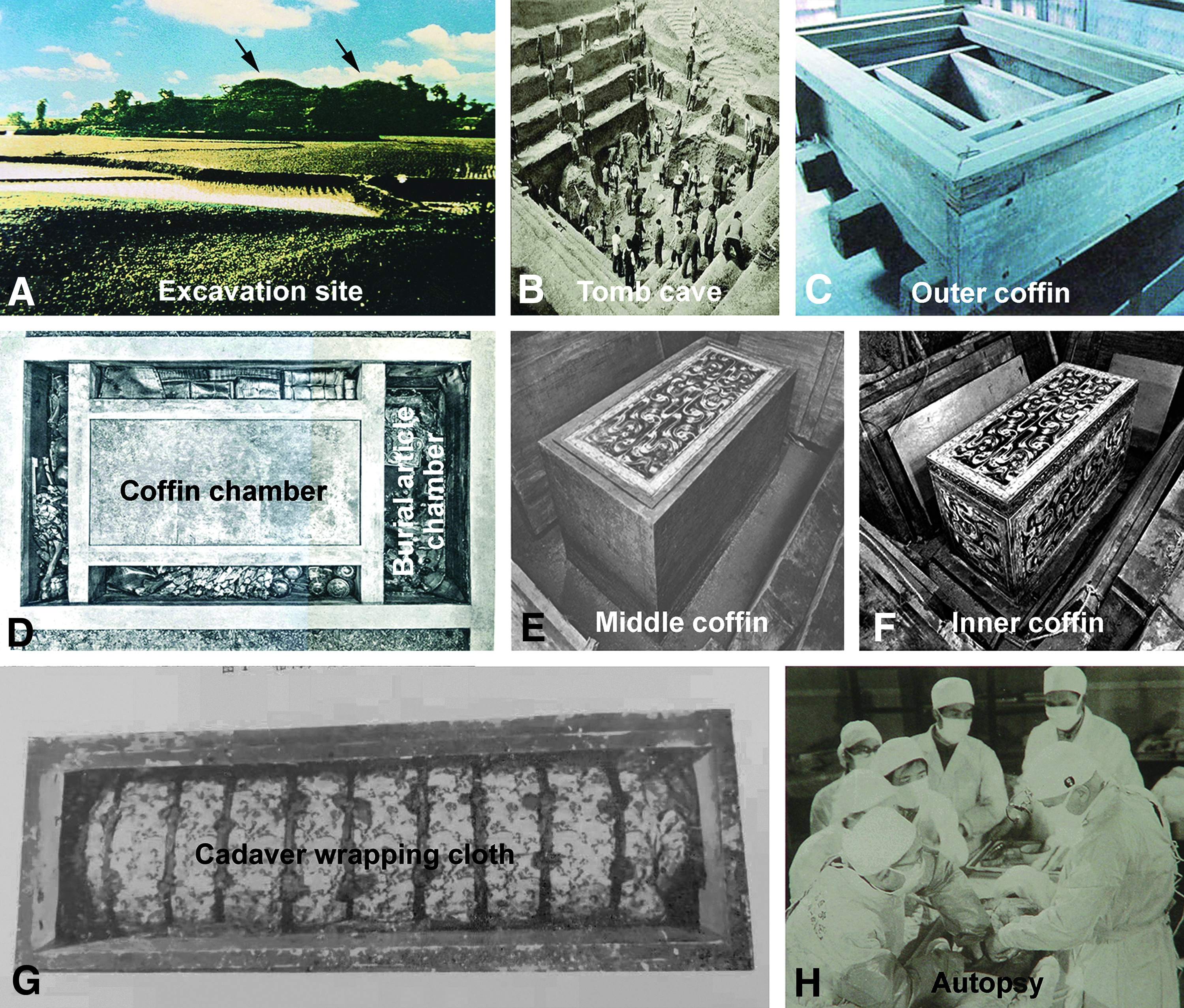
The excavation site, tomb structure, and burial conditions of the Mawangdui ancient cadaver.
Assessment of the Constituent Integrity of the Cadaver
Gross anatomic findings
The cadaver was identified as an adult female who died in her 50s, with a body height estimated to be ∼154 cm. All body parts including the head, neck, trunk, and limbs were intact, with the joints of the upper and lower limbs largely movable. The skin appeared deep brownish, covered the entire body surface, and had some extent of elasticity. Subcutaneous fat was palpable over most parts of the body. There were signs of early postmortem decomposition, including eyeball extrusion, mouth opening, tongue protrusion, and rectal prolapse. The hair on the head was well combed, covering the entire cranial vertex, and appeared firmly connected to the skin. It should be noted that a wig from another human individual was combed together with her own hair on the head.
The autopsy was performed by a group of experts including anatomists, pathologists, and clinicians. Major body cavities were opened, explored, and reclosed after the internal organs were removed and preserved separately in formalin fixative. Upon in situ exploration, all viscera and organs in the pelvic, abdominal, and thoracic cavities were intact and normally located anatomically. However, all organs had a certain degree of shrinkage, with the wall of the gastrointestinal ducts apparently thinned, relative to newly deceased cadavers. The brain was shrunken to occupy approximately a half of the cranial cavity, and lacked any identifiable morphological features, with brain tissue that felt ash-like fragile upon finger twisting.
To explore the vascular integrity, radiographic contrast was injected into the femoral artery of the cadaver before embalming, followed by whole body X-ray and angiography. The skeletal system was found to be completely intact, with even the smallest bony structures such as the nasal septum and sesamoid bones in the hand and foot visualized. The size and position of all bones appeared normal, although there were signs of decalcification, relative to age. All joints were arranged normally with the articular surface being smooth and clear. However, there was a narrowing of the articular cavity in some large joints (Fig. 2A–D). In angiography, the entire femoral artery and its major branches from the thigh to the foot were distinctly visualized without signs of contrast leakage. Localized widening and narrowing of the vascular diameter were seen along the distal segment of the femoral artery, at the popliteal artery, and at the anterior and posterior tibial arteries (see Fig. 1 in Ref. 8 ).
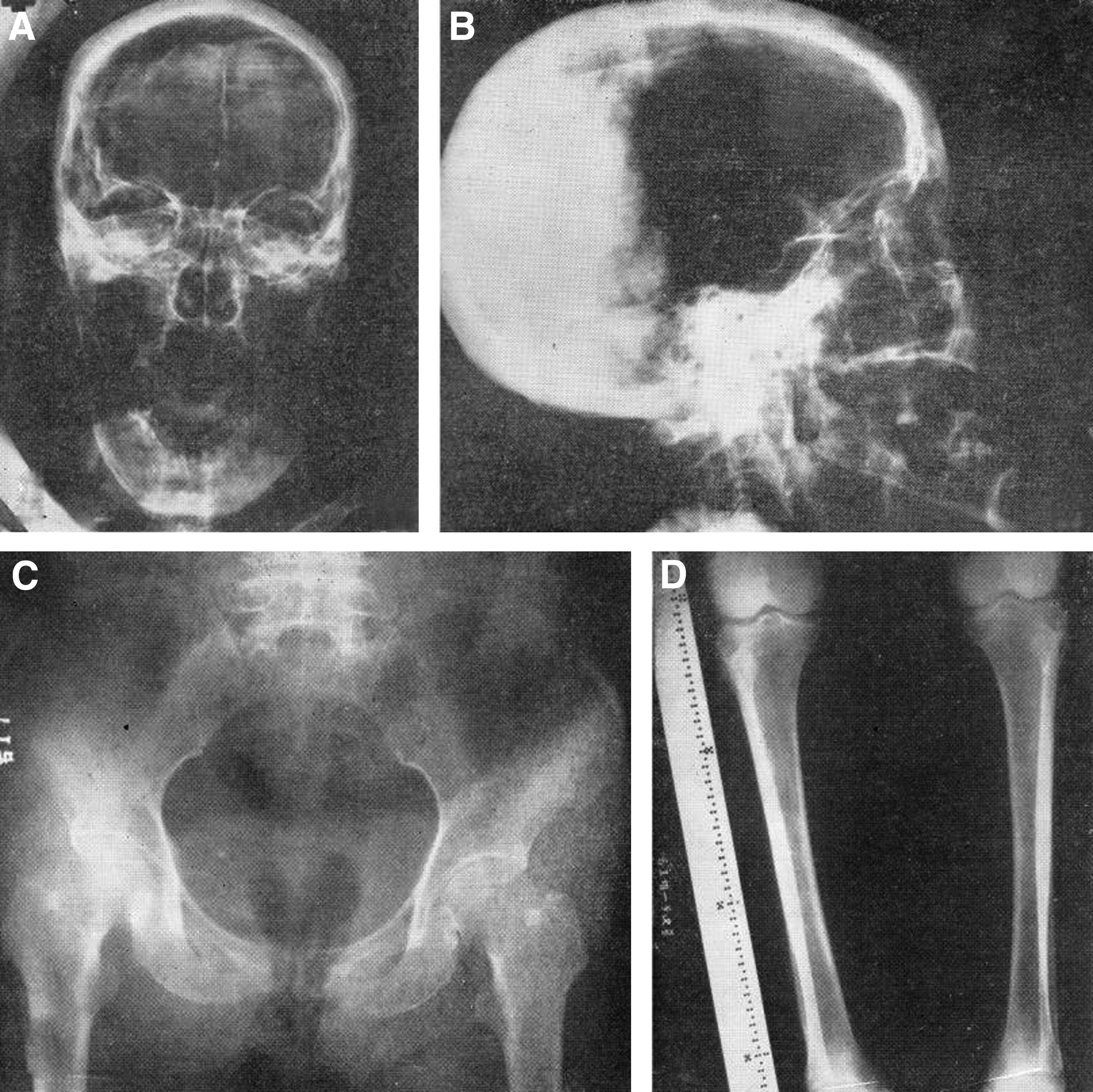
X-ray radiography of the Mawangdui tomb No. 1 cadaver.
Histological and cytological findings
Biopsies were obtained during autopsy dissection to determine the extent of histological integrity of various tissues, including the cutaneous, connective, muscular, and nervous tissues, as well as the cellular components thereof. The specimens were stained with the classic histological methods available at the time, with some samples also prepared for and examined with electron microscopy (EM). Samples from a modern-time cadaver were obtained and processed in parallel to serve as a reference in histological and cytological assessments.7,9,10
The preservation condition varied for the major types of tissues in the ancient cadaver. Overall, the connective tissue was the best preserved, especially regarding the extracellular fibrous components. Thus, collagen fibers were frequently observed in hematoxylin and eosin staining (H&E) of all tissues examined by light microscopy. In EM, the collagenous fibers had alternating dark and light bands that were further divisible into cross-striations indicative of considerable preservation of ultrastructure (Fig. 3A). The muscular tissues also appeared to be fairly well preserved, which was better seen in the striated skeletal muscles relative to the cardiac and smooth muscles. Thus, in EM preparations, the alternative striates of sarcomere of the skeletal muscles were identifiable, with the dark band (A band) remaining fairly distinct, although the light band (I band), H line, and Z line were disarranged or missing (Fig. 3B). In EM preparation of the costal cartilage, the nucleus of the lacunar chondrocyte showed dense material, whereas fibrous and vesicular profiles were present in the cytoplasm (Fig. 3C). In the EM preparation of the lumber nerve plexus, axon and surrounding myelin regions were identifiable, but myelin sheet organization was degraded (Fig. 3D).
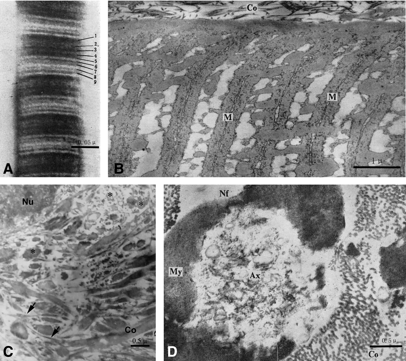
Electron microscopic assessment of tissue integrity in the Mawangdui tomb cadaver.
At the cellular level, a few types of cells in some tissues were well preserved morphologically based on classic histological stains. Thus, one of the best-preserved cell types was the chondrocytes (Fig. 4A). In cartilage sections with methylgreen–pyronin stain, the outline of cell bodies, nuclei, and plasma lemma were identifiable, although the nucleus appeared somewhat shrunken and the cytoplasm faintly stained with eosin. In Feulgen stain, the nucleus of the chondrocytes appeared purple-reddish. In H&E stain, the histological and cellular arrangements of the kidney and liver were not verifiable. However, in the EM preparation, cellular remnants of renocytes and hepatocytes were observed, including fragments of endoplasmic reticulum, broken plasma lemma, and nuclear membrane (Fig. 4B, C).

Electron micrographs of representative tissue cells in the Mawangdui tomb cadaver.
Macromolecular preservation
The extent of preservation of general classes of macromolecules, including proteinaceous, nuclear acidic, and lipidic and saccharidic components, was assessed in some tissues and cells of the ancient cadaver, using specific histological stains, biochemical assay, and other methods. X-ray diffraction was carried out for the cadaver's own hair and wig hair samples. The α-configuration of keratin in both the true and wig hairs appeared to be well preserved relative to modern-time human hair (Fig. 5A–C).7,11 The diazonium salt coupling reaction indicated that there existed proteinaceous products in muscle and collagen fibers, including sulfhydryl and amino groups (Fig. 5D). Lipidic remnant remained in some bodily cells according to Sudan III/IV stain. For instance, lipid drops of varying sizes and shapes stained in orange color were observed inside many chondrocytes (Fig. 5E). Also based on methylgreen–pyronin stain and Feulgen stain, there appeared to exist nuclear acidic elements in the nuclei of chondrocytes, as well as hyaluronic acid, chondroitin sulfate, and neutral and acidic mucopolysacchavide in the extracellular matrix of the hyaline cartilages (Fig. 5F). In addition, the signal of nucleic acids was detected in several tissue types according to ultraviolet absorption spectrometry, especially in the hepatic tissue sample (Fig. 5G, H).12,13
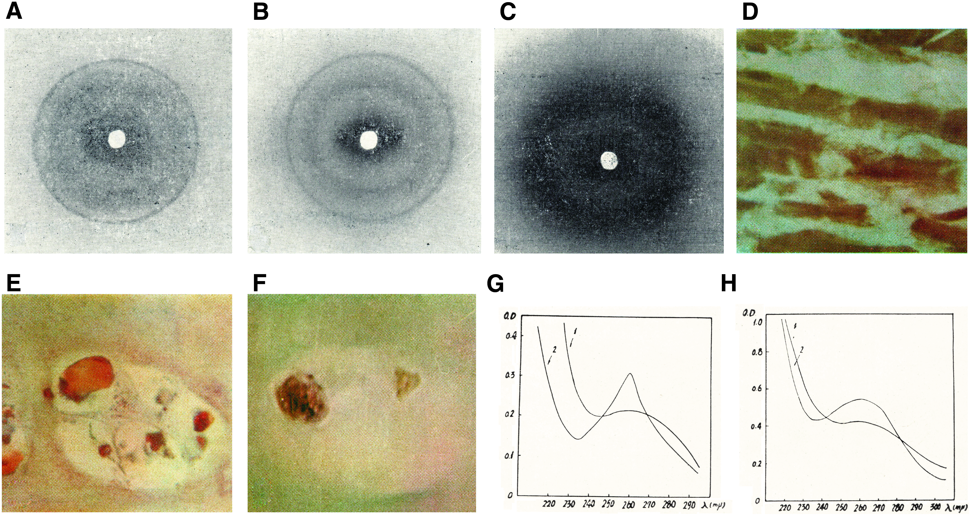
Assessment of the preservation of macromolecules in the Mawangdui tomb cadaver.
Forensic Pathological Evaluation
Systemic atherosclerosis and coronary atherosclerotic heart disease
During autopsy, the aorta and its major segments and branches to internal organs (heart, lung, kidney, liver, pancreas, intestines, and uterus) in the thoracic, abdominal, and pelvic cavities and to the upper and lower limbs were examined, with the arterial wall at multiple locations cut open and inspected. The internal, middle, and external layers (tunica intima, media, and externa) of the arterial wall were identifiable at all the mentioned branches, with the smooth muscle layer and the elastic laminas remained intact in cross-sectional view, although the innermost layer was incomplete or lost. Atherosclerotic plaques were found at many locations of the large to middle arteries including in the coronary arteries, whereas there were no plaques in the cerebral arteries (Fig. 6A). Severe atherosclerotic lesions were observed on the wall of the thoracic and abdominal aorta, renal arteries, common iliac arteries, and the uterus arteries, with areas of ulceration that occurred along the abdominal aorta (Fig. 6B).
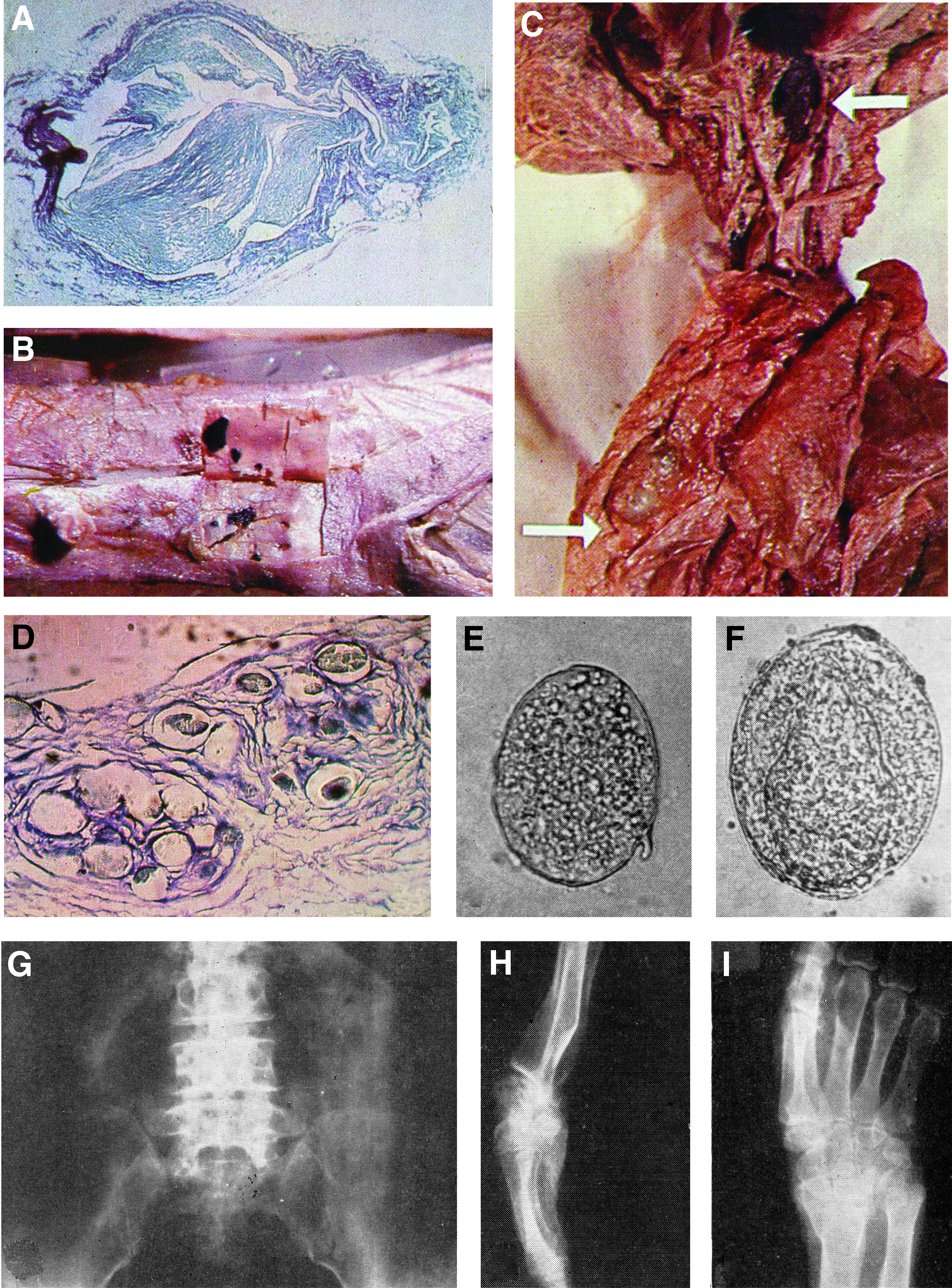
Forensic evidence for lady Xin Zhui's cardiovascular and digestive system diseases, and chronic osteoarticular injuries.
Coronary atherosclerosis can be graded into I–IV degrees, defined as the plaque lesions blocking the arterial inner cross-sectional area to ≤25%, 26%–50%, 51%–75%, and ≥76%, respectively. During forensic examination of the ancient cadaver, atherosclerosis was found in multiple sites of the coronary arteries. Specifically, the left coronary artery suffered more severe lesions than the right coronary artery, as were the anterior interventricular relative to the left circumflex branches. The plaque lesions along the first part (∼2.5 cm in length) of the left coronary artery were graded to III to IV degrees (Fig. 6A). The muscular wall was felt to be soft for most parts of the ventricles and the atriums, whereas there was a hardened area of the cardiac wall around the apex ∼3.5 × 2.8 × 1.1 cm 3 in size, which appeared to be protruded externally relative to the heart outline. Upon microscopic examination, the cardiac fibrous skeleton was completely maintained, and the laminar arrangement of the endocardium, myocardium, and epicardum remained identifiable. However, the nuclei and striation of the myocardial cells were rarely observed. Consistent with the autopsy findings, loci of myocardial necrosis and fibrillary scarring were evident around the apex in microscopic examination.
Gallstones and infection of Japanese schistosomiasis
Lady Xin Zhui appeared to have suffered from multiple gallstones and infection with Japanese schistosomiasis based on autopsy examination. Thus, there was dilation of the common bile duct as well as the intrahepatic bile ducts associated with thickening of duct walls. Multiple stones of soybean size with dark green coloration were found in the intrahepatic bile ducts. A stone measuring 1.4 × 0.7 × 0.8 cm 3 was located in the common hepatic duct, whereas another stone in size of 1.6 × 0.8 × 0.8 cm 3 was lodged at the Vater's ampulla. The gallbladder was enlarged, with its wall thinned. There was a membranous diaphragm between the body and ampulla parts of the gall bladder, with an aperture ∼0.8 cm in diameter, forming the so-called septate gallbladder (Fig. 6C). Based on the sandy texture, coloration, and X-ray radiographic features, the stones were considered to be likely the pigment type.
A notable finding from histological examination of the digestive system of the cadaver was the presence of pathological evidence of schistosomiasis infection. In sections prepared from the liver and the rectal wall, typical schistosome nodules consisting of eggs surrounded by fibrosis and granuloma were observed (Fig. 5D). Based on the morphology and size of the deposited eggs (Fig. 6E, F), and the tissue distribution pattern of schistosomiasis nodules, it was concluded that the subject had suffered with a fairly active infection of the Japanese schistosomiasis during her lifetime.
Osteoarticular abnormalities
Among the grave artifacts unearthed, two unique items were a wood cane and a bohua (silk painting, shown on the cover) displaying a female who walks with the help of a cane. Such information appeared to depict a scene of the daily life of the tomb occupant, who might have been at least somewhat handicapped. Consistent with the above speculation, during the autopsy examination of the ancient cadaver, it was noticed that her right lower limb was thinner (a smaller measured circumference) than the left limb. Furthermore, X-ray radiography revealed signs of chronic osteoarticular conditions that lady Xin Zhui might had suffered in her lifetime. Thus, there was significant narrowing of the intervertebral space between the fourth and fifth lumber vertebrates associated with hyperostosis, suggestive of right-posterior intervertebral disk herniation (Fig. 6G).
There was also radiographic sign of a healed radioulnar fracture that occurred above the right carpal joint. The bone fracture line seen in the distal radium was ∼3.5 cm above the joint surface, whereas the fracture line in the distal ulna was slightly proximal relative to the radial lesion. The fracture caused osteoarticular deformation and distortion in the forearm, including a dorsal rotation of the joint surface of radius and upward movement of the radial relative to the ulnar styloid process (Fig. 6H, I). These changes should have occurred during the healing process after the fracture. It is expected that these severe osteoarticular changes could significantly limit lady Xin Zhui's right hand movement and affect her daily life.
Discussion
Ancient human remains are considered as invaluable biological relics that can promote the understanding of socioeconomic and cultural development of humanity. The increasing use of modern biomedical technologies in paleoanthropological research has extended new cutting-edge approaches to explore human-related biomedical issues such as evolution, diseases, and medicine from a historic perspective. Various forms and types of human remains have been discovered worldwide and preserved in museums and institutes for public education as well as scientific research. Ancient human remains may be classified into mummified (dried), peat-tanned, adipocered, salted, iceman (frozen), and humid types.14–18 “Humid” cadavers are often unearthed from tombs and protected inside coffins. Several humid cadavers have been discovered in southern China, especially in areas alongside the Yangzi River. As already noted, the Mawangdui tomb No. 1 cadaver appeared to be remarkably well persevered from the gross anatomical, histological, cytological, and molecular levels. In fact, this unique human remains is not only the best preserved among the humid cadavers discovered in China so far, but also the cadaver with the longest burial time. Thus, the Mawangdui tomb No. 1 cadaver is considered archetypical for the humid cadavers. The Center for Preservation of Mawangdui Han Tomb Cadaver is dedicated to optimizing the methodology and condition for long time preservation of the humid type of human remains for public education and paleoanthropological research to the benefit of humanity.19,20
The Mawangdui tomb No. 1 cadaver has helped medical professionals to understand the occurrence of some human diseases in ancient Chinese people. Lady Xin Zhui appeared to have suffered from several disease conditions during her lifetime. The pathological examination suggests that she likely had severe atherosclerosis and biliary stones. Based on forensic findings, it was speculated that a biliocardiac attack might have caused Xin Zhui's death. Biliocardiac syndrome occurs in patients with biliary preconditions including calculous cholecystitis, which can trigger coronary artery contraction leading to ischemic cardiac stroke and death. The possibility of her acute death was also supported by the finding of undigested sweet melon seeds in the stomach and a pot of freshly prepared lotus root soup placed in the coffin, suggestive of consumption of a rich meal that could induce biliary duct spasms and a biliocardiac attack. The finding of her infection with Japanese schistosomiasis indicates that the epidemic of this parasite disease in southern China was active since 2000 years ago (which still persists today). The ill-healed upper limb bone fractures indicate a lack of sufficient medical knowledge at that time to properly manage the injury (such as external fixation that helps bone heal in correct position).
It is not clear as to why the Mawangdui tomb No. 1 cadaver was so rarely well preserved for >2000 years. However, a number of factors are considered key to prevent the body from decomposition. First, the tomb was deeply buried with the coffins placed >20 m from the ground in all directions. The tomb wall was built with white paste mud 100–130 cm in thickness, with a layer of charcoal 40–50 cm in thickness placed inside next to the mud wall. These can provide a water- and air-proof barrier that could also block microbial contamination from the soil outside the tomb. Second, the body was placed inside four sets of inner and outer wooden coffins, with the outmost coffin built of pine boards as thick as 90 cm. It is speculated that soon after burial, the tomb room would become an environment with constant low temperature and humidity, but under a hypoxic and sterilized condition, which would effectively and perhaps completely discontinue the process of postmortem decomposition, as such this humid cadaver could have remained largely intact over millenniums. It should be noted that in the coffin fluid, the concentration of some heavy metals including mercury was found to be considerably high. Mercury has an antibacterial effect, which might also have helped prevent biological degradation of the cadaver.7,21
Footnotes
Acknowledgments
The authors express their sincere gratitude and respect to Professor Long-Xiang Peng and other members of the original dissection team for their historic contribution in the examination and autopsy of the Mawangdui Han tomb No.1 cadaver, preparation and histopathological investigation of biopsied samples, and documentation of the initial forensic findings. This study was supported by the Project of Innovation-driven Plan of Central South University (Grant No. 2015CXS022), Key Project of China Hunan Provincial Science & Technology Department (Grant No. 2010SK2009), Fund for Humanities and Social Sciences of Ministry of Education of China (Grant No. 05JAZH024), National Key Technologies Research and Development Program of China (Grant No. 2012BAK14B03), and National Key Research and Development Program of China (Grant No. 2016YFC1201800).
Authors' Contributions
Article conceptualization was carried out by D.C. and X.G.L.; data assembly was performed by D.C., J.F.H., H.W., and X.S.W.; article composition was done by D.C., X.X.Y., and X.G.L.; and project supervision was undertaken by J.M.C., Z.Q.Y., and X.G.L.
Author Disclosure Statement
No conflicting financial interests exist.