
Abstract
The Mawangdui tomb No.1 cadaver, a female corpse from the Western Han Dynasty, was unearthed in 1972. Forensic examination at the time of discovery indicated fairly remarkable presence of bodily constituents at the anatomical, histological, and molecular levels. The cadaver was preserved in a formalin-based fixative afterward, and maintained in the Hunan Museum. To better protect this rare human corpse, a reappraisal of the status of preservation was carried out using noninvasive approaches, including X-ray radiography, gross anatomical examination, and histological, microbiological, and molecular analyses of sampled tissues. The cadaver remained essentially intact from a gross anatomical perspective, with radiography of the skeletal system and arterial contrast filling appeared comparable with the original documentation. The light microscopic features of the skin, cartilage, and skeletal muscle remained detectable, as were the stratified ultrastructure of the collagen and muscle fibers. The levels of nitrogen and amino acidic elements appeared elevated in the cadaver and liver preservation fixatives, with a higher calcium and phosphate concentration in the former. These findings suggest that there existed a certain degree of macromolecule degradation and bone decalcification in the cadaver, likely irrelevant to biological decomposition. The reappraisal also led to the implementation of stronger scientific measures to better protect the cadaver through a renovated Museum–University coadministrative management agreement.
Introduction
The excavation of the Mawangdui tombs accounts for one of the most significant archeological discoveries of the 20th century in China (https://en.wikipedia.org/wiki/Mawangdui). The female cadaver unearthed from tomb No.1, identified later as the remains of the wife of the first chancellor of the Kingdom of Changsha in Han Dynasty, was considered exceptionally rare but valuable from paleoanthropological and biomedical perspectives at the time of discovery.1–7 Protective measures were taken soon after the cadaver was found. The body was placed in ice chunks after removal from the inner coffin to minimize cadaver decomposition. Embalming was carried out with injections of a preservative containing formalin (4%–5% formaldehyde), ethanol (4%), and glycerol in 4:2:1 ratio into the femoral artery, trunk, limbs, and cranial, thoracic, and abdominal cavities, followed by immersion of the entire body in formalin.8–10 These initial treatments allowed a successful modern medical autopsy of the body 7 months later, which led to findings of a presence of remarkable bodily integrity from gross anatomical, histological, and pathological aspects never seen before among excavated ancient human remains.1,4,11 Follow-up research studies on tissue samples from the cadaver and other excavated items, including biological relics, corroborated the initially expected significance of the Mawangdui tombs in the fields of archeology and museology as well as insights regarding socioeconomics and ancient medicine in China.2,12,13
In 2002, a new building was completed to house the collections of cultural relics of the Hunan Museum. Meanwhile, many advances in scientific technology had occurred since the 1970s for biological and medical research, including paleoanthropological investigations. A decision was made to re-examine the cadaver to assess the structural integrity, to assure a safe transportation of it to the new storage location. It was also expected that the findings from a re-examination would help modify the preservation strategy, if needed, to maintain its value for future research as an ancient biomedical specimen. A team of experts in the fields of anatomy, pathology, microbiology, analytical biochemistry, surgery, and radiology was assembled to carry out the examination. Small amounts of tissue samples were removed from the cadaver and also obtained from the dissected organs that had been bottle-stored in formalin fixative since the original autopsy.
Materials and Methods
The examination of the cadaver was approved administratively by the Provincial and State Cultural Relics Bureaus in accordance with the law for the protection of cultural relics and the guidelines for sampling and repairing of biological relics set by the Chinese government. Thus, the cadaver was examined in a “noninvasive” manner, with minimal tissue samples obtained from unexposed locations or areas of the body, for routine and electron microscopic (EM) histological analyses and biochemical assays. 14
Gross anatomic examination
Under sterile conditions, the body was raised up from the storage coffin made of polymethyl methacrylate, and placed on a surgical table for examination. The entire body was visually inspected without removing the sutures holding together the cavities opened or the incisions made during the original autopsy 30 years ago. Skin, subcutaneous fat, and skeletal muscles over the body were palpated to assess the extent of elasticity. Limbs and joints were gently turned around to test the extent of mobility. The head and hair were examined visually and explored with caution using a pair of forceps. After the examination and X-ray radiography (see hereunder), the cadaver was wrapped with medical gauze around the chest, and the low abdomen to the hip–pelvis area (to cover the sutured incisions and also as a cosmetic consideration), and returned to the plastic coffin filled with newly prepared preservation fluid.
X-ray radiography
A mobile medical X-ray machine was used for radiographic documentation of the cadaver. X-ray exposures directed to the entire, or a part, of the region of the head, neck, trunk, upper limbs, and lower limbs were performed with the cadaver placed in the supine position as well as lying on the side. Film radiographs were analyzed in comparison with the original documents by experienced radiologists.
Tissue sampling and histological processing
To accurately evaluate the status of preservation of histological integrity, small amounts of tissue samples were incised from the cadaver during the gross anatomical examination. The specimens were limited to 1 cm 3 in size, and included the skin at the front part of the right leg, middle part of the rectus abdominis, a segment of the fifth rib, and its associated costal cartilage. A piece of liver was also removed from the liver specimen bottle-stored in formalin. A part of each specimen was dehydrated and embedded with paraffin following a standard protocol, with the other part embedded with Epon according to standard preparation procedures for transmission electron microscopy. The wax-embedded tissue blocks were cut into ∼5-μm thick sections mounted onto glass slides, further processed with hematoxylin and eosin (HE) or Massan stain. Semithin and ultrathin sections were prepared from the plastic tissue blocks, stained with lead and uranium, and examined on a Hitachi H-600 Transmission Electron Microscope.
Collection and analysis of body and organ preservants
After removal of the cadaver, the fluid in the plastic coffin was collected, with a part of it sampled. The fixative preserving the liver and other specimens was also sampled. These and a part of the new replacement preservation solution were subjected to parallel biochemical analyses. The samples were diluted 100 times in concentration, and assayed using a high-performance pH meter (Orion® 920A+; Thermo Fisher Scientific), spectrophotometer (Model 721; Shanghai Optical Instrument Factory, China), liquid chromatograph spectrometer (Model LC-6A; Shimadzu Corporation, Japan), and spectrophotofluorometer (Model RF-5301PC; Shimadzu Corporation). Readouts from the assays were obtained, including pH value and concentrations of nitrates, ions/electrolytes, organic acids, and amino acids.
Microbiological sampling and cultivation
The body and organ specimen preservatives were sampled under sterile conditions. Suspicious spots on the walls and bottom of the plastic coffin were also swap-sampled. Samples were placed in sterilized testing tubes, which were sealed and sent to microbiology laboratories for routine and special microbial cultures for the assessment of presence of aerobic and anaerobic bacteria, fungi, or spores in any of the cadaver/specimen preservation environments.
Data analyses and interpretation
Observations obtained during the gross anatomic examination were compared with the original descriptions documented in the first autopsy of the cadaver whenever available. X-ray radiographic, histological, and EM imaging data were also analyzed against the initial findings as recorded at the time of unearthing. Biochemical data were interpreted either as the assays reported or using the readouts from newly prepared (unused) preservation solution as a reference.
Results
Gross anatomic findings
All bodily parts of the cadaver including the head, neck, trunk, and limbs were completed by visual inspection. However, the skin coloration appeared somewhat paler relative to the initial state. Both the upper and lower limbs were laterally and anteriorly movable to some extent by gentle turning. All areas of the body surface were felt soft with some extent of elasticity when finger pressed. However, by finger twisting, the soft tissue (fatty and muscular) exposed on the edges of the original abdominal incision (along midline) was felt to be very fragile and became easily detached from the remaining proximal parts on the body.
X-ray radiographic finding
In comparison with the original X-ray radiographs, there were no impressive differences with regard to the structure and contrast of skeleton and individual bones. Thus, the osseous density seen in long bones appeared normal relative to age, and the bone cortex remained dense and distinctly delineated. Bony trabeculae were clearly seen at the ends of the long bones such as around the knee. Importantly, the contrast filling injected 30 years ago into the left femoral artery remained in situ along its course as well as in its branches at the segments of the popliteal, anterior/posterior tibial, and common peroneal arteries. No contrast leakage into extravascular tissue was detected in comparison with the original angiographic images of the same areas (Fig. 1A–C).

Comparable angiographic appearance of the left femoral artery between the first and second cadaver examinations.
Light microscopic findings
Skin
According to the original histological examination, the epidermis of the skin of the cadaver was essentially lost, whereas the dermis and subcutaneous structures were visible under the microscope, appeared to be largely consisted of connective tissue (Fig. 2A). In the Massan stain of the skin sample obtained for the reappraisal, collagen fibers sectioned transversely and longitudinally were displayed in the dermis. However, neither cellular nor vascular profiles were reliably detected (Fig. 2B, C).
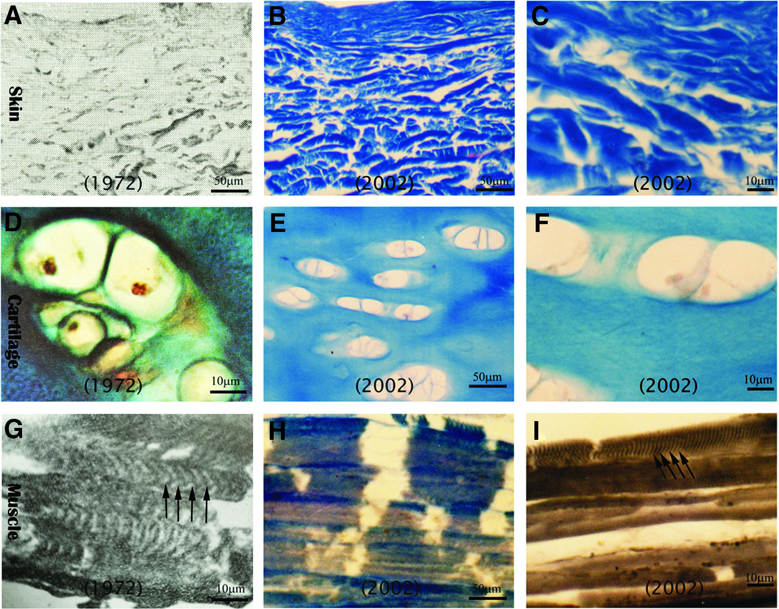
Comparison of light microscopic observations in representative tissues of the Mawangdui ancient cadaver between 1972 and 2002.
Cartilage
The chondrocytes were found to be the best preserved type of cells in the cadaver from the first autopsy. 11 For instance, in Azan trichrome stain, clusters of chondrocytes were labeled in the cartilage lacuna, with the nuclei appearing dark red, cytoplasm orange, and cartilage matrix blue (Fig. 2D). In comparison, the staining in the costal cartilage sections processed in this study was not as distinct as before. Thus, in the Massan stain, cartilage lacuna was identifiable, with remnants of chondrocytes lightly stained or unstained (Fig. 2E). There was also a reduction of the staining in cartilage matrix suggestive of the loss of collagen fibers (Fig. 2F).
Skeletal muscle
There was substantial preservation of the histological characteristics of skeletal muscles at the initial autopsy examination of the cadaver. 11 For instance, the transverse striates of the psoas major muscle were observed with iron hematoxylin stain under light microscope (Fig. 2G). In comparison, in Massan (Fig. 2H) and iron hematoxylin (Fig. 2I) stains, the rectus abdominis specimen obtained at the re-examination showed very few transverse striates. The muscle fibers remained individually visible and arranged as bundles. There were remains of connective collagen fibers between muscular fibers (Fig. 2H).
Light microscopic examination of other tissues obtained at the re-examination did not reveal remarkable changes. The rib sample was too fragile and soft (decalcified) to prepare ground bone sections for light microscopic examination. The liver sample was prepared into paraffin sections, but there was no clearly identifiable cellular profiles in HE stain (data not shown).
EM findings
Skin
The original EM study indicated that there was considerable ultrastructural integrity of the cutaneous tissues of the cadaver. 11 In particular, the collagen fibers appeared remarkably well preserved, with alternating dark and light bands visualized and subdividable into fine cross-striations at high resolution (Fig. 3A). At the reappraisal, a skin sample from the leg was obtained to assess the extent of preservation at EM levels. In comparison, bundles of collagen fibers in the dermis were visible, whereas it was fairly difficult to recognize the alternating dark and light bands in the majority of the fibers (Fig. 3B).
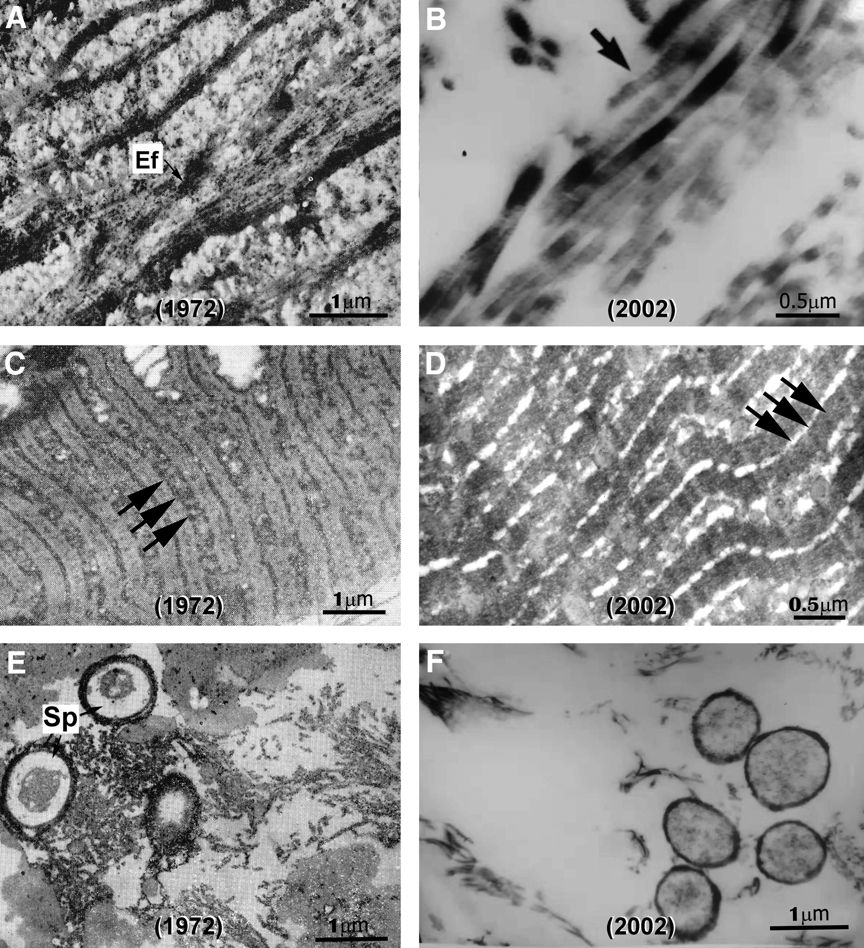
Comparison of electron microscopic findings from the skin, skeletal muscle, and liver specimens of the Mawangdui cadaver between 1972 and 2002.
Skeletal muscle
As with the collagen fibers in connective tissue, the remaining of the transverse striates in the muscular tissue were impressive at the original examination following the unearthing of the cadaver. 11 In comparison with electron micrographs from the initial study (Fig. 3C), a striated pattern remained visible at some locations along the muscle fibers (Fig. 3D). However, there was a large extent of tissue lysis and loss of structural integrity of remaining fibers. Overall, neither the nuclei of myocytes nor mitochondria were found (Fig. 3D).
Liver
As an example for the examination of the internal organs, samples from the bottle-preserved liver were prepared for light microscopic and EM studies, in comparison with the original findings on record. As already mentioned regarding the loss of cellular profiles at light microscopic levels, the change at the ultrastructural level was also dramatic in the hepatic tissue after 30 years storage. Originally the hepatocytes were identifiable, along with bacterial spores and collagen fibers (Fig. 3E). In contrast, no hepatocytes were observed in the resampled liver preparations, although there existed some fibrous elements probably representing the remnants of the collagen elements (Fig. 3F).
Chemical and biochemical findings from preservation fluids
The original solutions for preserving the cadaver in the plastic coffin and bottled organ specimens were formulated with neutral formalin diluted with distilled water, with a pH set at ∼5.5, and calcium concentration at 2.74 μg/mL. No amino acids or other organic chemicals were added in these solutions. Using analytical chemistry methods, the pH and concentrations of inorganic components in the body and liver fixative were assayed (Table 1). Specifically, the pH measured from the body preservative sample was 4.3, whereas that from the liver fixative was 2.54. Calcium and phosphate concentrations in the body fixative were 144 and 16.4 μg/mL, respectively, and those in the liver fixative were 1.85 and 7.2 μg/mL, respectively.
Amount of Elements in the Cadaver and Liver Preservation Fluids (μg/mL)
Likewise, the amounts of amino acids in the body and liver preservatives were measured as a part of the cadaver reappraisal (Table 2). Overall, 14 types of common human body amino acids were detected, and some reached impressive levels in the samples, including phenylalanine (Phe), serine (Ser), and glycine (Gly). In addition, the total nitrogen contents in the body and liver preservatives were 23.9 and 78.8 μg/mL, respectively.
Amount of Amino Acids in Cadaver and Liver Preservation Fluids
a.a., amino acids.
Microbiological examination results
Inoculations of samples from the cadaver preservation solution, organ preservation fixatives, and spot swapping on the walls of plastic coffin into culture medium did not yield a growth of flora of aerobic and anaerobic bacteria or fungi. As a result, no further microbiological studies were carried out with the collected samples.
Discussion
Many ancient human remains have been excavated and are being preserved as resources for public education and paleoanthropological research today and for generations to come.15–18 For those involved in ancient cadaver protection, it is a privilege but responsibility to find the best available approaches to preserve these precious biological relics. According to the varying forms of ancient human remains, different strategies and conditions could be used for storage and/or preservation purposes. For instance, mummies (dried corpses) might remain fairly stable in a temperature, humidity, and lighting controlled environment for a long period of time.19–21 How to preserve the “humid” or “wet” cadavers remains a great challenge, since many more factors need to be considered in formulating an effective preservation protocol.17,22 The Mawangdui ancient cadaver was remarkably well persevered at the gross anatomical, histological, cytological, and molecular levels at the time of discovery. 11 Not only because of its unique original buried and unearthed conditions, this ancient corpse has also been marked as a treasure among cultural relics of national interest (https://en.wikipedia.org/wiki/Mawangdui). No existing experience could be called upon to protect this extremely rare and valuable cadaver after its excavation. Thus, modern concepts about body embalming and tissue fixation were applied after it was initially examined. However, the effectiveness of such strategies remained unknown for as long as 30 years. The expectation that a reappraisal would be informative on assessing its preservation status was encouraged by the outstanding research carried out by the original expert team, who left precious data based on a thorough forensic examination. 8
Although only limited approaches were applied at the re-examination of the cadaver in abiding by the protective regulations for cultural relics, the data obtained are substantially informative. The gross anatomical examination suggests that the body has remained in shape and in structural texture largely comparable with the status at the time of excavation, whereas there existed a certain degree of skin decolorization likely due to the release of the dyeing materials (such as from the wrapping cloth) from the body. The X-ray radiography indicated that there were no noticeable changes in skeletal imaging features including bony structures. In particular, the arterial contrast filling remained in place and in clarity as it was seen 30 years ago. The light microscopic and EM findings suggest a preservation of basic histological organization of the cutaneous, muscular, and cartilage tissues, including some cellular profiles, of the cadaver. However, it should be noted that there was some loss of cytological properties with regard to staining features and ultrastructural details among all the examined tissue types. Specifically, the staining intensity of the chondrocytes, myocytes, and hepatocytes appears reduced relative to that observed at the first study. Furthermore, the striation pattern of the collagen and skeletal muscle fibers also appeared less distinct as it was reported after the initial autopsy studies. Overall, the light microscopic and EM observations indicate that there was a trend of degradation in the cadaver at subcellular and molecular levels between the first and second examinations.
Since the cadaver and separated organ specimens were stored in liquid preservatives, biochemical analyses of the preservative samples could provide some clues for potential molecular changes in the cadaver tissues. In a comparison between the cadaver body and liver preservatives that were the same in the original preparation, the calcium and phosphate concentrations in the former were remarkably higher. This finding suggests that there existed a release of calcium and phosphate elements from the body, likely from bones. This would point to a potential decalcification process in the bony skeleton, although the X-ray radiographic observation suggests that such a change did not occur to the extent enough to impact the imaging appearance of bone calcification. The readouts of the amino acid contents from the assayed body fixative are also important data from the reappraisal of the cadaver. Virtually all essential amino acids of the human body were detected in the sample, with the levels of Phe, Ser, and Gly particularly high. In addition, the concentration of the nitrogen content was also impressively high. Thus, these biochemical measurements pointed to a rise of organic components in the preservation fluid that would have been mostly likely derived from the cadaver tissues. Along with the light microscopic and EM findings of decreased clarity in tissue and cell morphology, it appears most likely that degradation of macromolecular constituents occurred in the cadaver and separated organ specimens.
Microbial contamination and infection are a risk for structural damage of biological relics and a particular concern in the preservation of the “wet” human remains. Suspicious coloration spots were found on the wall of the storage coffin before the cadaver re-examination, which raised a worry of potential mold growth and even fungi infection in the body. However, the microbiological culture studies with the samples collected on the cadaver and the wall of storage containers did not find evidence of bacterial and fungi growth. Accordingly, the formalin-containing preservant and the sealing methods appeared to be sufficient to prevent the proliferation of air-borne microbial species in the cadaver preservation environment. It also appears that macromolecular degradation in the cadaver was not related to biological decomposition, a process of molecular breakdown by microorganisms.
In summary, at the 30th anniversary after unearthing, a set of modern biomedical methods was used to examine the Mawangdui cadaver, organ specimens, and preservation solutions and environment. Relative to the documentation at the initial forensic study, the cadaver exhibited insignificant changes in gross anatomical and radiographic aspects. However, some deterioration in histocytological integrity and fairly evident ultrastructural decadence were observed in the examined skin and muscle samples. Concentrations of calcium, phosphate, and amino acids appeared increased in the cadaver and organ preservation fluids. Together, the findings suggest the existence of macromolecular degradation in the cadaver most likely resulted from abiotic decomposition, warranting further efforts to mitigate this process, including by modulation of the fixative formula and more frequent monitoring of chemical components in the cadaver preservation fluid.
Footnotes
Acknowledgments
The authors express their sincere gratitude and respect to Professor Long-Xiang Peng and other members of the original autopsy team for their historic contribution in examination and autopsy of the Mawangdui Han tomb No.1 cadaver, preparation, and histopathological investigation of biopsied samples, and documentation of the initial forensic findings. This study was supported by the Project of Innovation-driven Plan of Central South University (Grant No. 2015CXS022), Key Project of China Hunan Provincial Science & Technology Department (Grant No. 2010SK2009), Fund for Humanities and Social Sciences of Ministry of Education of China (Grant No. 05JAZH024), National Key Technologies Research and Development Program of China (Grant No. 2012BAK14B03), National Key Research and Development Program of China (Grant No. 2016YFC1201800).
Authors' Contributions
Project conceptualization was carried out by X.G.L., J.M.C., and Z.Q.Y.; gross anatomical study was done by X.G.L. and H.W.; light microscopic study was carried out by Q.L.C., H.W., and X.G.L.; electron microscopic study was performed by X.Y.W.; biochemical study was carried out by H.W. and X.S.W.; microbiological study was carried out by Z.D.X.; surgical consultation was done by H.H.T.; radiological consultation was done by G.C.P.; data assembly was performed by H.W., X.S.W., D.C., and J.F.H.; and article composition was carried out by H.W., X.X.Y., and X.G.L.
Author Disclosure Statement
No conflicting financial interests exist.