
Abstract

To the Editor:
Given the threats currently facing coral reef ecosystems, scientists the world over have attempted to gauge how corals respond to environmental challenges. 1 Although many such questions can be addressed in the field, laboratory studies must instead be undertaken when, for instance, the effect of a single stressor is to be modeled. 2 However, few studies have documented whether laboratory-raised corals perform similarly to wild conspecifics. 1 Not only will verifying whether or not this is the case advance coral ecophysiological research, but it will also have important implications for facilities seeking to rehabilitate and/or biopreserve reef corals. Please see our recent “Coral Hospital” article 1 in Biopreservation and Biobanking for details on this topic. As corals readily spawn in the husbandry facilities of Taiwan's National Museum of Marine Biology and Aquarium (NMMBA), we sought herein to exploit a published dataset 3 to determine whether released eggs of the reef corals Oxypora lacera and Echinopora gemmacea were morphologically similar to wild coral eggs. This question also stemmed from our observation that the growth rates of cultured corals are generally lower than those of wild corals.
Egg ultrastructure was observed with transmission electron microscopy (Fig. 1; O. lacera TEM images only), and cultured eggs of both species were smaller and possessed (1) thicker microvilli layers and (2) higher yolk content. Furthermore, the cultured parent colonies possessed higher Symbiodiniaceae dinoflagellate densities and chlorophyll a concentrations. The seawater in NMMBA, as is the case in most other aquaria worldwide, is filtered through sand, and the consequently lower external food supply could account for the majority of these differences. It is worth noting that the dinoflagellate endosymbionts, which are transmitted horizontally in these species, can increase transfer of lipid material to their coral hosts under nutrient-deprived conditions 4 ; perhaps, then, the increased amount of yolk in the cultured coral eggs is a result of nutrient stress. This, as well as their smaller sizes, could suggest that aquarium-reared corals are less fit than their wild counterparts, although future work must be done to confidently verify this. For now, such differences lead us to recommend that those conducting laboratory-based experiments carefully consider how well the physiology of cultured corals, as well as their gametes, approximates that of their in situ counterparts, particularly in cases wherein seawater has been partially filtered (leaving corals to rely predominantly on autotrophy). Such considerations will be especially critical for those aiming to raise or rehabilitate corals in officinarum for later outplant onto local reefs. 1
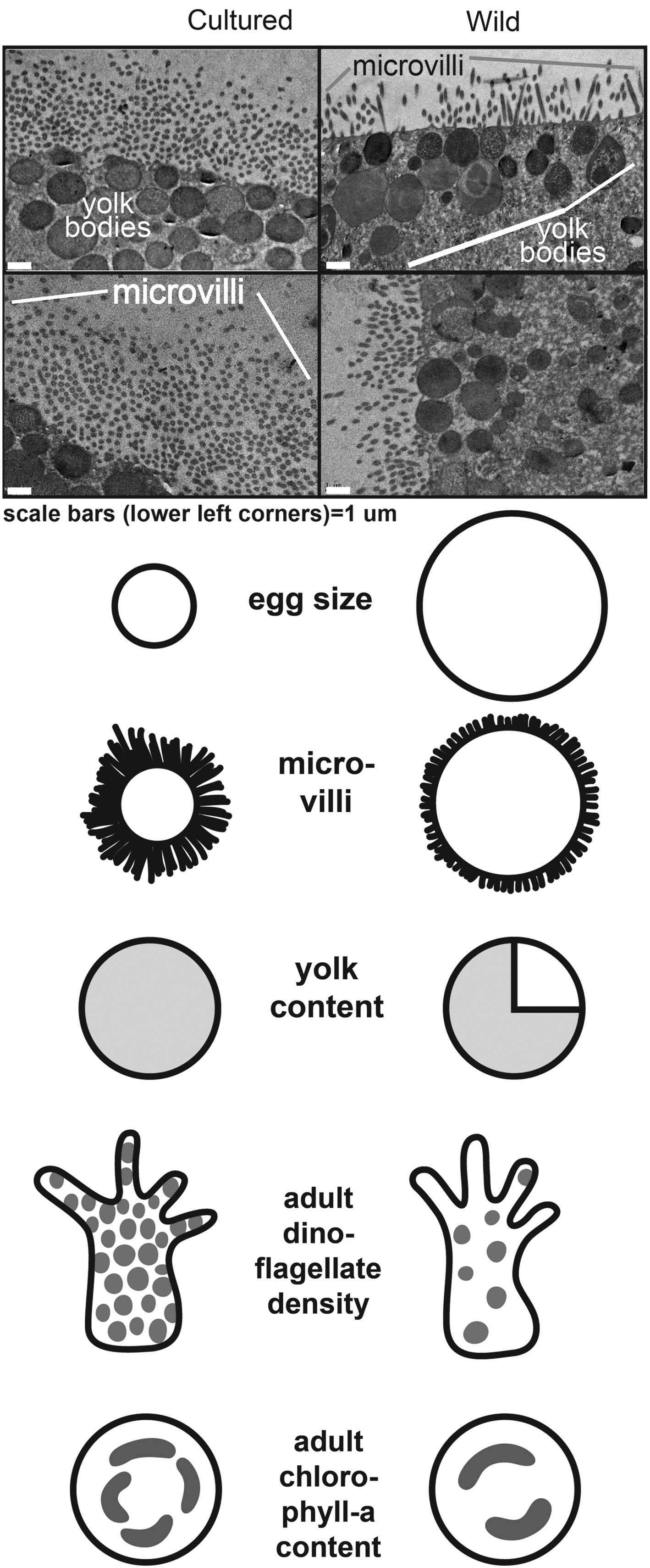
Images of representative eggs from cultured and wild corals (Oxypora lacera) and a summary of the physiological differences between them. Source of non-microscopic images: National Museum of Marine Biology and Aquarium (NMMBA) editorial team.