
Abstract
Background:
The growing interest in mesenchymal stromal cells (MSCs) in equine medicine, together with the development of MSC biobanking for allogeneic use, raises concerns about biosafety of such products. MSCs derived from umbilical cord (UC) carry an inherent risk of contamination by environmental conditions and vertical transmission of pathogens from broodmares. There is yet no report in the scientific literature about horses being contaminated by infected MSC products, and no consensus about systematic infectious screening of umbilical cord-derived mesenchymal stromal cells (UC-MSCs) to ensure microbiological safety of therapeutic products.
Objectives:
To develop a standard protocol to ensure UC-MSC microbiological safety and to assess the risk of vertical transmission of common intracellular pathogens from broodmares to paired UC-MSCs.
Study Design and Methods:
Eighty-four UC and paired peripheral maternal blood (PMB) samples were collected between 2014 and 2016. Sterility was monitored by microbiological control tests. Maternal contamination was tested by systematical PMB PCR screening for 14 pathogens and a Coggins test. In case of a PCR-positive result regarding one or several pathogen(s) in PMB, a PCR analysis for the detected pathogen(s) was then conducted on the associated UC-MSCs.
Results:
Ten out of 84 UC samples were contaminated upon extraction and 6/84 remained positive in primo culture. The remaining 78/84 paired PMB & UC-MSC samples were evaluated for vertical transmission; 37/78 PMB samples were PCR positive for Equid herpesvirus (EHV)-1, EHV-2, EHV-5, Theileria equi, Babesia caballi, and/or Mycoplasma spp. Hepacivirus was detected in 2/27 cases and Theiler Diseases Associated Virus in 0/27 cases (not performed on all samples due to late addition). All paired UC-MSC samples tested for the specific pathogen(s) detected in PMB were negative (37/37).
Main Limitations:
More data are needed regarding MSC susceptibility to most pathogens detected in PMB.
Conclusions:
In-process microbiological controls combined with PMB PCR screening provide a comprehensive assessment of UC-MSC exposure to infectious risk, vertical transmission risk appearing inherently low.
Introduction
In equine veterinary medicine, the use of mesenchymal stromal cells (MSCs) for regenerative purposes is expanding steadily, with clinical safety now established, whether used in an autologous or allogeneic setting. 1 However, the biosafety infectious profile of MSCs has been poorly reported so far, although they carry a potential infectious risk originating from the donor or the biomanufacturing process and cannot be completely sterilized. Compared with autologous use, the risk is higher with allogeneic MSCs since products manufactured from a single donor can be administered to several recipients. Reports about iatrogenic contamination with blood-derived products exist.2,3
The European Medicines Agency (EMA)'s Committee for Medicinal Products for Veterinary Use (CVMP) developed an expert group for novel therapy-related issues named ADVENT (Ad Hoc Expert Group on Veterinary Novel Therapies), aiming to provide guidelines strengthening quality and security. It recently released two question and answer documents relative to sterility 4 and freedom from extraneous agents 5 of allogeneic MSC products, with recommendations based on product specificities and epidemiological considerations. These guidance documents cover biosafety aspects that must be considered when manufacturing stem cell-based products for allogeneic use, according to the requirement to test veterinary medicinal products for potential infectious contaminants specified in Directive 2001/82/EC, Regulation (EU) 2019/6. 5
Similarly, in the United States, cell-based drugs must be reviewed and approved by the Food and Drug Administration following Center for Veterinary Medicine's (CVM) guidance before they can be legally marketed. 6 This article provides a precise description of a protocol defined on a risk-based analysis, integrating the existing regulatory recommendations.
It is acknowledged that the various anatomical structures from which MSCs can be isolated (UC, bone marrow, and so on) do not present the same risk: human umbilical cord-derived mesenchymal stromal cells (UC-MSCs) are considered less likely to contain infectious agents than MSCs derived from adult tissues. 7 No data have been published in the equine veterinary context yet. Equids also present a high risk of environmental contamination during UC sampling. Therefore, we conducted a systematic evaluation of bacteriological and fungal changes upon initiation of biomanufacturing and before freezing of master bank MSC units. Although the equine epitheliochorial placentation reduces risk of transplacental transmission of common pathogens, vertical transmission has been demonstrated for some of them, such as piroplasmosis or classical abortive viruses like Equid herpesvirus (EHV)-1 and equine arteritis virus,8,9 or recently hepacivirus, 10 whereas evidence-based information is missing for others.
To assess the risk of vertical contamination of UC-MSCs, we carried out a systematic screening on peripheral maternal blood (PMB) samples from healthy broodmares whose UC was collected for MSCs. Fifteen equine intracellular agents of interest 11 were selected based on four criteria adapted to the French epidemiological situation: endemic microorganisms (Babesia caballi, Theileria equi, EHV-1, EHV-2, EHV-4, and EHV-5, Rhodococcus equi, Anaplasma phagocytophilum, Borrelia burgdorferi, and Leptospira interrogans), notifiable diseases of importance and high pathogenicity (equine infectious anemia and equine viral arteritis), emerging pathogens (hepacivirus and Theiler Diseases Associated Virus [TDAV]), and identified risks of unknown relevance (Mycoplasma spp.).
All those data will support a better assessment of UC-MSC-specific infectious risk for allogeneic use and will help in defining the donor qualification strategy.
Materials and Methods
Experimental cohort
This protocol was conducted on 84 foals between breeding seasons during 2014 and 2016. Broodmares were stationed at the experimental farm of French Horse and Riding Institute (IFCE, Chamberet, France) or in privately owned high-standard French stud farms and benefitted from medical follow-up by qualified equine veterinarians. Two weeks before expected parturition, broodmares were examined for clinical signs of systemic or urogenital disease. In the absence of abnormalities, mares were considered healthy and prepared to foaling according to farm protocols, with adequate monitoring.
Tissue and biological fluid collection
PMB samples were collected immediately after foaling (one tube with EDTA and one tube without anticoagulant). Trained staff were in charge of the collection of umbilical cord tissue (UCT) (n = 80) or umbilical cord blood (UCB) (n = 4), according to standardized protocols that have been proven harmless to both foal and mare. 12 UCT samples were serially washed with sterile physiological solution and conditioned in storage medium complemented with 2% (v/v) antibiotic solution (penicillin 10,000 U/mL—streptomycin 10 mg/mL—amphotericin B 25 μg/mL). UCB samples were collected through clean venipuncture in 250 mL collection bags with anticoagulant, serving as containers for shipment. Within 24–48 hours after foaling, all samples were shipped to the biomanufacturing laboratory using single-use material to ensure temperature between +2°C and +12°C. Umbilical samples were processed and PMB tubes redirected to control quality laboratory for pathogen screening according to flowchart (Fig. 1a).
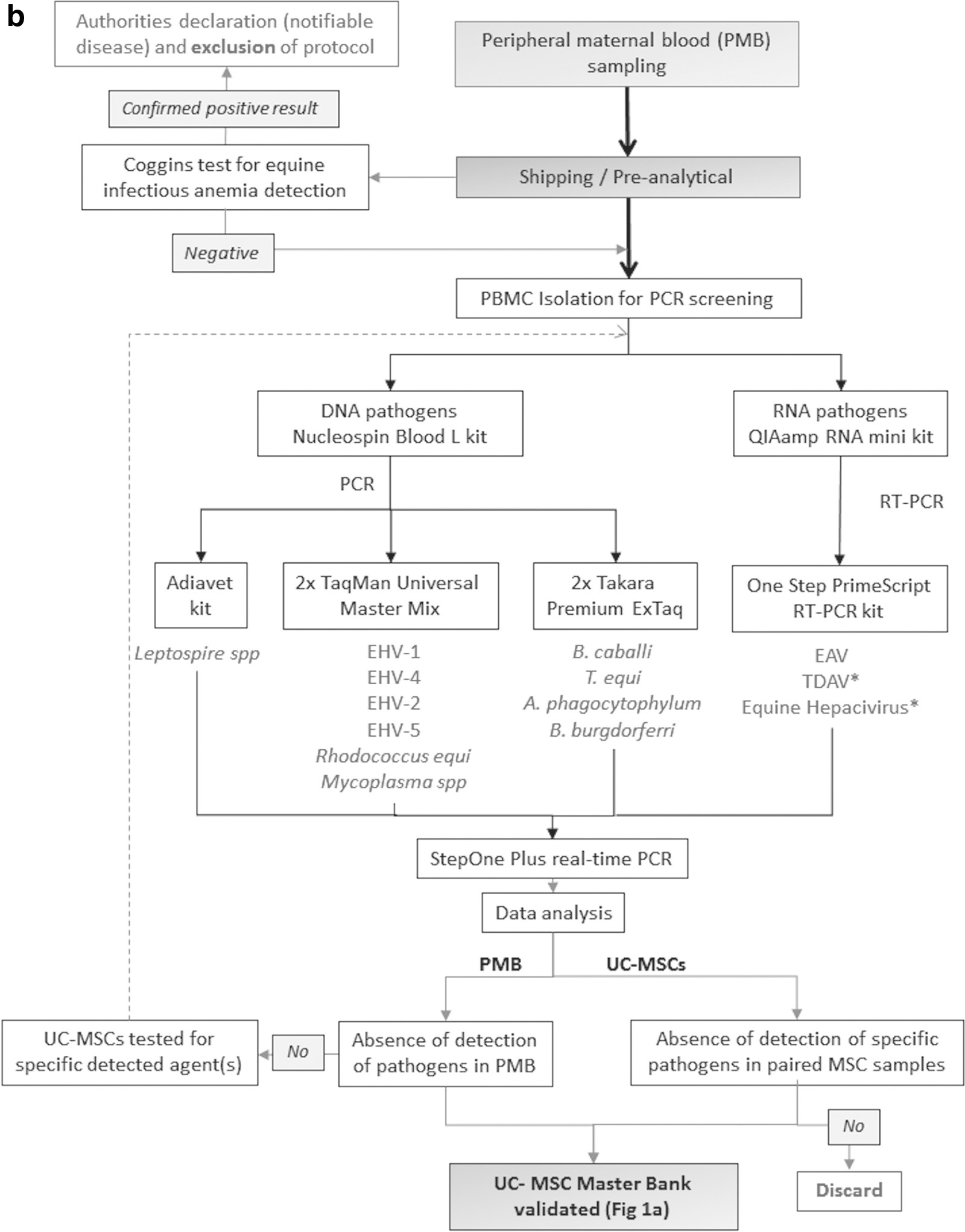
Flowchart describing
UC-MSC isolation and culture
Processing of UCT
UCT were submitted to serial washings using sterile NaCl 0.9%, soaked in povidone iodine for 15 minutes, and dissected with single-use material to extract the conjunctive tissue surrounding umbilical vessels (Wharton's jelly). Dissected tissue was minced and digested for ∼1 hour with collagenase type I (Sigma–Aldrich) at 37°C, to allow cell extraction.
The suspension of digested tissue was sampled for environmental contamination testing (see During MSC Extraction section). Suspension of digested tissue was plated into Falcon® cell culture flasks with growth medium, consisting of DMEM supplemented with 100 U/mL Penicillin/Streptomycin, 2 mM L-glutamine, and 10% (v/v) HyClone® fetal calf serum.
Processing of UCB
As previously described, 13 whole UCB samples were loaded onto a Ficoll (1.077 g/mL density) solution. After density gradient centrifugation at 900 g for 30 minutes at room temperature, the cellular suspension was sampled for environmental contamination testing (see During MSC Extraction section). Mononuclear cells were removed from the interphase and washed twice with Dulbecco's phosphate-buffered saline (D-PBS), plated at a defined seeding density (150,000 cells/cm2) into Falcon cell culture flasks, and cultured as for UCT-MSCs.
Culture and storage of isolated UCT-MSCs & UCB-MSCs before screening
After 72 hours, nonadherent cells were eliminated by changing the medium. After enzymatic detachment with trypsin 0.05%/EDTA 0.02% in PBS, adherent cells were reseeded and allowed to expand until 80% confluence, harvested, washed to eliminate antibiotics, and sampled for microbiology testing (see After MSC Expansion section). Finally, UC-MSCs were conditioned for freezing at 1.5 × 10⁶ cells/unit in DMEM supplemented with 10% (v/v) FBS and 10% (v/v) DMSO and stored at −196°C. Specific units were frozen for future PCR screening.
All cell culture reagents were purchased from Pan Biotech (PAN-Biotech, Aidenbach, Germany) unless otherwise specified.
Environmental contamination testing
Environmental contamination was monitored throughout the manufacturing process to detect microorganisms coming from raw material (UC sampling conditions) or biomanufacturing process (laboratory contamination) as described in Figure 1a.
During MSC extraction
Samples from digested tissue (UCT) or cellular solution (UCB) were tested with a broad-spectrum test (brain heart infusion [BHI]; bioMérieux, Marcy l'Etoile, France), incubated for 7 days at 37°C, and checked daily for visual turbidity.
After MSC expansion
Aliquots from prefreezing cell suspension were also tested with a broad-spectrum test (BHI; bioMérieux), incubated for 7 days at 37°C, and checked daily for visual turbidity. Moreover, on day 5, a sample from BHI suspension was plated on Columbia Agar +5% sheep blood (bioMérieux) and examined for bacterial colony growth proximity (LVD69, Marcy l'Etoile, France). Since 2015, an additional aliquot had been seeded on Sabouraud Dextrose Chloramphenicol Agar (bioMérieux) incubated at +20°C for 10 days.
Results were considered positive when microorganism development was visually detected. Further identification of colonies on these plates was not pursued.
PMB screening and analysis on paired UC-MSCs
All 78 samples that were free of detectable fungal or bacterial contamination were submitted to a Coggins test and PCR screening on PMB. Hepacivirus and TDAV PCR were developed after the beginning of the study and performed on only 27 PMB samples. When positive results were obtained for one or several agents, paired UC-MSCs were retrieved from nitrogen storage, thawed, washed from cryoprotectant, conditioned at 1–2.5 × 10⁶ cells/mL in a DNA/RNA stabilizing RNAse-free solution, and tested for the specific detected pathogens (Fig. 1b).
PCR tests
Concerning DNA pathogens, nucleic acids were extracted from 2 mL of each sample with the Nucleospin Blood L kit (Macherey-Nagel, Düren, Germany), then eluted in a final volume of 200 μL, and stored at −80°C until used. Concerning RNA viruses, nucleic acids were extracted from 140 μL of each sample with the QIAamp® RNA viral Mini Kit (Qiagen, Courtaboeuf, France), then eluted in a final volume of 50 μL, and stored at −80°C until used. Five microtiter of extract was used for PCR tests, except for for EHVs, R. equi, B. caballi, and T. equi where 2.5 μL of extract was used.
All primers and probes used are presented Table 1. Real-time PCRs for EHV-1, EHV-4, EHV-2, EHV-5, TDAV, equine hepacivirus, B. caballi, T. equi, R. equi, Mycoplasma spp., A. phagocytophilum, and B. burgdorferi were developed as previously described14,15 based on the standard model AFNOR NF U47-600-2.16,17 Each reaction was processed in a total volume of 25 μL containing Taqman® Universal PCR Master Mix (Life Technologies, Saint Aubin, France) for EHV-1, EHV-4, EHV-2, EHV-5, R. equi, and Mycoplasma spp. or Takara Premix Ex Taq for B. caballi, T. equi, A. phagocytophilum, and B. burgdorferi, and in a total volume of 25 μL of One Step Prime Script RT-PCR kit (Perfect Real time) for the TDAV and equine hepacivirus. L. interrogans kit analysis was used as described by the manufacturer (Adiagene, Ploufragan, France) and EAV PCR test performed as described previously. 18 Quantitative PCR assays were performed on StepOnePlus™ Real-Time PCR systems (Life Technologies). Controls in each series included extraction positive and negative controls; PCR positive and negative. An internal standard (HPRT1 gene, TaqMan gene expression assay; Life Technologies) was amplified to attest good extraction and absence of inhibitors. Data were analyzed manually using the StepOne™ software, version 2.2.2 (Adiagene, Ploufragan, France). All the steps were carried out by referring to the quality criteria of the AFNOR U47600 standard.
Sequences of Primers and Probes Used in This Study
F, forward; R, reverse; S, sense.
EAV, equine arteritis virus; EHV, equid herpes virus; NA: not available (Diagnostic kit); TDAV, Theiler's disease associated virus.
Coggins test
A Coggins test was performed on all 78 PMB serum samples according to AFNOR NF-U47-00219 by the agar immunodiffusion technique with protein p26 incubated at 21°C + 4 for 20 hours.
Results
Broad-spectrum environmental screening
Ten of 84 UC samples were positive (turbid BHI broth) during the MSC extraction phase. After cellular culture expansion with antimicrobial complementation, 4 samples turned out negative, but 6/84 remained positive (BHI & Sabouraud cultures), and were therefore excluded.
Detection of microbial genome by PCR in PMB
PCR screening was performed on 78/84 PMB samples, and 37/78 were positive for a least one agent. Among the systematically tested pathogens, EHV-1, EHV-2, EHV-5, T. equi, and B. caballi, Mycoplasma spp. were detected at least once throughout the study, with respective prevalence of 2/78, 22/78, 26/78, 7/78, 1/78, and 1/78. Co-infections were frequent (Fig. 2). Hepacivirus tested positive in 2/27 cases and TDAV in 0/27 cases (not included in Fig. 2 due to late addition to protocol).
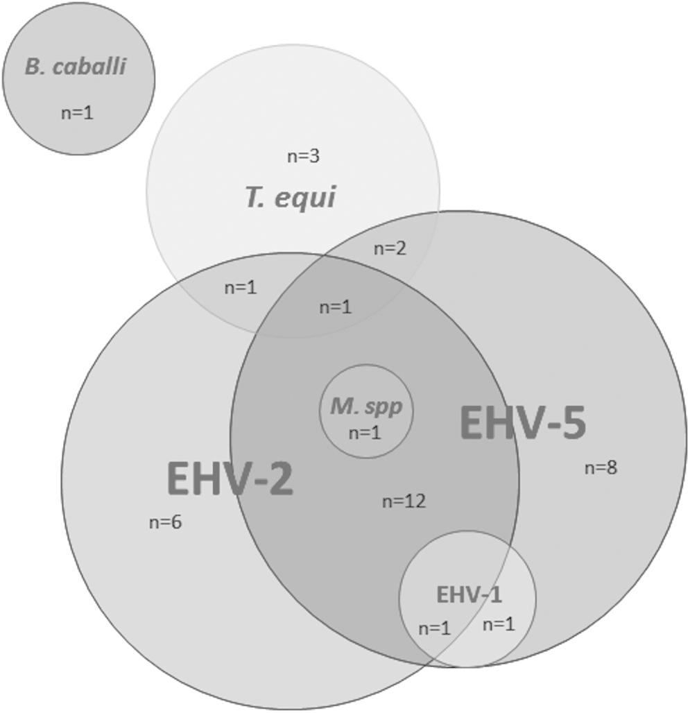
Pathogens detected in peripheral maternal blood by PCR: n indicates the number of positive samples. Co-infections appear with the overlap of bubbles. B. caballi, Babesia caballi; EHV-1, equid herpesvirus 1; EHV-2, equid herpesvirus 2; EHV-5, equid herpesvirus 5; M. spp, Mycoplasma spp.; T. equi, Theileria equi.
Absence of detection of equine infectious anemia by Coggins test in PMB
All PMB samples tested (78/78) were negative for equine infectious anemia.
Absence of detection of intracellular pathogens in UCT and UCB-MSCs
For every PMB sample displaying a positive PCR result regarding one or several extraneous agent(s) (37/78), paired UC-MSC units were tested negative for the specific relevant pathogen(s) (37/37). No hepacivirus was detected in the two UC-MSC samples associated with PCR-positive PMB.
Discussion
UC (tissue and blood) can suffer environmental contamination from the female genital tract and foaling conditions. To assess this risk, broad-spectrum bacteriological and fungal cultures were performed. Inherent to sampling procedures, UCT appear more prone to external contamination than UCB, 12 although the great number difference between UCT (n = 80) and UCB (n = 4) samples in our study does not allow statistical confirmation of this. However, we highlight the importance of an early broad-spectrum test to detect contaminated material.
Donor qualification is a key element of allogenic products. The use of specific pathogen-free (SPF) herds whose infectious status is known and controlled is usually recommended. For example, the American Association of Equine Practitioners (AAEP), regarding licensed equine plasma, recommends testing donor animals against equine infectious anemia, piroplasmosis, dourine, glanders, and brucellosis. However, considering current epidemiological French status free of the last three diseases, as monitored by the RESPE (French Epidemiological Network of Equine Diseases), we decided not to include them in our panel. SPF herds are difficult and costly to operate in practice for allogenic UC-MSCs and could also raise ethical concern, hence the choice of UC collection upon usual foaling. Like in human public cord blood banking, a comprehensive qualification strategy addressing both broodmare infectious status and collected products is needed. Broodmares should be free of active infection and vaccinated, even though vaccination does not offer a full protection against infection as illustrated by EHV-1 vaccination failing to completely suppress cell viremia and its consequences. 20 Samples must therefore be screened for an extensive panel of infectious agents on a risk-based analysis defined by their pathogenicity and prevalence.
Although most of the studies involving equine MSCs include a screening panel, only a few of them document the detection techniques, the list of pathogens, and the corresponding results. Previous studies reported the presence of EHV in half bone marrow samples 21 and 71% of UCB samples without maternal infectious status. 22 Our study provides screening results of a larger cohort, together with all the details about the detection methods. In our methodology, we tested PMB and paired MSC products as recommended by EMA, 5 rather than an aliquot of UCT/UCB itself because an uneven pathogen distribution may decrease detection sensitivity. PCR techniques (excluding Coggins for equine infectious anemia) seem to be the most suitable methods at this stage. Indeed, the serological methods often used for screening are not recommended because the presence or absence of the genome of the pathogen is the only information of interest for the qualification of the samples. If other molecular biology methods allowing the direct detection of pathogens, such as DNA ship approaches or whole-genome sequencing, will present an alternative tomorrow, their use remains limited today, especially because it is difficult to know what to do when unknown agents are detected. Regular updating of a well-defined list for PCR screening remains an appropriate tool. The defined panel of pathogens is adapted to diseases observed in France, considering current knowledge and epizootic data, and its evolutionary aspect is illustrated by the recent addition of hepacivirus and TDAV as a guarantee of performance and security. It aims to be scalable, including other emerging pathogens in the future, like equine pegivirus 1 or maybe Neospora caninum and Coxiella burnetii recently found in equine aborted fetuses, 23 suggesting vertical transmission. 24
Our study showed the presence of various intracellular pathogens in PMB of clinically healthy broodmares, undetected in each UC-MSC paired sample. Two PMB samples were detected with EHV-1 together with gamma-herpesviruses (GHVs) EHV5 (n = 1) and EHV2-EHV5 (n = 1). This is consistent with the conventional thought that subclinical EHV-1 infections are common. 9 The absence of clinical signs has been confirmed in our study, both in broodmares and newborn foals, and in other horses from the same barns. Subclinical infection of apparently healthy broodmares must therefore be considered, and risks of exogenous or endogenous (stress-induced viral reactivation) introduction of EHV-1 should be closely monitored in the context UC-MSC banking, especially as susceptibility of equine blood-derived MSCs for EHV-1 has been demonstrated in vitro, 25 even if no EHV-1 sequence could be detected in paired UC-MSC samples in our study. In a similar approach, despite EHV-4 low prevalence (confirmed by its absence of detection), we recommend testing this agent whose variants present genetic similarities with EHV-1, since it is regularly found in respiratory diseases and is sporadically responsible for abortion (1–2 cases reported in France per year, RESPE data).
PMB screening revealed high levels of equine gamma-herpesviruses EHV-2 (22/78) and EHV-5 (26/78) with 15/78 co-infected mares, consistent with a high percentage of EHV-2 and EHV-5 prevalence in France. 15 GHV genome was previously found in 4/8 bone marrow samples and 5/7 cord blood samples.22,21 No GHV could be detected by PCR on any UC-MSC paired sample, arguing for a possible “privileged” status of umbilical tissue cord regarding GHV distribution. 26 Our results are consistent with another study showing that some mares carrying GHV in PMB can deliver a negative fetus/foal or placenta. 27 EVH-5 is a ubiquitous virus found in healthy and diseased horses. Even if EHV-2 pathogenicity is considered weaker, this virus also remains an infectious agent for the equine species. 28 Considering this and the high prevalence of GHV in our population, we recommend future systematic PCR screening of UC-MSCs for EHV-2 and EHV-5 without a need of PMB screening for those pathogens, even if UC-MSCs appear free of equine GHV contamination for the moment.
No genome of B. caballi and T. equi was detected in UC-MSC samples paired to positive PMB despite transplacental transmission to the fetus as previously described. 29 Susceptibility of MSCs to those two intraerythrocytic parasites is currently unknown (although a multiplication phase inside leukocytes has been described for T. equi). Since bone marrow is suspected to be a reservoir site of these protozoans, UC might be a safer source of MSC than bone marrow regarding these pathogens. 30
Mycoplasma spp. genome was detected in one PMB sample and not in the paired UC-MSCs. Hepacivirus and TDAV detection in equine commercial serum-based products 3 motivated development of detection techniques during this study, although their pathogenicity is not well understood. PCR screening was performed on the last 27 samples. The prevalence of equine hepacivirus in PMB samples in our study (2/27) correlates with results reported by another study on 1037 French horses (6.2%). 31 More data are needed to assess tropism of this emerging threat toward UC-MSCs.
Endemic pathogens were unsurprisingly detected in PMB, but interestingly undetected in paired UC-MSCs. Because of the high prevalence of asymptomatic carriers among the equine population in France, including broodmares, it seems reasonable not to exclude these donors based on a broad-spectrum PCR screening. However, to fully investigate vertical transmission of these pathogens, an experimental protocol should include analyses on PMB, raw UCB or UCT, and processed UC-MSCs. Also, UC-MSC samples were only tested for specific agents detected in paired PMB samples, postulating that an agent undetected in PMB would not be present in UC-MSCs. However, we may underestimate the frequency of other routes of infection of UC-MSCs (like presence of GHV in the female genital tract 32 ).
In conclusion, this article provides a first comprehensive assessment of the biosafety of equine UC-MSCs for allogeneic use and cell banking. Biobanking is a dynamic process in veterinary therapy and requires a new regulatory status to ensure equine patients against iatrogenic contamination. A decisional algorithm as described in our study could be applied, considering the specific risk associated with each agent and based on screening results from both broodmare and paired UC-MSCs.
Authorship
S.M. and S.P. conceived and designed the study. Data collection was performed by M.D. and C.R. C.R. and N.S. performed MSC isolation and culture. PCR analysis and development were performed by A.L., L.L. and S.P. A.J.-S. and L.W. supervised sampling of the first umbilical cords provided and reviewed the process. The article was drafted by M.D., C.R., S.M., and S.P., and reviewed by all authors before submission.
Ethical Animal Research
Research Ethics Committee was not requested for this study.
Owner Informed Consent
All mare owners received detailed procedural information. Specific prior written consent from owners, including a retraction delay period, was obtained.
Footnotes
Acknowledgments
We wish to thank Dr. Romain Paillot (LABÉO Frank Duncombe) for his kind contribution to this article, Dr. Samuel Buff and the Biological Resources Center (CRB CryAnim, member of Infrastructure CRB-Anim, ANR11-INBS-0003) for providing the logistical support for the storage of biological samples, Cédric Dubois (IFCE Chamberet) for collecting umbilical cord samples, Claire Milard, Karine Groud, and Brigitte Chataigner (LVD 69) for performing broad-spectrum environmental contamination testing and reviewing this part of the article.
Author Disclosure Statement
S.M. is a current and principal shareholder of Vetbiobank. C.R. and N.S. are current employees of Vetbiobank. The remaining authors declare that the research was conducted in the absence of any commercial or financial relationships that could be construed as a potential conflict of interest.
Funding Information
Vetbiobank partially funded the study by supplying all cellular products and covering for microbiological and PCR screening costs.