
Abstract
The choice of a suitable preservation method is critical for long-term microorganisms' viability. The strains should be preserved for long periods using reliable and reproducible methods that minimize genotypic and phenotypic variations and viability losses. The methodologies are usually designed for a better performance in isolated microorganisms. However, atypical primary isolates of Cryptococcus neoformans or Cryptococcus gattii, such as mixed species or even different species of a species complex, are a challenge for long-term preservation and taxonomic review studies. The aim of this study was to evaluate which of the four preservation methods tested presented better performance in the preservation of simulated coexistence strains of C. neoformans and C. gattii. Two environmental strains, one C. gattii and one C. neoformans, were mixed in vitro to test four different preservation methods (freezing at −20°C, −80°C, −196°C, and freeze-drying). The colony-forming units from each preservation method were evaluated, and colonies were randomly selected and cultivated in canavanine glycine bromothymol blue (CGB) agar to evaluate the amounts of CGB-positive (C. gattii) and CGB-negative (C. neoformans) colonies resulting from each preservation method after 1 week, 15 days, 1 month, 6 months, and 1 year. According to our results, cryopreservation at −20°C demonstrated was preferable for C. neoformans species, and further studies after long-term storage are necessary. Recovery of yeast cells after freeze-dried preservation in skim milk is better for both species. Ultrafreezing methods evaluated (−80°C and −196°C) also showed better results in the maintenance of C. gattii. Freeze-drying should be preferred for the maintenance of multilineage isolates from the C. neoformans and C. gattii species complexes.
Introduction
Cryptococcosis is a life-threatening mycosis, frequently causing meningoencephalitis. Only two of the species in the basidiomycetous yeast genus Cryptococcus primarily cause human diseases. These are the members of the Cryptococcus neoformans and Cryptococcus gattii species complexes, which are responsible globally for 15% of deaths in persons with AIDS in the form of cryptococcal meningitis. 1 Those sibling species not only differ in their serological, epidemiological, and ecological features but also on clinical presentations, antifungal susceptibility, and therapeutic outcomes.
Therefore, mixed infections should be taken into account when developing therapeutic strategies against these pathogens. 2 Mixed infections in humans are not very well understood, but they are more common than previously thought, occurring in almost 20% of patients diagnosed with cryptococcosis. 2 These mixed infections are composed of different mating types, serotypes, species, and/or genotypes. 2
The epidemiology of C. neoformans is well characterized and this organism causes diseases mainly in immunocompromised hosts. C. gattii, to the contrary, has historically been considered as a pathogenic agent of apparently immunocompetent patients. 2
The maintenance and preservation of fungal cultures are essential for the development of systematic and biodiversity studies. 3 As fungi are a very diverse group, several cultivation and preservation methods are required to ensure the viability, morphological, physiological, and genetic integrity of the cultures over time. 4 Thus, many studies were carried out with the objective of evaluating efficient methods for the preservation of fungi.5–10
Freezing methods are versatile and widely applicable. With freeze-drying (lyophilization), the fungal cultures are frozen and subsequently dried under vacuum. Freeze-drying and freezing below −139°C are good methods for permanent preservation. 10 However, cryopreservation and lyophilization may result in deleterious impacts on viability, stability, and functionality of the microorganisms. Thus, achieving a balance between stabilization and cell damage is critical in pursuing an efficient cell preservation strategy. 11
It is extremely important to protect the cells against ice crystal formation within the cell interior in cryogenic temperatures. 12 Several studies evaluating cryoprotectants have been published.12–15 Both cryopreservation and lyophilization have advantages and disadvantages, and the response to preservation varies according to the species. 16 As such, a number of studies have been conducted to compare the different methodologies applied to certain species.17–19
A pure culture theoretically should contain a single microorganism species, and the major objective of the preservation methods are designed to meet this demand. However, one isolated colony does not always mean a single species. In the case of coinfections and environmental isolations, coexisting sibling species can be present in a single colony, and the presented characters are determined by the predominant species.
The Collection of Reference Fungi in Sanitary Surveillance (CFRVS/Fiocruz) and the Collection of Pathogenic Fungi (CFP/Fiocruz) have maintained cryopreserved and freeze-dried original clinical isolates from patients infected by different lineages of cryptococcosis agents (about 2%), as well as environmental isolates cohabiting both species complexes: C. neoformans and C. gattii.
Divergent results in different periods have been observed in the analysis of some clinical and environmental isolates in our laboratory routine, such as divergent phenotypic characteristics in serological tests that pointed to the coexistence of the two species complexes. Besides, microevolution during cryptococcosis infection has been suggested and shown to occur during long-term storage and repeated passage in the laboratory. 2 Therefore, the maintenance and stabilization of mixed cultures are important for future studies on ecology interactions and pathogenesis of sibling species, and can be more challenging than the maintenance of pure cultures.20,21
The present study was carried out to determine the best preservation method among the four most used ones in fungal culture collections (freezing at −20°C, −80°C, and −196°C and freeze-drying) to ensure phenotypic (thermotolerance, melanin, and capsule) and genetic (polymerase chain reaction [PCR]-fingerprinting) stability of the isolates to enable studies over time that allow a better understanding of the population structure of the agents of cryptococcosis.
Methods
Microorganisms
To determine the most effective preservation method to maintain the maximal genetic diversity of the original mixed culture, two environmental strains, one C. gattii—VGII (CFRVS 40324) and one C. neoformans—VNIV (CFRVS 40323), isolated, both from a Guettarda acreana tree hollow in the Amazon region, Brazil, 22 were mixed in vitro to test the different preservation methods at a ratio of about 1:1. Although Fortes et al. 22 only mention one isolate, two other strains were isolated from the same hollow, and these were characterized, deposited on Fiocruz/CFRVS and used in this study.
Preservation methods
An aliquot of 100 μL for both strains of the stock culture maintained in Fiocruz/CFRVS in glycerol at −196°C was thawed at 37°C in a warm water bath for 30 minutes and inoculated separately into 9.9 mL of yeast extract-malt extract broth (Difco Laboratories). The tubes were homogenized and incubated at 25°C for 48 h. The cells were washed twice with 0.5 mL of sterile purified water to remove residues from the culture medium. 23 Final cell concentrations were adjusted to 0.3 McFarland in water, this suspension was diluted in water (1:10), and another dilution was performed directly on the cryoprotectant (1:100). Skim milk 20% was used as the cryoprotectant for freeze-drying and frozen at −20°C, and 15% glycerol for freezing −80°C, and −196°C (liquid nitrogen). The suspensions were maintained in constant agitation in an ice bath and the cryotubes and ampoules (freeze-drying) were filled with 600 μL of the suspensions.
The preservation methods tested were the most used ones in fungal culture collections (freezing at −20°C, −80°C, −196°C, and freeze-drying), which are also part of the routine in the CFRVS and CFP. Forty cryotubes or ampoules were prepared for each method according to the general protocols for freezing and lyophilization respectively, with modifications, established by Nakasone et al. 4 The cryotubes were kept at 4°C for about 30 minutes, and then cooled at −20°C for 20 minutes. Forty cryotubes were kept at −20°C and 80 were transferred to an ultrafreezer −80°C for 90 minutes. Forty cryotubes were preserved at −80°C and the other 40 were transferred immediately to liquid nitrogen (Fig. 1). Cultures were thawed rapidly by placing the vials in a warm water (37°C) bath for 30 minutes. The ampoules were partially closed with a rubber cap and transferred to the ultrafreezer −80°C for at least 24 hours. After this period, the ampoules were freeze-dried (Liotop, K105). After lyophilization, ampoules were stored at −20°C.
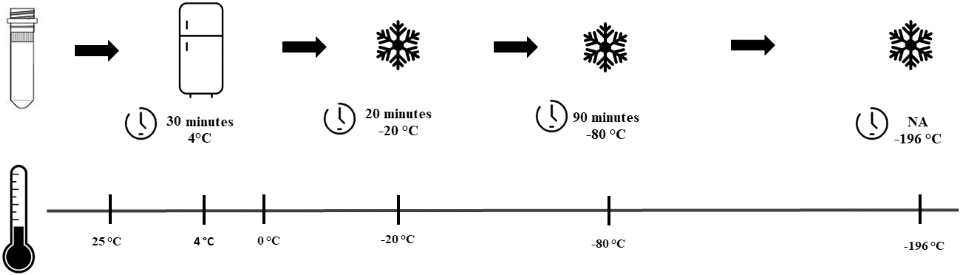
Step-down temperature treatments scheme. NA, not applicable.
Viability tests
To verify the viability before and after preservation of C. gattii and C. neoformans suspensions separately, as well as mixed, 100 μL of the final suspensions were inoculated into three yeast-malt agar (Difco Laboratories) Petri dishes. The plates were incubated at 25°C for 48 h for subsequent counting of colony-forming units (CFU) per 100 μL, and the mean was calculated. In addition, for suspension containing both species, the sample size was calculated for each of the triplicate counts and colonies were randomly selected and cultivated in
Thermotolerance tests
A cell suspension was adjusted to the standard 0.5 McFarland scale (1–5 × 10⁶ CFU·mL−1), and serial dilutions were prepared at the concentrations of 105 and 104 CFU·mL−1. Two microliters of each concentration was inoculated onto Sabouraud dextrose 2% (SD2; Difco Laboratories) Petri dishes in different spots and incubated at 25°C, 37°C, and 40°C for 72 h. The Petri dishes at different temperatures were observed for the presence or absence of cell growth. All plates with no growth were reincubated at 25°C for 24 to 48 h to observe the presence of cell growth. The well-characterized high (CDCR265) and low (CDCR272) virulent strains from the Vancouver Island outbreak were used as control.25,26 The thermotolerance was analyzed visually where: (−) no growth; (+) growth at one inoculation point; (++) growth at two inoculation points; and (+++) growth at three inoculation points.
Melanin production
Similar methodology to that described for thermotolerance was used for the melanin production. However, the Niger seed agar (NSA) medium was used and the Petri dishes were incubated for 5 days. The high (CDCR265) and low (CDCR272) virulent strains were used as control. The melanin production on NSA was analyzed visually and a scale from (−) negative to (++++) high melanin production were applied, where: (−) did not produce apparent melanin with white coloration; (+) produced a beige coloration; (++) light brown; (+++) brown; and (++++) dark brown.
Capsule size
After growth of 48 h at 25°C on SD2, a cell suspension adjusted to the standard 0.5 McFarland scale (1–5 × 10⁶ CFU·mL−1) was prepared. Capsule production was induced by inoculating 0.1 mL of the suspension in SD2 broth, pH 7.0, diluted 1:10 with 50 mM morpholino propanesulfonic acid (MOPS) buffer (Sigma Aldrich, USA), pH 7.3. The tubes were incubated at 36°C for 48 h, without stirring. 27
After the incubation period, ∼10 μL of the culture was placed on a microscopic slide with a drop of 1% nigrosin in glycerol; covered with a clear glass cover for observation under light field optical microscope (Zeiss, Axio Scope.A1) and 40 × magnification. The capsule formation was indicated by a transparent halo around the cell, in contrast to the dark background of the preparation. The thickness of the capsule was measured as the distance between the boundary of the cell (cell wall) and the transparent border produced by nigrosin. At least 40 cells were evaluated.
PCR-fingerprinting
The molecular typing of Cryptococcus strains was performed according to the methodology described by Meyer et al., 28 which is based on random amplification of DNA fragments using the microsatellite-specific primer M13 (5′GAGGGTGGCGGTTCT 3′). The PCR fingerprinting amplicons were visualized on 1.4% agarose gels under UV light after running at 70 V to 14 cm. This methodology was used to observe the band pattern visually and to analyze possible differences between preservation methods. The strains, C. neoformans (CFRVS 70301—WM 629) and C. gattii (CFRVS 70302—WM 178), were used as molecular type VNIV and VGII, respectively.
Statistical tests
The number of colonies cultivated on CGB medium was calculated using the Kruskal–Wallis test, followed by Dunn's multiple comparisons test to calculate statistical differences between the methods used. Wilcoxon test was applied to compare the proportion CGB+ and CGB- over the times with the different methods. In all cases, p values ≤0.05 were considered significant. All data were analyzed, and plots were performed using GraphPad Prism version 8.02 for Windows, GraphPad Software, La Jolla, CA.
Results
The mean of viable cell counts, before preservation, in skim milk of C. gattii and C. neoformans separately were 47 CFU × 100 μL−1 and 50.33 CFU × 100 μL−1, respectively. In glycerol, they were 27.66 CFU × 100 μL−1 and 32.66 CFU × 100 μL−1, respectively. After mixing the species, the viable count was 40 CFU × 100 μL−1 in skim milk and 32 CFU × 100 μL−1 in glycerol. Of the total colonies recovered on CGB, after mixture, C. neoformans was 45.8% in skim milk and 54.7% in glycerol; C. gattii presented 54.1% and 45.3%, respectively. The viability counts of C. neoformans, C. gattii, and mixed cells, before preservation, in both skim milk 20% and glycerol 15% suspensions did not show significant variations (Fig. 2).
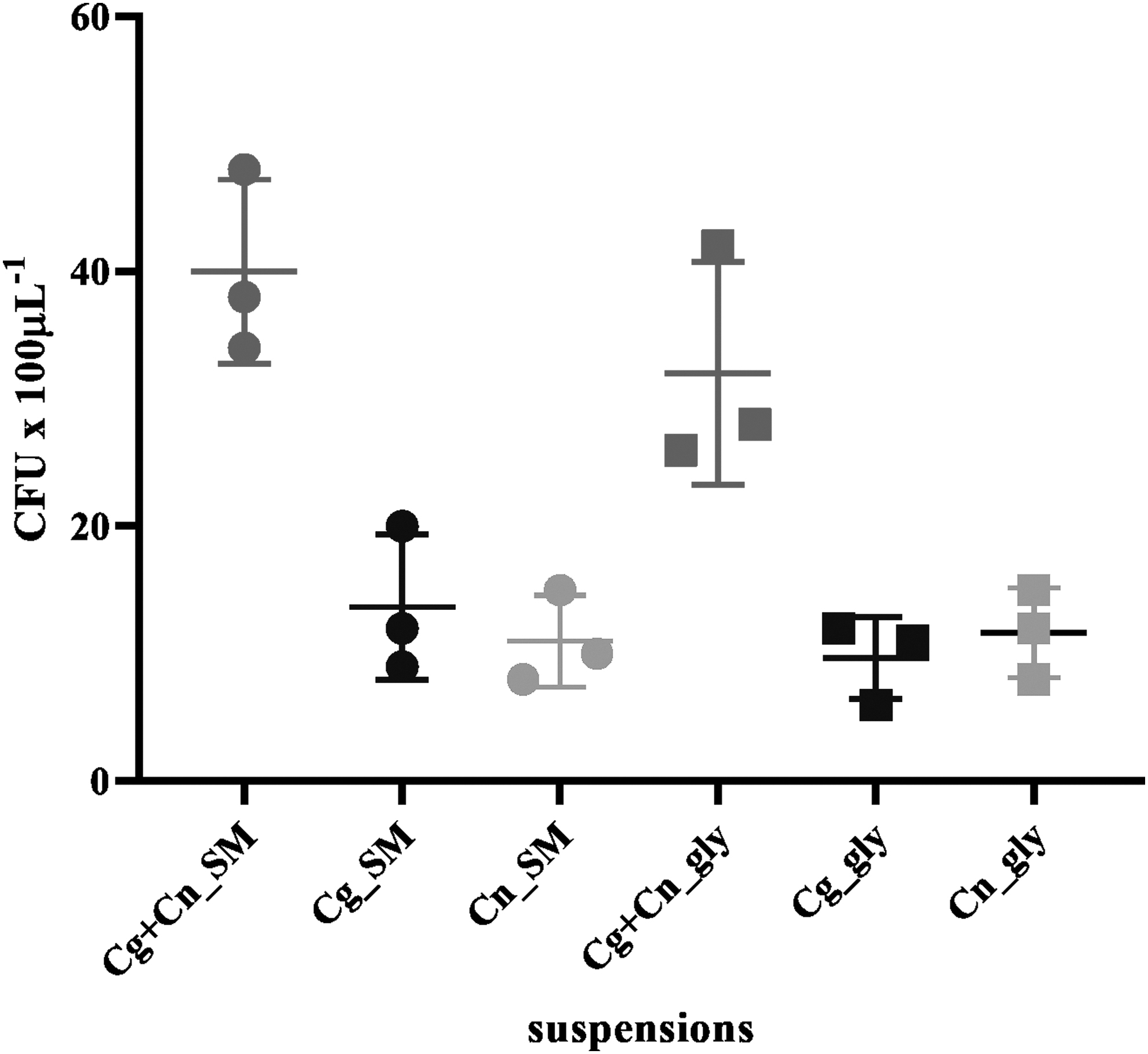
Colony Forming Units of C. gattii – CFRVS 40324 (Cg) and C. neoformans – CFRVS 40323 (Cn) separately and mixed (Cg+Cn) in 20% skim milk (SM) and 15% glycerol (GLY) before preservation.
In the first week, the viability of cells, after preservation, demonstrated that skim milk methodologies presented better recuperation of the species, freeze-drying did not result in a decrease, and cryopreservation at −20°C presented a loss of 41%. The methodologies that used glycerol as cryoprotectant had a large decrease, ultra-cryopreservation at −80°C decayed 62% and liquid nitrogen 73% (Fig. 3).
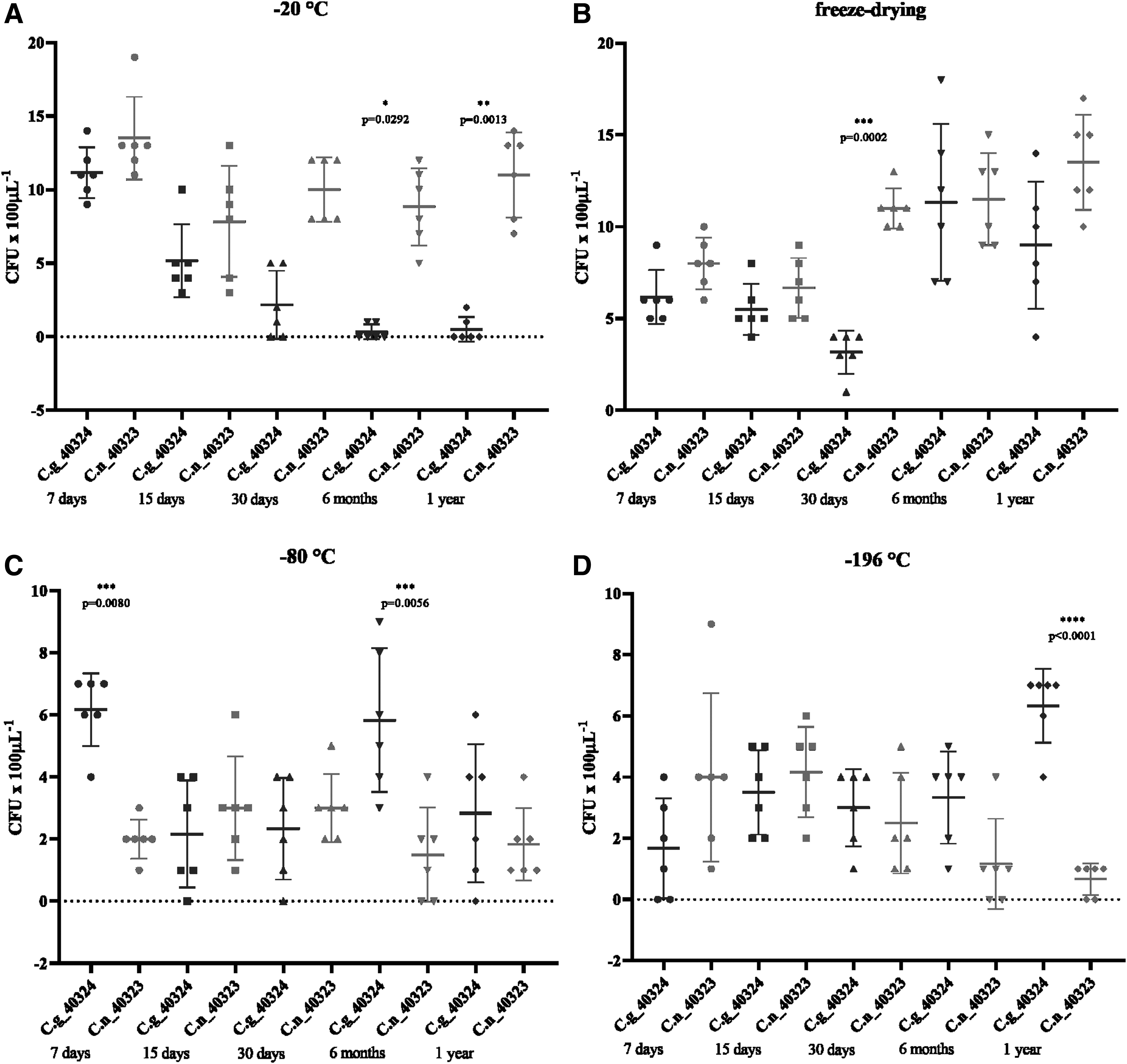
Cell counts of C. gattii – CFRVS 40324 (C.g) and C. neoformans – CFRVS 40323 (C.n) colonies in different periods and methods of preservation; at −20 °C
The recovery of C. gattii and C. neoformans colonies varied according to the methodology used and the period studied. The recovery of C. neoformans colonies was significantly higher than C. gattii colonies after preservation over 30 days at −20°C (p = 0.0292 in 6 months; p = 0.0013 in 1 year). Freeze-drying showed a good performance in maintaining the proportion of both species, however, a discrepancy was observed only at 30 days (p = 0.0002). Ultra-cryopreservation at −80°C favored C. gattii recovery in 7 days (p = 0.0080) and 6 months (p = 0.0056). Despite ultra-cryopreservation at −196°C showing the highest decrease of CFU, this methodology maintained the proportion between both strains. A discrepancy was observed only after 1 year of preservation (p < 0.0001) (Fig. 3).
After 1 year of preservation, the strains of C. gattii and C. neoformans preserved together were reisolated, and phenotypic and genotypic tests were performed to evaluate possible changes concerning the different preservation methods used. Both strains of C. gattii and C. neoformans, as well as the controls, were considered +++ growth at 25°C and 37°C, since they presented growth at the three different concentrations. There was no growth at 40°C and the result remained the same after reincubation of these plates at 25°C. All strains showed high melanin production at 25°C, being considered positive (+++) (Table 1). The result was the same for the strains incubated at 37°C, excepting the control C. gattii (CDCR272) that showed beige coloration being considered (+). No strain grew at 40°C (Table 1).
Molecular and Phenotypical Analysis of the Cryptococcus neoformans (CFRVS 40323) and Cryptococcus gattii (CFRVS 40324) Strains After 12 Months Preservation
CFRVS, collection of reference fungi in sanitary surveillance; LN, liquid nitrogen (−196°C); lyo, lyophilization; PCR, polymerase chain reaction.
The only phenotypical test which demonstrated differences between before and after preservation was capsule size. The median capsule sizes varied significantly, both between the C. gattii group (p = 0.0010) and the C. neoformans group (p = 0.237). For multiple comparisons test, only the group C. gattii showed statistical significance, between C. gattii_−20 vs. C. gattii_lyo (p = 0.0008) and C. gattii_lyo vs. C. gattii_−80 (p = 0.0172) (Fig. 4).
Capsule size of C. gattii – CFRVS 40324 (C.g) and C. neoformans – CFRVS 40323 (C.n).
No difference was observed in the PCR-fingerprinting analysis of the strains preserved by different methods (Fig. 5), where CFRVS 40324 was compatible with the molecular type VGII and CFRVS 40323 was VNIV (Table 1).
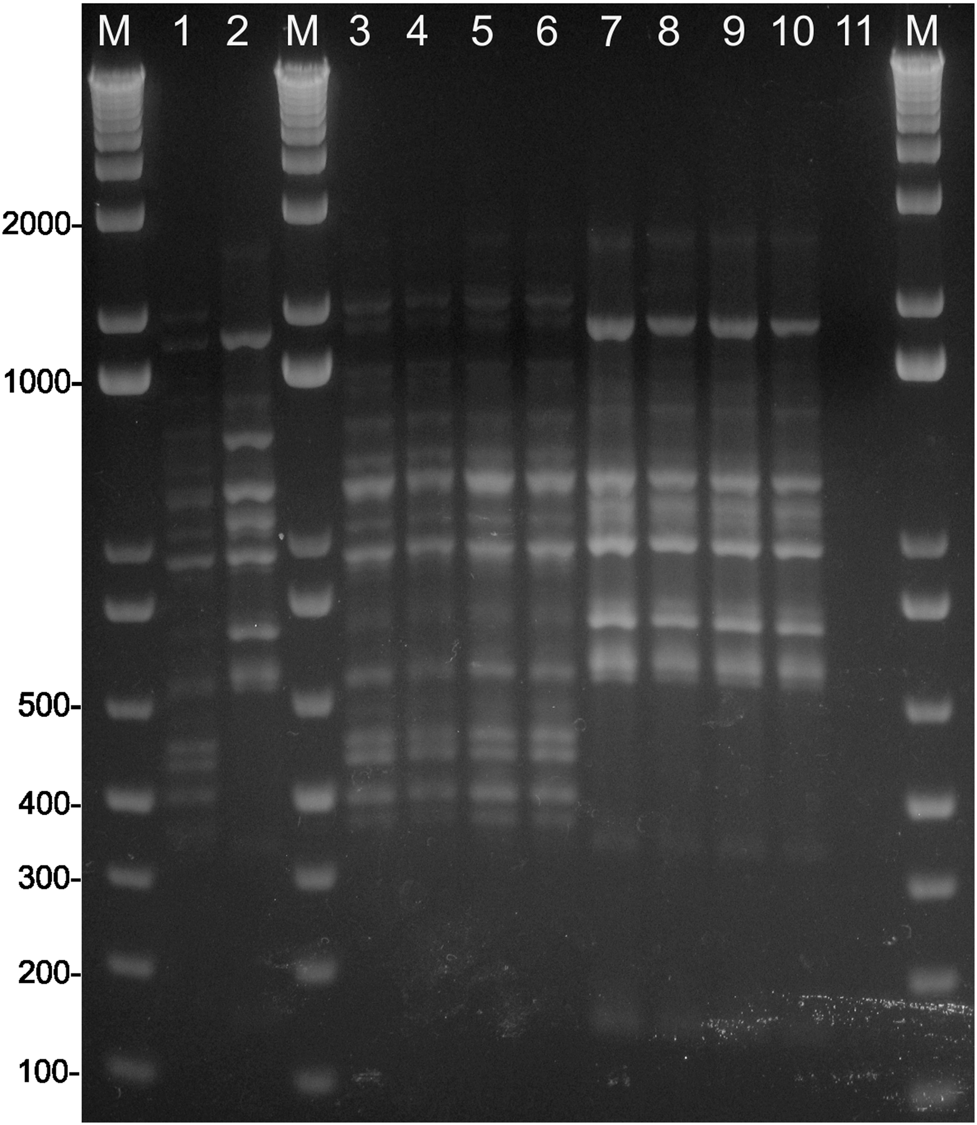
PCR-fingerprinting patterns obtained with primer M13. Lanes: 1—molecular type VNIV (CFRVS 70301—WM 629), 2—molecular type VGII (CFRVS 70302—WM 178), 3—CFRVS 40323 (−20°C), 4—CFRVS 40323 (freeze-dried), 5—CFRVS 40323 (−80°C), 6—CFRVS 40323 (−196°C), 7—CFRVS 40324 (−20°C), 8—CFRVS 40324 (freeze-dried), 9—CFRVS 40324 (−80°C), 10—CFRVS 40324 (−196°C), 11—blank, M—1 kb plus ladder. PCR, polymerase chain reaction.
Discussion
The agents of cryptococcosis, members of the C. neoformans and C. gattii species complexes, are traditionally divided in eight major molecular types. These molecular types differ in their epidemiological, ecological, and molecular characteristics,29,30 antifungal susceptibility, 30 clinical presentations, and therapeutic outcomes. 31 Infections caused by C. gattii often have a worse prognosis than those caused by C. neoformans, 32 but in the regions where both species complexes are endemic, mixed infections are not rare. Desnos-Ollivier et al. showed that almost 20% of cryptococcosis cases were associated with multiple strains of the fungal pathogen C. neoformans, however, the authors point out that the clinical routine, where analysis is of only one colony from only one anatomical site, would make it difficult to find mixed infections; and that the scarcity of reports can occur due to technical bias. 2 Thus, it is important to maintain the original isolates in long-term storage for future studies on mixed infections and its clinical outcomes, and also for the monitoring of virulence factors, such as those presented in this work (thermotolerance tests, melanin production, and capsule size).
The freezing at −20°C method obtained a good total cell recovery, although the recovery of both species C. gattii and C. neoformans differed one month, indicating C. neoformans is more resistant to this method (Fig. 3). Freeze-drying showed the highest total cell viability counts and for the maintenance of viability for both species. The ultrafreezing methods (−80°C and −196°C) showed good results in the maintenance of proportion of both species, despite the low overall viability counts. C. gattii overcame C. neoformans viability count in 1 week and 6 months at −80°C and 1 year at −196°C (Fig. 3). Considering that the differences observed at only one point can be a consequence of a bias in randomly picked colonies, preservation at −20°C and freeze-drying using skim milk 20% were shown to favor C. neoformans species after long-term storage. Miyamoto-Shinohara et al. stated that the presence of extracellular polysaccharides in freeze-dried bacteria is disadvantageous for long-term survival. C. neoformans and C. gattii have a capsule composed primarily of a high molecular weight polysaccharide. In this study, C. gattii presented capsule sizes larger than C. neoformans, which could explain the predominance of C. neoformans using skim milk as cryoprotectant. 33 It is important to note that C. gattii presented the favored viability with the use of glycerol when compared to C. neoformans. Even so, viability counting after preservation using glycerol demonstrated a higher loss of cells. This could be related to the amount of cells used in this study, which is smaller than the concentration of at least 107 CFU × mL−1 recommended. 34 In the CFRVS collection, the cryopreservation methods with glycerol at 15% do not show significant decrease in viability after 7 days of preservation, and it is usually without logarithmic loss. Another possible reason for the high cell loss using glycerol is that the mode of action of these different cryoprotectants can influence the cell viability, since the skim milk acts extracellularly and the glycerol intracellularly, with the last one possibly causing higher cellular damage. 12 However, future studies on cryoprotectants should be performed to confirm this theory. All the preservation methods did not interfere with the virulence factors thermotolerance and melanin, but the differences were observed in the C. gattii capsule size specifically between the freeze-drying methodology and freezing at −20°C and −80°C. Although freezing and lyophilization methods are the most suitable for strains' maintenance, neither of the storage techniques guarantees total stability for the strains 35 and thus microevolution processes can occur causing changes in strain characteristics even during preservation.
According to our results, freeze-drying should be preferred for the maintenance of multilineage isolates, but it requires specialized and expensive equipment, 10 which are not often present in the research laboratories. Homogeneity is an important factor to be considered in the production of microorganisms lots, especially in culture collections. Despite the care taken in the lots production used in this study, such as constant agitation in ice bath, apparently there was some deviation in freeze-drying lots, because recovery was better after 6 month and 1 year than in the previous analyses.
Freezing at −20°C is low-cost and used to keep a few organisms for as long as 1–2 years. 36 At this storage temperature, the preservation period varies depending on the cryoprotectant used. 34 For example, although preservation at −20°C did not perform satisfactorily in the present study, adjustments, such as the use of combined cryoprotectants, should be evaluated, especially in facilities where more than one methodology can be used. Reductions in labor costs, operation time, and the space required for the containment of a large number of cultures are also desirable. 37 Nevertheless, such a universal method has not been created up to now. Therefore, it is highly desirable to develop new, or improve the current preservation methods, combining advantages and eliminating disadvantages of individual techniques. 10
Cryopreservation at −20°C favors C. neoformans isolates and further studies on long-term storage conditions are necessary. The methods that used skim milk (freezing at −20°C and freeze-drying) had a smaller loss of the initial suspension, while the ultrafreezing methods had, despite the significant loss over the initial suspension, stable counts for both species. Adjustments in the concentration of cells and composition of the cryoprotectant can improve the performance of these preservation methods.
The present work mainly concluded that freeze-drying should be preferred for the maintenance of multilineages isolates of the C. neoformans and C. gattii species complexes. This is a new and important study in the field of the cryptococcosis agents, which includes virulence factors as essential characteristics to be preserved for future studies that will be of great significance for the survival of the yeasts in the mammal host.
Footnotes
Authors' Contributions
C.R.S.N. and L.T.: Conception of the study and writing of the article; C.R.S.N., A.S.S.S., and R.M.G. participated in the design; M.S.L. writing of the article.
Acknowledgments
We thank Prof. Wieland Meyer and Prof. Rodrigo de Almeida Paes for the revision of the article.
Author Disclosure Statement
No conflicting financial interests exist.
Funding Information
This work was supported by “Conselho Nacional de Desenvolvimento Científico e Tecnológico” [grant numbers 442837-2014-3 to L.T.].