
Abstract
Aim:
This study compared growth kinetics of human amniotic fluid stem cells (hAFSCs) in different maternal age groups and two different media of AmnioMAX and Dulbecco's modified Eagle's medium (DMEM).
Materials and Methods:
Three milliliters of amniotic fluid (AF) was provided from 16 pregnant women who were referred for amniocentesis from 16 to 18 weeks of gestation. Mothers were divided to 20–29 (n = 5), 30–39 (n = 5) and 40–49 (n = 6) years old age groups. AF was immediately centrifuged and the cell pellet was cultured. Cells were characterized morphologically, by flow cytometry and for osteogenic and adipogenic inductions. Population doubling time (PDT) and growth kinetics were determined. AFSCs cultured in AmnioMAX were compared in various age groups. A comparison of growth kinetics of AFSCs cultured in AmnioMAX and DMEM from 40 to 49 years old pregnant women was undertaken.
Results:
AFSCs were adherent to culture flasks and were spindle shape, and positive for osteogenic and adipogenic inductions and for expression of CD73, CD90 and CD105 markers, but negative for CD34 and CD45. PDT among 20–29, 30–39, and 40–49 years old women was 30.9, 38.3, and 43.9 hours, respectively showing a higher cell proliferation in younger ages. When comparing AmnioMAX and DMEM, PDT was 53 and 96.9 hours, respectively denoting to a higher cell proliferation in AmnioMAX.
Conclusions:
Higher proliferation and plasticity of hAFSCs were noted in AmnioMAX and in younger mothers' samples. These findings can be added to the literature and open a new avenue in regenerative medicine, when hAFSCs are targeted for cell therapy purposes.
Introduction
Mesenchymal stem cells (MSCs) are multipotent undifferentiated cells with self-replicating ability that have been isolated from various tissues including bone marrow, 1 adipose, 2 Wharton's jelly, 3 dental pulp, 4 and amniotic fluid (AF). 5 MSCs have self-renewal, differentiation, migratory, anti-inflammatory, and immunosuppressive characteristics, which are needed for cell-based therapies.6,7 Human gestational tissues have been demonstrated to be a rich source of different stem cell populations. AF has been proposed as an alternative source of stem cells for tissue regeneration. Mid-trimester and term human amniotic fluid stem cells (hAFSCs) are available in large quantities and have been well characterized as multipotent cells that can be a resource for clinical transplantation and allow their future therapeutic use as an “off-the-shelf” cell source in regenerative medicine.8,9
hAFSCs collected during pregnancy were demonstrated to have remarkable plasticity, and immunomodulatory effects and have the potential for the treatment of various diseases especially in neonates when autologous cell therapy is required; even immediately after birth.5,9 They also have a pluripotent potential property identical to embryonic stem cells (ESCs) or induced pluripotent stem cells (iPSCs) that can be reprogrammed to a pluripotency potential without any genetic manipulation. 10 The use of AFSCs does not lead to ethical concerns that were reported for ESCs and do not have the risks of teratoma formation seen in the case of ESCs and iPSCs. 9
AFCs can also be banked easily and used for both allogeneic and autologous transplantation. 11 Corcelli et al. used a mouse model of hypoxic-ischemic encephalopathy and illustrated that injection of hAFSCs had neuroprotective effects. 12 Otani et al. demonstrated that hAFSCs could successfully migrate to the brain and induce functional recovery by preserving neurons and oligodendrocytes, and by reducing neuroinflammation. 5 George et al. illustrated that hAFSCs facilitated functional and structural improvement in a rat model of chronic kidney disease. 9
Intravascular injection of hAFSCs revealed a cardioprotective effect and improvement in cell survival and decrease in the infarct size. 13 Therefore, based on clinical application of hAFSCs, characterization and assessment of growth kinetics of these cells seem necessary and this study aimed to determine the growth kinetic of hAFSCs in pregnant women in different age groups and two different routine culture media.
Materials and Methods
Isolation of hAFSCs
AF samples were provided from women who referred to the Department of Obstetrics and Gynecology of Shiraz University of Medical Sciences, Shiraz, Iran for prenatal diagnoses and amniocentesis at 16–18 weeks of gestation, and no abnormal findings were recorded in their reports. The subjects were 16 pregnant women from three different age groups of 20–29 (n = 5), 30–39 (n = 5), and 40–49 (n = 6) years. A written informed consent was provided from each woman and institutional ethical approval for the study. Three milliliters of AF were obtained from each woman and was immediately transferred in a 15 mL falcon on dry ice to the Stem Cell Technology Research Center, Shiraz University of Medical Sciences, Shiraz, Iran.
Culture of hAFSCs
The samples were centrifuged at 200 g for 10 minutes at room temperature (RT), the supernatant was discarded, and the remaining pellet was suspended in AmnioMAX -II medium (Gibco®, ThermoFisher Scientific, Germany) as a complete fully-supplemented medium developed for culture of hAFSCs. The cell suspension was transferred to T25 culture flasks containing media and incubated in an incubator at 37°C with 5% CO2 and saturated humidity. The first media change was conducted after 5 days to remove nonadherent cells. Then, the medium was replaced every 3 days and cell harvesting was performed when they reached 80% confluence using Trypsin-EDTA, and the cells were then routinely subcultured at 1:3 dilution.
hAFSCs cultured in AmnioMAX were compared in different age groups. Cells from 40 to 49 year old women, as the oldest age group, which were expected to have the least proliferation rate, were used to compare AmnioMAX and low glucose Dulbecco's modified Eagle's medium (DMEM; Sigma-Aldrich) supplemented with 10% fetal bovine serum (FBS; Gibco Life Technologies, Italy), 5 ng/mL human basic fibroblast growth factor (R&D Systems), 1% penicillin/streptomycin (Sigma-Aldrich), and 1% L-glutamine (Sigma-Aldrich). All experiments were undertaken on cells at the fourth passage (P4).
Cell characterization
Morphologic characterization
hAFSCs from all samples at all passages were morphologically evaluated under an inverted microscope (Olympus, Japan).
Characterization by flowcytometry
A total of 1 × 10⁵ hAFSCs were used to evaluate the expression of CD73, CD90, and CD105 as mesenchymal markers and CD34 and CD45 as hematopoietic markers using flowcytometry (Partec, Münster, Germany) as described by Corcelli et al., 2018. 12
Characterization by in vitro osteogenic differentiation and Alizarin Red staining
hAFSCs were seeded at a density of 1.5 × 10⁴ cells in six-well plates and expanded in DMEM. Osteogenic induction was undertaken by culturing the hAFSCs with DMEM supplemented with 10% FBS, 0.05 mM ascorbic acid (Sigma-Aldrich), 10 mM β-glycerophosphate (Sigma-Aldrich), and 100 nM dexamethasone (Sigma-Aldrich) for 21 days, while replacing the media every 3 days. To visualize calcium sediments, cultures were stained with Alizarin Red (Sigma-Aldrich) and visualized under an inverted microscope.
Characterization by in vitro adipocyte differentiation and Oil Red O staining
hAFSCs were seeded at a density of 1.5 × 10⁴ cells in six-well plates and expanded in DMEM. At 80% confluence, the medium was changed to an adipogenic induction medium that contained DMEM supplemented with 10% FBS, 1% penicillin/streptomycin, 1 mM dexamethasone (Sigma-Aldrich), 10 μg/mL insulin (Sigma-Aldrich), 0.5 mM 3-isobutyl-1-methylxanthine (Sigma-Aldrich), and 200 μM indomethacin (Sigma-Aldrich). hAFSCs were cultured in the supplemented media for 3 weeks. Adipogenic differentiation was verified on day 21 using an Oil Red O staining (Sigma-Aldrich) to demonstrate the intracellular lipid accumulation. In summary, hAFSCs were washed twice in phosphate-buffered saline (PBS), fixed with 4% formaldehyde for 10 minutes at RT, rinsed once with 3% isopropanol, and stained with Oil Red O staining solution. Thereafter, the cells were rinsed with distilled water and visualized under the inverted microscope.
Cell viability
Trypan blue exclusion test (0.4% trypan blue in PBS) was conducted for each passage of hAFSCs to assess the number of cells with intact membranes (viable cells), with the percentage presented as the number of the viable cells divided by the total number of cells.
The population doubling time
To quantify hAFSCs, the cells at the fourth passage were seeded in 24-well culture plates (1 × 10⁵ cells per well). The cells were enumerated until 8 days by trypsinization (three replicates for each time point) and the mean number of the cells at each time point was traced using GraphPad Prism (version 6.01; GraphPad Software, Inc., San Diego, CA). The cells were stained by trypan blue (Sigma-Aldrich) and counted by a hemocytometer using a light microscope. PDT was defined as the time needed in culture to double the cell number and was determined by the following formula as described before,
4
where T was the incubation time in hours, Xb was the cell number at the beginning of the incubation time, Xe was the cell number at the end of the incubation time, Ln = loge, v was the volume of media, n1 to n4 were the frequency of cell count up to four times, and e = the Euler's number.
Growth kinetics
The growth curve was compared for different age groups and for DMEM and AmnioMAX culture media among the 40–49 years age group.
Statistical analysis
Statistical Package for the Social Sciences (SPSS, version 20, Chicago, IL) was employed for the statistical analyses. The mean ± standard error of the counted cells was compared using the Mann–Whitney U test and a p ≤ 0.05 was regarded statistically significant.
Results
Cell characterization
Morphologic characterization
hAFSCs at all passages in all samples were adherent to culture flasks and were spindle shaped in morphology visualized under the inverted microscope (Fig. 1A–D).
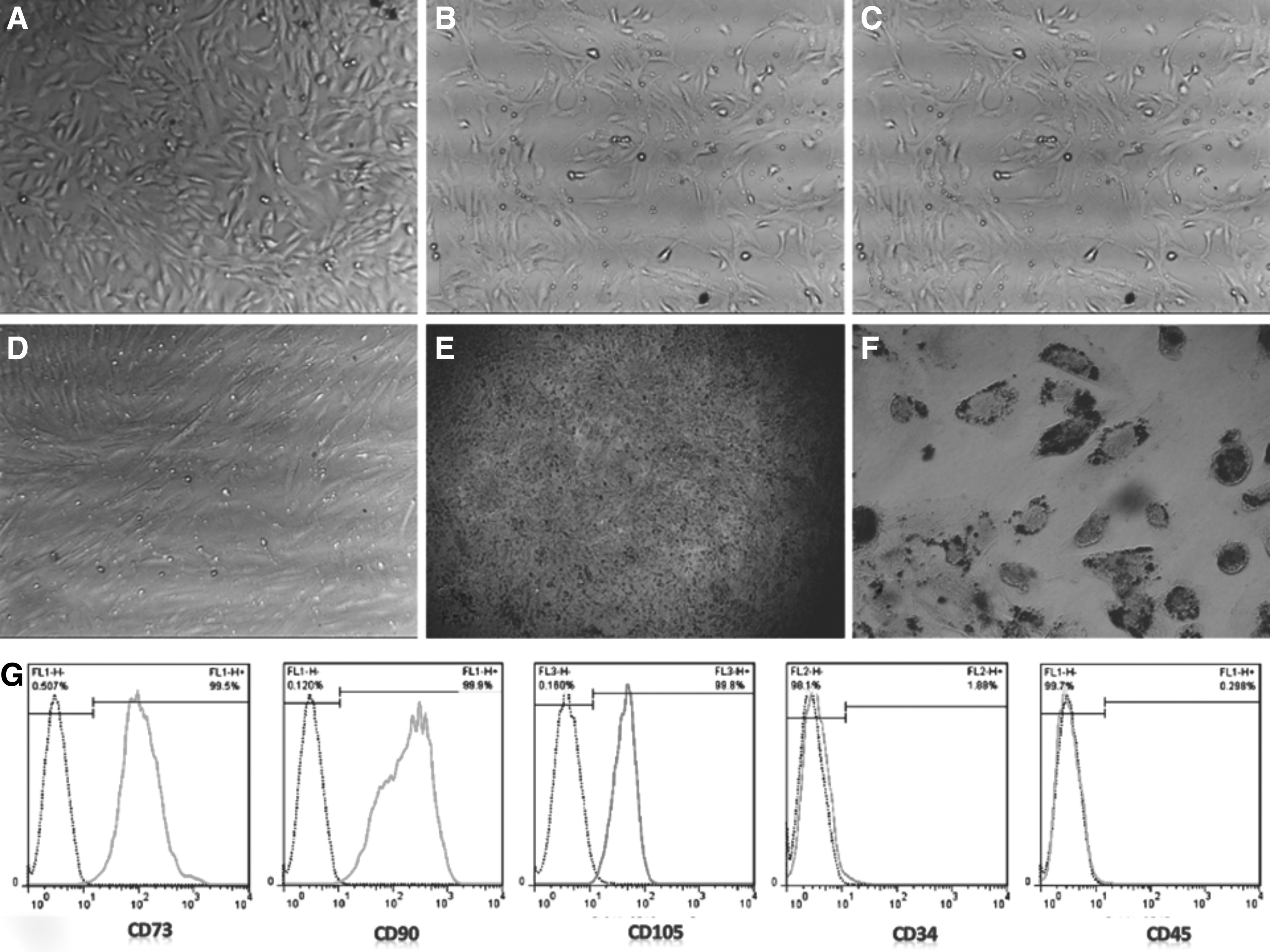
Morphology of human AFSCs isolated from AF.
Osteogenic and adipogenic characterization
Staining of hAFSCs by Alizarin Red indicated positive osteogenic differentiation by presence of calcium deposits appearing in red color in the cells (Fig. 1E). Staining of hAFSCs by Oil Red O revealed positive adipogenic induction by presence of oil droplets in red color in the cells (Fig. 1F).
Characterization by flowcytometry
The hAFSCs were positive for expression of CD73, CD90, and CD105 mesenchymal surface markers, and were negative for expression of CD34 and CD45 hematopoietic surface markers (Fig. 1G).
PDT
The PDT value of hAFSCs isolated from 20 to 29, 30 to 39, and 40 to 49 years old pregnant women that were cultured in AmnioMax was 30.9, 38.3, and 43.9 hours, respectively. The findings showed that the highest proliferation of AFSCs was visible among the 20–29 years old group and the least among the 40–49 years old group. The PDT of hAFSCs recovered from 30 to 39 years old women was more than 40–49 years old group and less than 20–29 years old subjects. The lag phase in the three age groups was 1 day and the maximum proliferation was among 20–29 years old women on the 6th day and for 30–39 and 40–49 years old subjects was on the 7th day (Fig. 2; p < 0.0001).
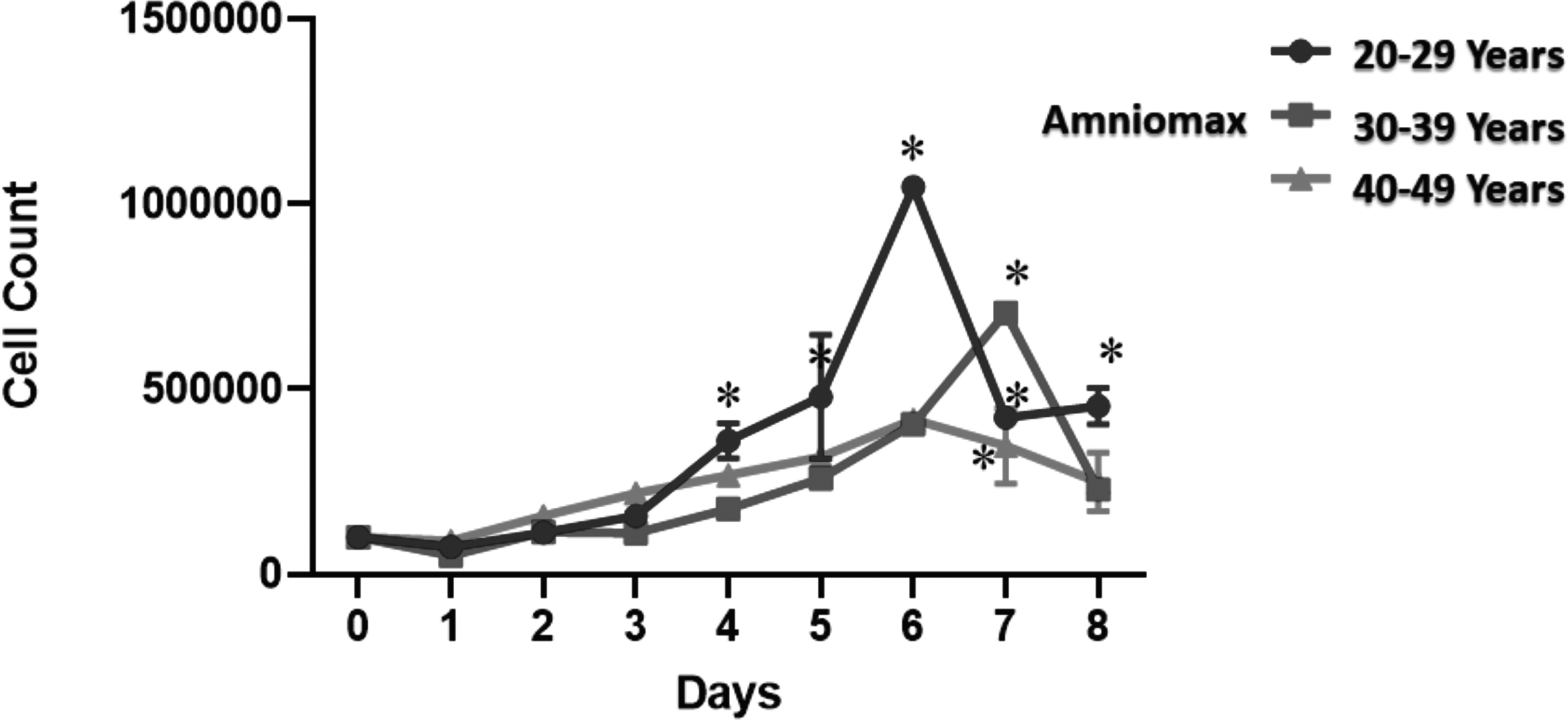
Comparison of hAFSCs isolated from AF of 20–29, 30–39, and 40–49 years old pregnant women cultured in AmnioMax media. *p < 0.0001.
When comparing proliferation of AFSCs in AmnioMAX or DMEM among 40–49 years old women, a significant statistical difference was noted between the groups, while PDT in DMEM was 96.9 hours and in AmnioMAX was 53 hours, denoting a higher proliferation rate of hAFSCs in AmnioMAX in comparison to DMEM (Fig. 3; p < 0.001).

Comparison of the growth kinetics of AFSCs isolated from 40 to 49 years old pregnant women cultured in AmnioMax and DMEM. *p < 0.001. DMEM, Dulbecco's modified Eagle's medium.
Discussion
MSCs are present in the stroma of several tissues including AF, while AF contains a subpopulation of stem cells able to be differentiated into adipocytes, chondrocytes, osteocytes, and other mesodermal derivatives.14–19 Differentiation of ovine AFSCs showed successful induction to adipocytes, osteoblasts, and chondrocytes. 20 Shaw et al. compared early and late second trimesters for potential differentiation of hAFSCs and showed that hAFSCs in early second trimester had more potential for differentiation properties. 21 We also reported here the successful differentiation properties of hAFSCs to osteocytes and adipocytes.
Regarding the mesenchymal property of AFSCs, Alessio et al. compared AFCs with bone marrow-derived stem cells in terms of phenotype potential and found positive expression of CD29, CD44, CD73, CD90, and CD146 and negative expression of CD14, CD45, CD117, and CD144, demonstrating that AFSCs had similar mesenchymal features as bone marrow-derived stem cells.14,15 Filioli Uranio et al. compared canine amnion- and umbilical cord-derived MSCs and successfully characterized the mesenchymal properties of these cells. 22
Characterization of ovine AFSCs by Pei et al. showed positive expression of mesenchymal markers and negative expression of hematopoietic markers. 20 Spitzhorn et al. in AFSCs isolated from the AF of both amniocentesis and caesarean section illustrated the mesenchymal properties of cells regarding the morphology, differentiation potential, and expression of mesenchymal surface markers. 23 hAFSCs isolated in our study from different age groups of pregnant mothers were positive for CD73, CD90, and CD105 as mesenchymal markers and negative for CD34 and CD45 as hematopoietic markers, confirming the abovementioned studies.14,15,20–24
hAFSCs have been shown to have self-renewal ability and a high growth rate, making them a good candidate for cell transplantation. 25 Varghese et al., in a systematic review on 41 articles regarding factors affecting adipose tissue stem cell viability and function, demonstrated a decreased proliferation and differentiation potential of adipose tissue-derived stem cells (AdSCs) with an increase in age. 26 Naz et al. by comparing dental pulp stem cells (DPSCs) derived from permanent and deciduous teeth observed a shorter PDT for DPSCs isolated from deciduous teeth confirming DPSCs in younger age to have a higher proliferation capacity. 27
Aged AdSCs illustrated senescence features in comparison to cells isolated from young donors, together with a reduction in viability and proliferation. Also, a significant reduction was noticed for differentiation potential of aged AdSCs compared with young AdSCs. 28 Filioli Uranio et al. compared early-passages of MSCs derived from canine amniotic membrane and MSCs derived from umbilical cord matrix at early (35–40 days) and late (45–55 days) gestational ages and reported similar proliferation and differentiation potential of these cells in both gestational ages and believed that these cells can be a good candidate in canine regenerative medicine. 22 In our study and consistent with previous findings;26–28 hAFSCs isolated during 16 to 18 weeks of gestational age demonstrated a significantly higher cell proliferation among 20–29 years old pregnant women in comparison with other age groups (30–39 and 40–49 years old women). Among 30–39 years old subjects, the proliferation was even more than 40–49 years old women.26–28
To determine the appropriate conditions for in vitro expansion of cells, Kobayashi et al. compared DMEM and AmnioMAX in culture of dermal papilla cells and showed AmnioMAX to be more efficient in cultivation. 29 Bratka-Robia et al. similarly recommended AmnioMAX for successful culture of dermal papilla cells. 30 These findings are in agreement with our results illustrating a higher proliferation and plasticity of hAFSCs in amnioMAX medium. There were also limitations in our study including the sample size from healthy pregnant women and AF from different age groups, which were overcome by extending the study period.
Conclusion
Our findings noted for the mesenchymal properties of hAFSCs, a higher proliferation and plasticity of these cells when recovered from younger mothers and a higher proliferation when cultured in AmnioMAX. As samples from AF can be easily prepared during amniocentesis, and hAFSCs can be isolated and expanded in culture for several passages and the ethical and safety of tumorigenic concerns inherent with ESCs do not exist, hAFSCs can be a good source for regenerative medicine purposes. These findings can be added to the literature and open a new window in regenerative medicine, when hAFSCs are targeted for cell therapy purposes.
Footnotes
Acknowledgment
The authors would like to thank Islamic Azad University for financial support of this study.
Author Disclosure Statement
No conflicting financial interests exist.
Funding Information
This study was funded as master thesis by Department of Biochemistry, Science and Research Branch, Islamic Azad University, Shiraz, Iran (Grant number IAU-9391-58-3280).