
Abstract
Human umbilical vein endothelial cells (HUVECs) have great potential in tissue engineering, regenerative medicine, and clinical applications. There is an ever-increasing demand to provide living HUVECs and HUVECs-hydrogel constructs to end users when needed in cell-based therapy and clinical applications. However, current methods to provide living cells and their constructs are mainly continuous culture and cryopreservation, which are high-cost, labor-intensive, time-consuming and inflexible. The research about hypothermic storage of HUVECs and their hydrogel constructs is still limited. Here, we studied the cell survival of HUVECs without encapsulation (W/O Encap) or with encapsulation (alginate, alginate with carboxymethyl chitosan [CMCH]) at 4°C and 25°C during 7 days, respectively. Also, we explored the optimal CMCH concentration for hypothermic storage, which is 0.5% (w/v) at 4°C and 25°C. Moreover, we evaluated the cell attachment after hypothermic storage. Our results enable the hypothermic storage of HUVECs and HUVEC-hydrogel constructs, and facilitate their application in tissue engineering and clinical medicine.
Introduction
Human umbilical vein endothelial cells (HUVECs) have extensive applications in tissue engineering, cell therapy, and many other fields due to their excellent performance in establishing functional vasculature, supporting blood perfusion, promoting wound healing, and so on.1–5 In addition, HUVECs can detect local blood signals, regulate blood vessel tone, and improve hormone transport, thus having great potential in biomedical applications.6–9 In recent years, cell-hydrogel constructs also have drawn attention and play critical role in tissue engineering, cell delivery, regenerative medicine, and treatment of various diseases.10–13 Therefore, there is an ever-increasing demand to provide sufficient cells and cell-hydrogel constructs to end users when needed for clinical applications.
At present, commonly used approaches to provide living cells are as follows: continuous culture and cryopreservation. For continuous culture, 37°C and 5% CO2 humidified incubators are necessary, and the cells need to be passaged and cultured when reaching high confluency. This method needs expensive instruments and requires complex operations.14,15 For the cryopreservation method, the cells were preserved at subzero temperature (e.g., −20°C, −80°C, or −196°C in liquid nitrogen). 16 The cells will suffer from solute damage and mechanical damage during the cooling and warming processes. The cryoprotective agents have cytotoxicity and the ice crystals are harmful to cell membranes. 17 Besides, liquid nitrogen containers are needed to store the cells. 18 The disadvantages of continuous culture and cryopreservation are that they are high-cost, labor-intensive, and time-consuming. Moreover, they rely on laboratory conditions and sophisticated instruments to a large extent, thus making it difficult to transfer cells between different places to support clinical needs. 19 Hence, a convenient and simple approach for preserving cells flexibly is urgently needed.
Recently, hypothermic storage has attracted attention. 20 It is an attractive method to preserve cells between 0°C and 37°C.21,22 The commonly used temperatures are refrigeration (2°C–8°C) and ambient temperatures, which facilitate the short-term storage and transportation of cells and cell-hydrogel constructs.23,24
Alginate is a natural polysaccharide and has been previously used for hypothermic storage of human adipose-derived stem cells, embryonic stem cells, and mouse hepatocytes.25–27 It has been reported that it can protect against mechanical stress and osmotic shock and can stabilize the cell membrane. 28 Moreover, carboxymethyl chitosan (CMCH) is a water-soluble derivative of chitosan and has been used to maintain quality of many fruits and vegetables. It shows good antibacterial activity and can inhibit respiration rate and water loss in fruits and vegetables, thus extending their lifetime.29,30 As far as we know, research concerning hypothermic storage of HUVECs and HUVEC-hydrogel constructs are still limited. Also, there are few studies about the effect of combining alginate and CMCH on hypothermic storage of biosamples.
In this work, we study the cell viability of HUVECs without encapsulation (W/O Encap) or with encapsulation (alginate, alginate and CMCH) at 4°C and 25°C during 7 days, respectively. Also, we compare cell viability when encapsulated with alginate with different CMCH concentrations (0.1%, 0.2%, 0.5%, 1%, and 2% [w/v]). We also assess the cell attachment after hypothermic storage. This work provides a novel approach for hypothermic storage of HUVECs and HUVEC-hydrogel constructs, and facilitates the application of them in cell delivery, tissue engineering, and clinical applications.
Materials and Methods
Chemicals
All chemicals were purchased from Sigma (St Louis, MO) unless otherwise stated.
Cell culture
HUVECs were seeded in a T25 culture flask (Eppendorf, Hamburg, Germany) and were cultured in DMEM (Hyclone, Logan, UT) containing 10% (v/v) fetal bovine serum (FBS; Hyclone). Cells were incubated in a 37°C and 5% CO2 humidified incubator. When the cells reached ∼80%–90% confluence, they were washed with phosphate-buffered solution (Hyclone), and were detached using 0.25% trypsin-EDTA (Gibco, Carlsbad, CA) for 5 minutes. After stopping digestion with DMEM containing FBS, cells were collected by centrifugation at 1000 rpm for 5 minutes and were resuspended with corresponding solutions for subsequent experiments.
Encapsulation and storage of HUVECs
The microcapsules were produced by the electrostatic spraying method. An electrostatic spraying system consisted of a high voltage generator (Dongwen High Voltage Power Supply Co., Ltd., Tianjing, China), a syringe pump (High-Precision Programmable Syringe Pump, WK-101P; Nanjing Anerke Electronics Technology Co., Ltd., Nanjing, China), a needle (diameter of 400 μm), an iron support, and a gelling bath (0.15 mol/L CaCl2 solution). All the solutions were filtered with a 0.22-μm pore size filter before use, and all the accessories were sterilized with 75% (v/v) alcohol or exposed under UV for 30 minutes before use.
The collected HUVECs were mixed with 500 μL 1.5% (w/v) sodium alginate (Aladdin, Shanghai, China) solution or 1.5% (w/v) sodium alginate containing different concentrations (0.1%, 0.2%, 0.5%, 1%, and 2% [w/v]) of CMCH (Macklin, Shanghai, China) with a cell density of 8.0 × 10⁶ mL−1, then the solutions were introduced into 5 mL syringes and pumped with a flow rate of 10 μL/min. The voltage applied between electrodes was 10 kV, and the distance between the needle tip and surface of CaCl2 solution was 3 cm. Microdroplets of mixture solutions were generated at the needle tip and were sprayed into CaCl2 solution to form microcapsules. Later, the microcapsules were collected into a 2 mL cryovial tube (Corning, CA) containing 1 mL culture medium. Meanwhile, the cells without encapsulation were resuspended with 1 mL culture medium and were added into a 2 mL cryovial tube.
All the groups were placed at 4°C and 25°C for 1, 3, 5, and 7 days, respectively. The differential interference contrast (DIC) images of cells and microcapsules were obtained by an inverted microscope, and the diameters of microcapsules were counted using Image-Pro Plus 6.0.
Evaluation of cell viability
Fluorescence staining was used to evaluate the cell viability. After storing at 4°C and 25°C at each time point, HUVEC-laden microcapsules and the cells without encapsulation were stained with an acridine orange/ethidium bromide (AO/EB) staining kit (KeyGen Biotech Co., Ltd., Nanjing, China) (1:1 v/v) for 2 minutes in the darkroom at room temperature. The fluorescence images of cells and microcapsules were acquired by an inverted fluorescence microscope (Ti-U; Nikon, Japan). The live cells were stained green and the dead cells were stained red. Moreover, cells in the microcapsules were released using 75 mmol/L sodium citrate (Sangon Biotech, Shanghai, China) to obtain viability. The cell viability was calculated using the following equation:
Evaluation of cell attachment
To evaluate cell survival after hypothermic storage, HUVECs encapsulated with 1.5% (w/v) alginate and 0.5% (w/v) CMCH at 4°C and 25°C after 7 days were released by 75 mmol/L sodium citrate, respectively. The cells were seeded in 24-well plates and were cultured in a 37°C and 5% CO2 humidified incubator overnight. The DIC images of cells were obtained using an inverted microscope.
Statistical analysis
All data are expressed as means ± standard deviation, and each experiment was repeated at least three times. Statistical analysis was performed with a Student's two-tailed unpaired t-test (p < 0.05 was regarded as statistically significant).
Results
Fabrication of HUVEC-hydrogel constructs
As shown in Figure 1, HUVEC-laden microcapsules were produced by an electrostatic spraying method. The mixed solution of HUVECs and alginate solution was loaded into the syringe that fixed on the syringe pump. Then the high voltage generator was turned on, and the microdroplets were sprayed into the CaCl2 solution. The microdroplets of sodium alginate can quickly react with calcium ions to form microcapsules. The production of microcapsules encapsulated by alginate and CMCH solution can follow the step as mentioned above. Later, the cells and microcapsules were loaded into cryovials containing 1 mL culture medium, and were stored at 4°C and 25°C for 1, 3, 5, and 7 days, respectively.
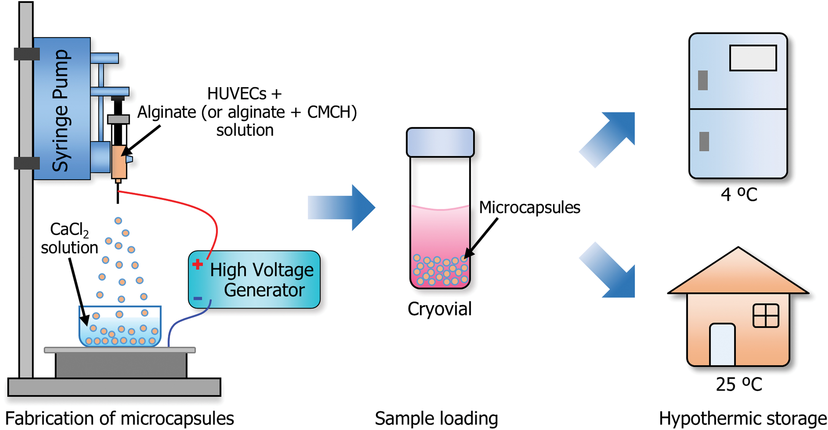
Schematic illustration of fabrication of HUVEC-hydrogel constructs and their hypothermic storage. HUVECs, human umbilical vein endothelial cells. Color images are available online.
Morphologies of HUVEC-laden microcapsules
The morphologies of HUVECs without encapsulation and the HUVECs encapsulated by alginate (or alginate with CMCH) hydrogels are shown in Figure 2. HUVECs without encapsulation were dispersed in culture medium. The microcapsules were almost in spherical shapes, and the size of microcapsules was uniform. HUVECs were uniformly dispersed in the microcapsules. The diameter of microcapsules produced by alginate was 516.3 ± 11.8 μm, and the diameter of microcapsules produced by alginate and CMCH was 528.9 ± 15.4 μm.
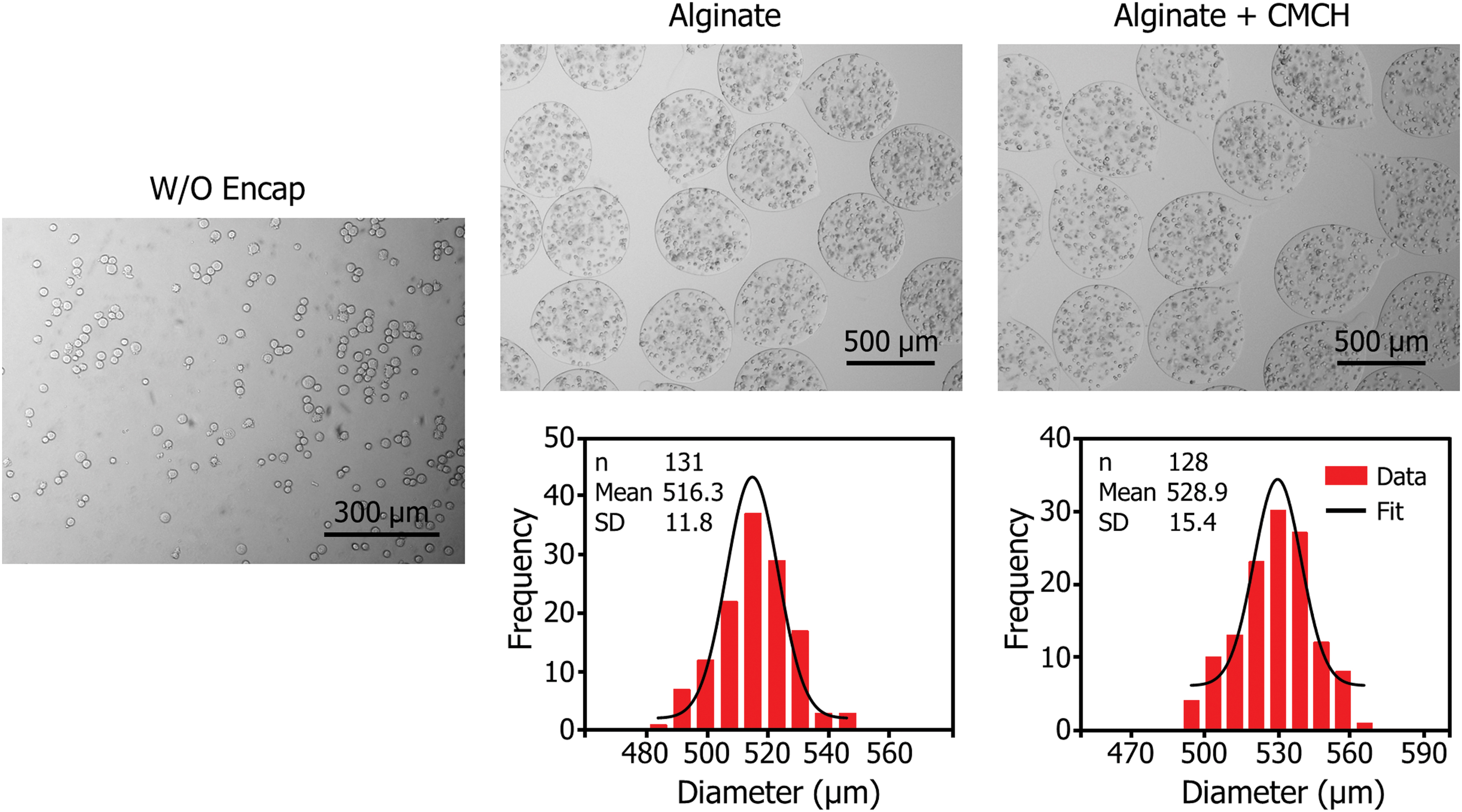
Typical DIC images of HUVECs suspended in culture medium, and DIC images and size distribution of HUVEC-hydrogel constructs. DIC, differential interference contrast. Color images are available online.
Cell viability after hypothermic storage
Cell viability of HUVECs and HUVEC- hydrogel constructs after storing at 4°C and 25°C are shown in Figure 3. According to the results, cells encapsulated with alginate and 0.5% (w/v) CMCH showed higher viability than cells without encapsulation at each time point. In addition, cells encapsulated with alginate and CMCH can survive >7 days under 4°C (cell viability of 81.1%) and 25°C (cell viability of 84.9%).
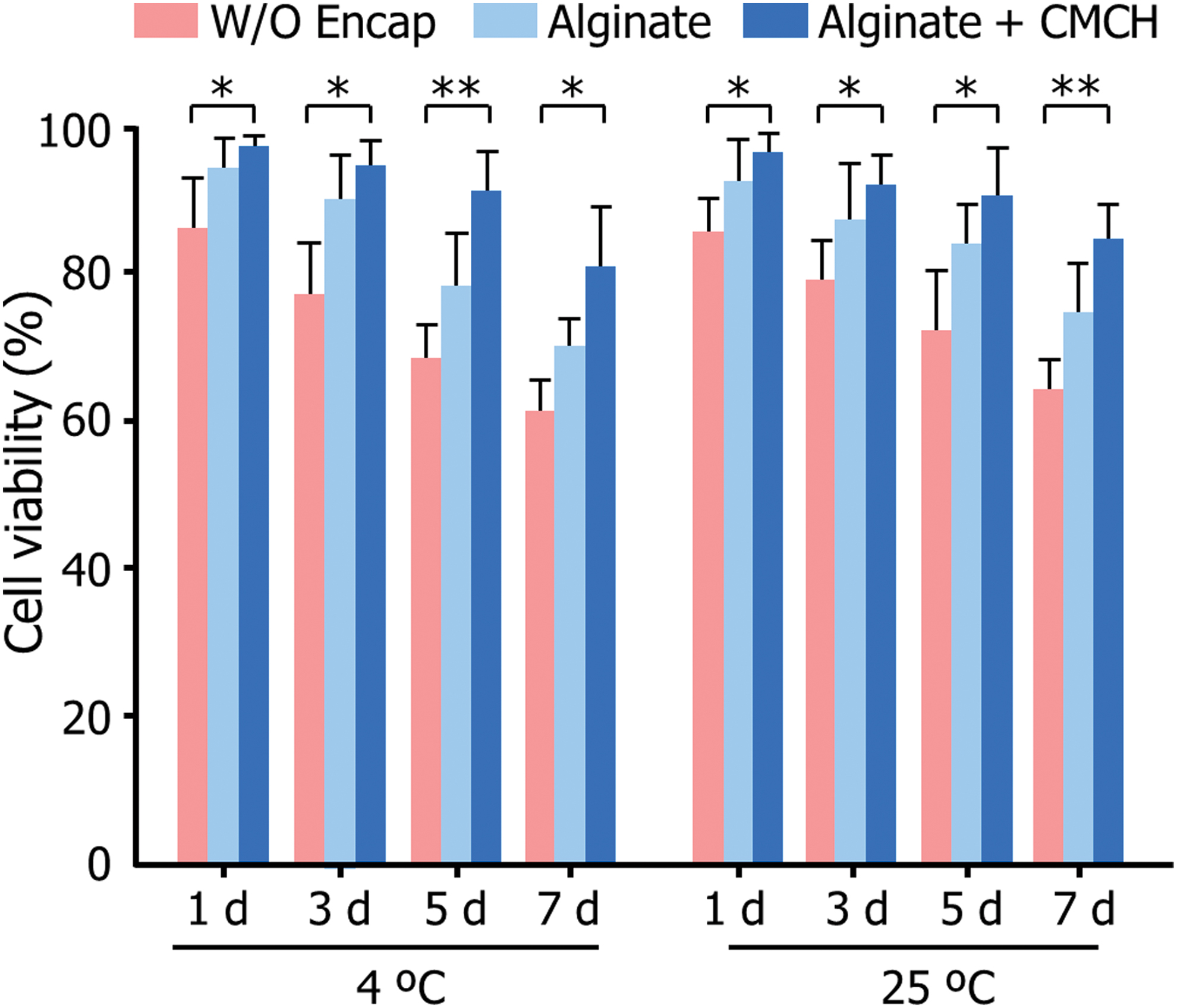
Cell viability of HUVECs and HUVEC-hydrogel constructs after storing at 4°C and 25°C for 1, 3, 5, and 7 days, respectively. (*p < 0.05, **p < 0.01). Color images are available online.
Fluorescence images are also shown in Figure 4. The results were in accordance with the cell viability shown in Figure 3. The cells showed good survival at 4°C and 25°C at 7 days when encapsulated by alginate with 0.5% (w/v) CMCH.

The AO/EB staining images of HUVECs and HUVEC-hydrogel constructs (1.5% [w/v] alginate and 0.5% [w/v] CMCH) after storing at 4°C and 25°C for 1, 3, 5, and 7 days, respectively. AO/EB, acridine orange/ethidium bromide; CMCH, carboxymethyl chitosan. Color images are available online.
The effect of concentration of CMCH on cell viability
To study whether the concentration of CMCH influenced the cell viability, experiments of alginate with 0.1%, 0.2%, 0.5%, 1%, and 2% (w/v) CMCH were performed (Fig. 5). From the results, we can see that the cell viability is CMCH concentration dependent. The cell viability increased when the CMCH concentration increased until 0.5% (w/v). In addition, the cell viability decreased when the CMCH concentration was larger than 1% (w/v). The results suggested that the optimal concentration of CMCH is 0.5% (w/v).
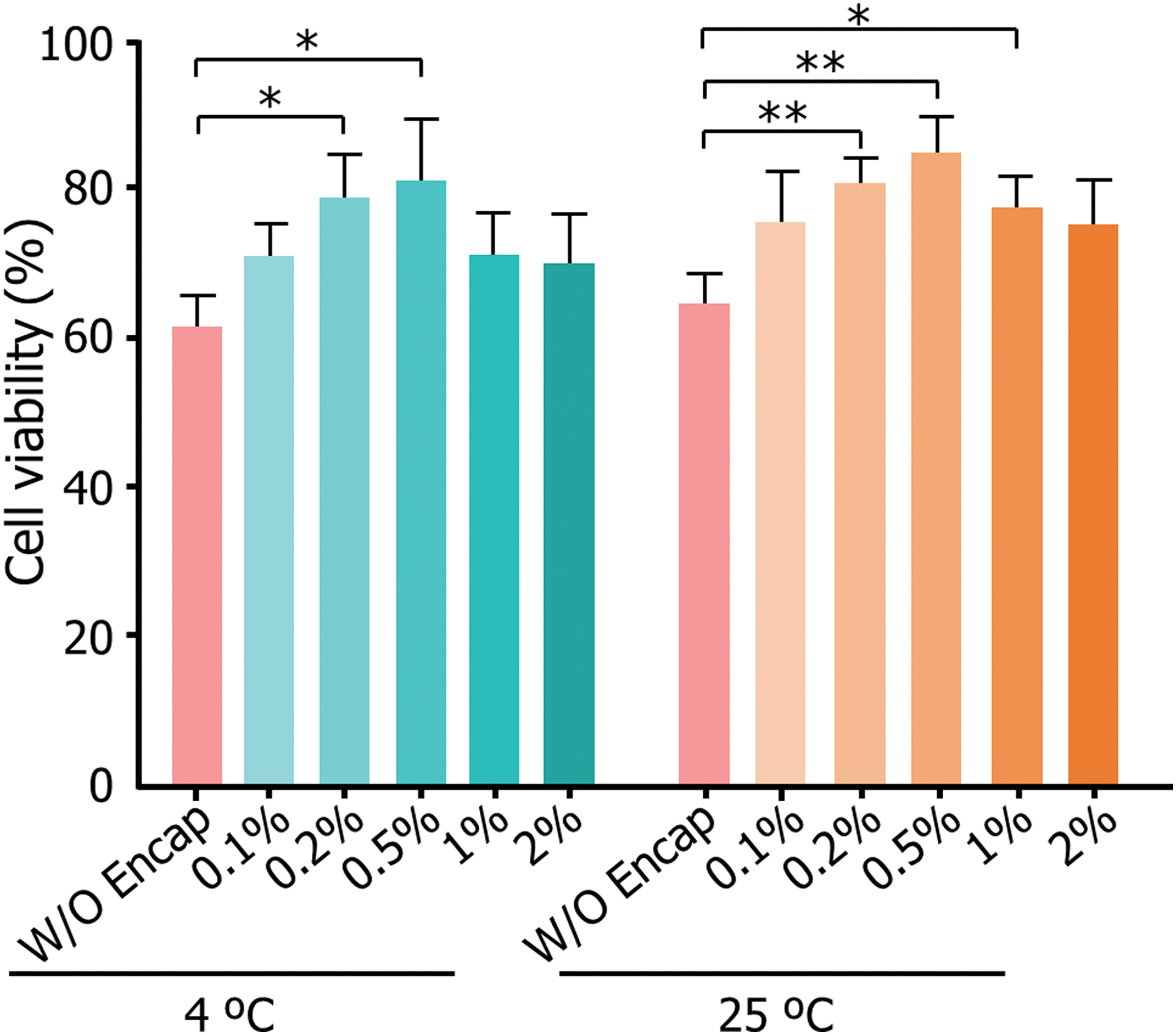
Cell viability of HUVECs encapsulated with alginate and different CMCH concentration (0.1%, 0.2%, 0.5%, 1%, and 2% [w/v]) after storing at 4°C and 25°C for 7 days. (*p < 0.05, **p < 0.01). Color images are available online.
Cell attachment
Cell attachment of HUVECs encapsulated with 1.5% (w/v) alginate and 0.5% (w/v) CMCH at 4°C and 25°C after 7 days are shown in Figure 6. The results indicated that after hypothermic storage for 7 days, HUVECs encapsulated with 1.5% (w/v) alginate and 0.5% (w/v) CMCH showed good cell survival and cell attachment.

Cell attachment of HUVECs encapsulated with alginate and 0.5% (w/v) CMCH after storing at 4°C and 25°C for 7 days.
Discussion
HUVECs and HUVEC-hydrogel constructs have wide applications in cell-based therapy and clinical needs. Traditional storage methods have many disadvantages. For example, continuous culture requires expensive instruments and complex operations. Moreover, the cryopreservation method can lead to solute damage and mechanical damage of cells during the cooling and warming processes. Compared with these methods, hypothermic storage has unique features. It does not rely on laboratory conditions and sophisticated instruments, resulting in low-cost, labor and time savings, and more flexible transfer of cells between different places when needed for clinical applications. Therefore, we studied the hypothermic storage of HUVECs and HUVEC-hydrogel constructs.
In this work, we have analyzed the cell viability without encapsulation or with encapsulation (alginate, alginate with CMCH) at 4°C and 25°C, respectively. The results show that the cells have higher survival when encapsulated in constructs, and the addition of CMCH can improve the effects of hypothermic storage. This is probably because alginate and CMCH can stabilize the cell membrane, protecting against osmotic shock, and they can also reduce respiration rate and water loss, thus increasing cell survival.
The cell viability in the study is CMCH concentration dependent. At 4°C, the cell survival rises as the CMCH concentration varies from 0.1% to 0.5%. However, when the concentration of CMCH increases to 1% and 2%, the cell viability slightly decreases comparing to 0.5% (w/v) CMCH. Similar results were found at 25°C. This may result from the high CMCH concentration, which is harmful to cell membranes and the influence on the metabolism of cells. The optimal concentration of HUVECs encapsulated by hydrogels is 1.5% (w/v) alginate and 0.5% (w/v) CMCH at 4°C and 25°C. In addition, the cells released from constructs can maintain the ability of cell attachment after hypothermic storage for 7 days. The storage within 7 days may not represent long-term preservation effects, but is sufficient to support short-term transport between laboratory preparation and clinical application. Furthermore, long-term hypothermic storage of HUVECs and HUVEC-hydrogel constructs should be studied in future work.
In summary, our results promote the hypothermic storage of HUVECs and HUVEC-hydrogel constructs, and will facilitate the development of cell delivery and biomedical applications.
Footnotes
Author Disclosure Statement
No conflicting financial interests exist.
Funding Information
This work was supported by the National Key R&D Program of China (Nos. 2018YFC0115500 and 2018YFE0194500), and the National Natural Science Foundation of China (No. 11627803). It was partially performed at the USTC Center for Micro- and Nanoscale Research and Fabrication.