
Abstract
The cryobanks of agouti somatic tissues represent a promising tool for the conservation of this species and of those that are phylogenetically related and endangered. For these purposes, one strategy to guarantee the quality of samples after warming would be to choose the appropriate tissue vitrification technique. Therefore, we evaluated the effects of two different techniques, direct vitrification in cryovials (DVC) and solid-surface vitrification (SSV), on the preservation of ear somatic tissues derived from agoutis kept in a scientific center of creation. Noncryopreserved somatic tissues were used as controls. Although SSV reduced the thickness of the dermis and cartilage (p < 0.05), the epidermal thickness of these samples was observed to be similar to controls (p > 0.05). Notably, the number of fibroblasts was not altered with either technique. However, both vitrification methods led to an increase in the number of perinuclear halos, with a particularly strong increase observed in DVC-derived fragments (p < 0.05). Compared with the DVC group, SSV showed a larger number of normal chondrocytes and smaller number of degenerate chondrocytes. Furthermore, the number of empty lacunae in SSV-derived fragments remained similar to controls (p > 0.05). In summary, SSV was found to be a more efficient method for vitrifying agouti somatic tissues compared with DVC. These results are important for the proper formation of agouti somatic banks, an essential step in the study of biological resources in this species.
Introduction
The term agouti is the name common to all mid-sized rodent species belonging to the genus Dasyprocta, family Dasyproctidae, and order Rodentia. Among these, Dasyprocta leporina has attracted considerable attention as a species of interest in wildlife conservation research, 1 especially in some Central and South American countries where it can normally be found. 2 In these countries, this species plays an important role in seed dispersal 3 and in maintaining plant diversity. 4 Moreover, as its population is at a lower risk of extinction, 5 D. leporina can be used as an experimental model for endangered species of the same genus.
Species conservation is an essential task for maintaining biodiversity. 6 One of the conservation strategies that can be employed involves the use of biological sample banks. 7 Tissue cryobanks, containing samples such as somatic tissues derived from skin and cartilage, allow genetic material from an individual or a population to be stored for long periods of time. 8 When properly stored, these samples can be employed for species multiplication through cloning by somatic cell nuclear transfer, 9 or nuclear reprogramming studies through pluripotency-induced cell production. 10
In general, the choice of cryopreservation technique is the first step for the establishment of cryobanks. 11 Although slow freezing has been used, 12 the use of vitrification has increased in wild species such as collared peccaries 11 and jaguar. 13 The reasons for using the latter technique compared with slow freezing include lower cost and reduced ice crystal formation during cryopreservation. 14 Consequently, different vitrification methods have been considered.14,15
In recent years, direct vitrification in cryovials (DVC) and solid-surface vitrification (SSV) have been studied,11,13 with the appropriate vitrification method depending on the tissue and species of interest.16,17 Therefore, we aimed to evaluate the effects of DVC and SSV methods on the preservation of agouti ear somatic tissues.
Materials and Methods
All protocols were approved by the Ethics Committee of Animal Use of the Federal Rural University of the Semi-Arid (CEUA/UFERSA; 23091.001074/2015-81) and Chico Mendes Institute for Biodiversity Conservation (ICMBio; 48635-1). The reagents, media, and solutions used in this study were obtained from Sigma-Aldrich (St. Louis, MO), Gibco-BRL (Carlsbad, CA), and Labimpex (São Paulo, SP, Brazil).
Skin biopsy and experimental design
The animals were previously anesthetized by intramuscular administration of 15 mg/kg ketamine hydrochloride (Ketalar; Pfizer, São Paulo, Brazil) and 1 mg/kg xylazine hydrochloride (Rompun; Bayer, São Paulo, Brazil). 18 Ear margin tissues derived from four agoutis (24–36 months) were obtained from the Center for Wild Animals Multiplication (CEMAS/UFERSA, 1478912) using pliers and transported to the Laboratory of Animal Biotechnology (UFERSA) in Dulbecco's modified Eagle medium (DMEM) supplemented with 2% antibiotic-antimycotic solution (penicillin G, streptomycin, and amphotericin B) at 37°C for 1 hour. The identification information of ear sections used for experiments was recorded in the management systems of this species, so these ear tags were made to identify the approximate age of the individuals.
Tissues measuring 1–2 cm2 were washed in 70% ethanol and with the help of a scalpel blade and a cutting mold 18 small fragments (9.0 mm3; 3 × 3 × 1 mm) per individual were obtained and equally and randomly distributed into three groups. The first group consisted of fresh controls (noncryopreserved fragments), the second group of samples was submitted for DVC, and the third group of samples was submitted for SSV. After 2 weeks, DVC- and SSV-derived fragments were warmed, morphometric characterization and histological analysis of the epidermis, dermis, and cartilage of fresh and vitrified fragments were performed.
Somatic tissue vitrification
Eighteen tissue fragments were equally and randomly allocated for each treatment (six fragments per group). A vitrification solution (VS) composed of DMEM supplemented with 20% ethylene glycol (EG), 20% dimethyl sulfoxide (DMSO), 0.25 M sucrose (SUC), and 10% fetal bovine serum (FBS) was used for both DVC and SSV techniques. 11 Both methods were conducted as previously described for somatic tissues derived from collared peccaries 11 and jaguar. 13
For DVC, six fragments were immediately transferred to three cryovials containing 2.0 mL VS and kept for 15 seconds at 25°C. Next, the cryovials were stored in liquid nitrogen. For SSV, six fragments were exposed to 1.8 mL VS for 5 minutes, after which tissues were dried on absorbent paper. Subsequently, the fragments were individually placed on a metal cubic surface partially immersed in liquid nitrogen. Fragments were then transferred to three cryovials for storage in liquid nitrogen.
After 2 weeks of cryostorage for both groups of vitrified tissues, cryovials were maintained for 1 minute at 25°C and then immersed at 37°C water bath. For the removal of cryoprotectants, fragments were washed three times for 5 minutes in DMEM supplemented with 10% FBS and decreasing concentrations of SUC (0.50, 0.25, and 0.0 M).
Evaluation of somatic tissues using morphometry and classical histology
Eighteen fragments of each agouti derived from control and vitrified groups were fixed in 4% paraformaldehyde for a period of 7 days and dehydrated in a graded series of ethanol solutions, and then embedded in paraffin. 11 Sections of 7.0 μm in thickness were stained with hematoxylin and eosin. Morphometric and histological analyses were performed using a light microscope (Olympus CX 31 RBSFA, Tokyo, Japan) and ImageJ software (U.S. National Institutes of Health, Bethesda, Rockville) in 400 × magnification. For all analyses, 20 images per animal were acquired for each group, totaling 80 images per group.
For skin, the following parameters were examined: epidermal and dermal thickness, number of perinuclear halos in the epidermis, and number of fibroblasts in the dermis. 19 For cartilage, the following parameters were examined: cartilage thickness, number of normal and degenerate chondrocytes, and number of filled and empty lacunae. 20 Normal chondrocytes were considered to have a defined nucleus and delimited cytoplasm, whereas degenerates were those with undefined nucleus and cytoplasm associated with an empty lacunae.
Statistical analysis
Data generated from the four agoutis were expressed as the mean ± standard error (one repetition per animal) and analyzed using GraphPad (Graph-Pad Software Incorporation, La Jolla, CA). Significance was set at p < 0.05. All results were verified for normality by the Shapiro–Wilk test and for homoscedasticity by Levene's test. Data obtained from morphometric analyses were analyzed by analysis of variance (ANOVA) followed by Tukey's test. Cell and perinuclear halo numbers were analyzed by the Kruskal–Wallis and Dunn's tests.
Results
The morphological features of nonvitrified somatic tissues (control) and of tissues after vitrification using DVC or SSV are shown in Figures 1 and 2. Although SSV reduced the thickness of the dermis (Fig. 3B) and cartilage (Fig. 3C), epidermal thickness was observed to be similar to that of the controls (Fig. 3A). Despite changes in tissue thickness, the number of fibroblasts was not affected in either technique (Fig. 4).
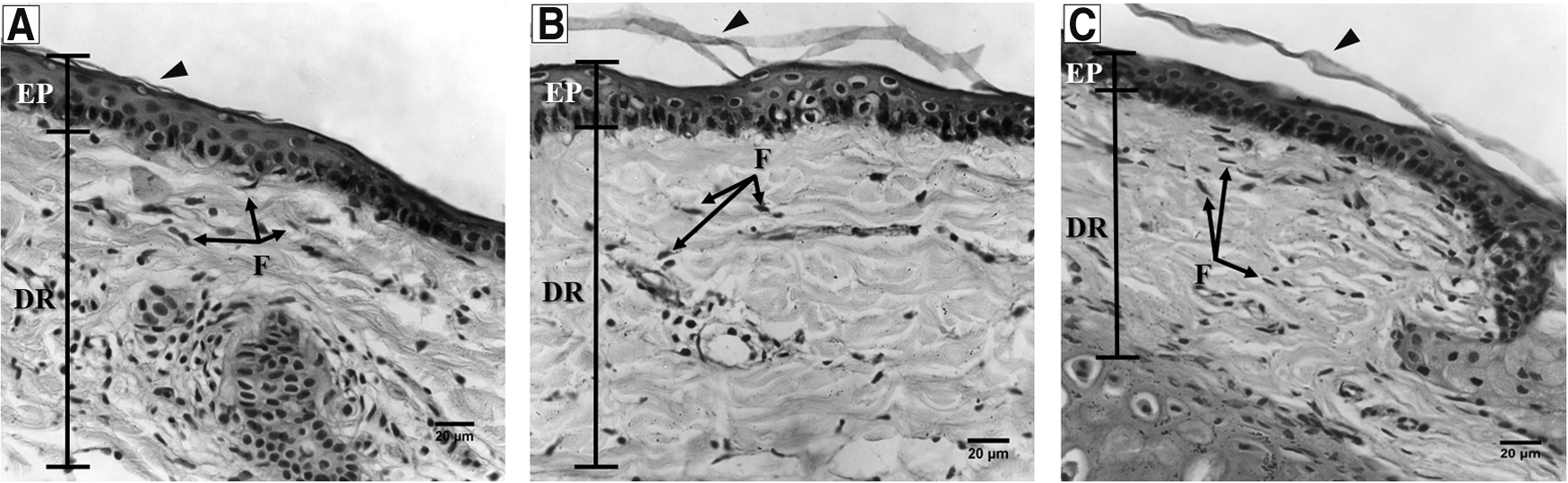
Morphological characterization of ear skin layers of fresh agouti

Morphological characterization of cartilage of agoutis in fresh

Morphometry of the ear skin and cartilage of agoutis.
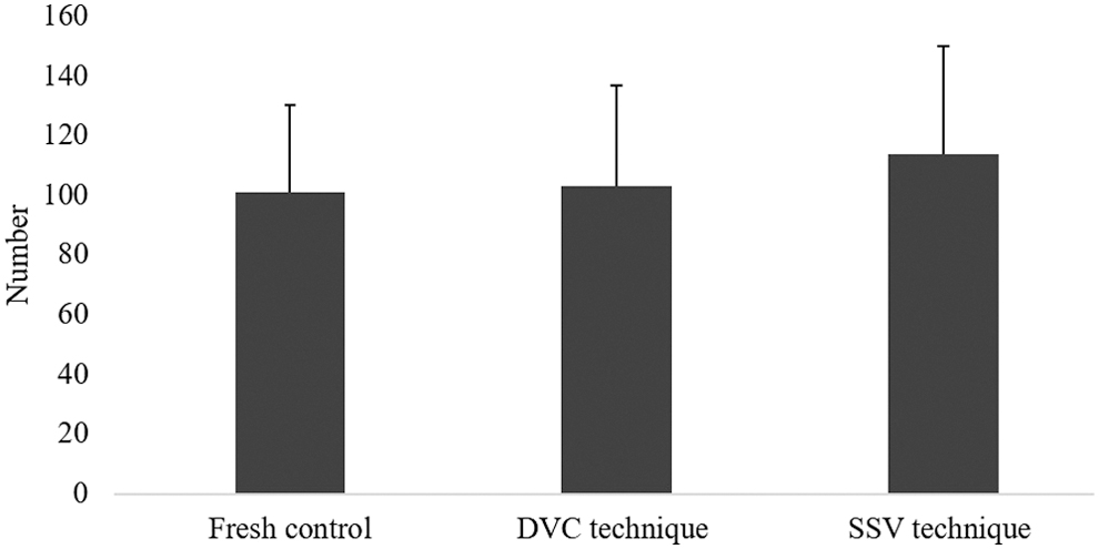
Number of dermal fibroblasts from ear skin of fresh agouti, DVC technique, and SSV technique.
Both DVC- and SSV-treated groups exhibited increased numbers of perinuclear halos (DVC: 17.3 ± 12.9 and SSV: 13.1 ± 10.9) compared with controls (5.3 ± 6.4), with a particularly sharp increase observed in DVC-derived fragments (Fig. 5A–D). In addition, SSV showed a larger number of normal chondrocytes and a smaller number of degenerate chondrocytes compared with those of the DVC group (Table 1). Finally, both techniques reduced the number of filled lacunae (Table 1); however, only SSV maintained a similar number of empty lacuna-like control fragments.
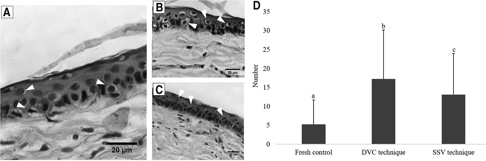
Morphological characterization of perinuclear halo of the ear skin of agoutis in fresh control, DVC technique, and SSV technique.
Cell Number Quantification and Lacunae of the Ear Skin and Cartilage of Agoutis After Cryopreservation Using Different Techniques
Different letters show differences statistically significant in the same column (p < 0.05).
DVC, direct vitrification in cryovials; S.E., standard error; SSV, solid-surface vitrification.
Discussion
In this study, we observed that compared with DVC, SSV is a promising technique for the conservation of somatic tissues derived from agoutis. This was demonstrated through the analysis of several tissue parameters, such as epidermal morphology, perinuclear halo quantification, characterization of normal and degenerate chondrocytes, and the quantification of empty lacunae. In general, two factors contribute to the success of this technique; rapid cooling rates due to direct contact with the metal surface, and good heat conduction that ensures vitreous state formation, 21 which are associated with preventing the exposure of tissue to VS at the time of warming.22,23
No retraction was observed in the epidermis of SSV-derived fragments, possibly because these fragments comprised a more resistant layer, composed of epithelial tissue, and characterized by limited extracellular matrix and the presence of juxtaposed cells bound by intercellular junctions. 24 However, in the DVC-derived fragments, epidermal thickness was increased. This was likely due to the method of vitrifying samples in cryotubes, where the process occurs more slowly, resulting in the requirement of more time for the vitrification of the solution contained in the cryotubes. 25 In addition, in DVC treatment, the samples are exposed for a longer time with large amounts of VS. In contrast, in SSV samples are not in contact with a large amount of cryoprotectants that decrease toxicity due to exposure during drastic temperature changes. 14 Moreover, it was possible to observe a reduction in the thickness of the dermis and cartilage of the fragments preserved by SSV when compared with DVC and control groups. This phenomenon is explained by the technical procedure itself, which results in tissue constriction during vitrification 21 and this reduction would likely influence the quantity and viability of cells to be employed in future biotechniques. 16
The formation of perinuclear halos, which are indicative of decreased cell viability and apoptosis,19,26 was found in greater quantities in the DVC group. Although cryoprotectants promote protection against cryoinjury by preventing the formation of ice crystals, at high concentrations they are toxic to cells. 27 As DVC-derived tissues are exposed to VS during warming, higher cellular degeneration observed in this group is probably due to increased toxicity of the VS at higher temperatures.14,28
Studies have shown that the ideal combination of intracellular and extracellular cryoprotectants is able to reduce the formation of intracellular ice crystals, thereby facilitating cell membrane maintenance and minimizing damage from the cryopreservation process. 11 DMSO is a highly membrane permeable cryoprotectant with low cell toxicity 29 that can be used alone or in combination with other substances, such as EG, 30 which has a higher permeability. 31 The addition of SUC and FBS as extracellular cryoprotectants aims mainly to help reduce osmotic pressure and shock, 32 as well as provide membrane protection for increased stability during cryopreservation, 16 thus helping in maintaining cellular quantity.
With both techniques, it was possible to observe the maintenance of similar numbers of fibroblasts compared with that of the controls. Therefore, these cells are ideal for the recovery of genetic material, for use in biotechniques such as somatic cell nuclear transfer, 33 and in cell pluripotency induction studies. 34 Thus, with the storage of these tissues, these cells can later be used in in vitro cultures for biotechnological applications. 35
Another cell type is the chondrocyte, the main cell type present in cartilage.36,37 Through histological evaluation of this region, it was possible to identify viable and nonviable chondrocytes based on their morphology. 38 Chondrocytes fill the lacunae of cartilaginous tissue, with some lacunae containing up to two cells, as well as some lacunae being empty. 39 The presence of empty lacunae occurs when some chondrocytes contract and detach from the cartilaginous matrix due to the cryopreservation process, or due to degeneration and cell death. 20 Compared with the DVC group, SSV had a positive effect, resulting in fewer empty lacunae. The larger number of empty lacunae found in the DVC group may be related to the time of tissue exposure to the cryoprotectant as a function of fragment size, where the short exposure time may not have been sufficient for the total permeabilization of the substance in the tissue. 40
In summary, SSV was found to be a more efficient method for vitrifying agouti somatic tissues compared with DVC. This is due to the SSV method, in comparison with DVC, maintaining epidermal thickness, a larger number of normal chondrocytes and a smaller number of degenerate chondrocytes. In addition, the number of fibroblasts and lacunae in SSV-derived fragments remained similar to controls. These results are important for the proper formation of agouti somatic banks, an essential step in the study of biological resources in this species. Finally, with the future establishment of in vitro culture in agoutis, this tool will serve to obtain new parameters of the effects of tissue cryopreservation on cell quality.
Footnotes
Author Disclosure Statement
No conflicting financial interests exist.
Funding Information
This study was supported by Brazilian Council of Scientific Development (CNPq) and Coordenação de Aperfeiçoamento de Pessoal de Nível Superior—Brasil (CAPES, Financial Code 001).