
Abstract
The integrity of blood plasma/serum (P/S) specimens can be impacted by preanalytical handling and storage conditions that result in thawed-state exposures (> −30°C). We recently reported a simple dilute-and-shoot, intact-protein liquid chromatography/mass spectrometry (LC/MS) assay called ΔS-Cys-Albumin that quantifies cumulative exposure of P/S to thawed conditions based on the change in relative abundance of the oxidized (S-cysteinylated) proteoform of albumin (S-Cys-Albumin) in the native sample to that of an aliquot of the sample intentionally driven to its maximum oxidation state. Herein, we evaluated the effect of prestorage delay and initial storage temperature on sample integrity by applying the ΔS-Cys-Albumin assay to a set of plasma samples (n = 413) collected under a single clinical study but from 12 different collection sites. Major differences (p < 0.0001) were observed between different groups of samples with modestly inconsistent initial handling conditions (i.e., initial processing of whole blood to plasma and placement at −80°C completed in under 3 hours, 3–13 hours, and over 17 hours). ΔS-Cys-Albumin was significantly inversely correlated with delay time at 4°C before centrifugation and total delay before final storage at −80°C (p < 0.0001). Samples from two collection sites had much lower ΔS-Cys-Albumin values relative to samples from other sites, in accordance with the fact that they were stored at −20°C for an average of 7.6 months before shipment to the central repository for final storage at −80°C. Based on the rate law for S-Cys-Albumin formation in plasma ex vivo, the average time that each plasma specimen had been exposed to the equivalent of room temperature (23°C) was back calculated from the measured ΔS-Cys-Albumin values. A survey of clinical analytes in P/S whose measured concentrations are sensitive to the initial handling/storage conditions documented in this study is provided and the ramifications of the plasma integrity findings from this multisite clinical study are discussed.
Introduction
In the past two decades, significant advances have been made in bringing the issue of research biospecimen integrity to light.1–15 Numerous studies have revealed that the preanalytical variables (PAVs) pervading the collection, processing, and storage of biospecimens may potentially generate samples with unknown or unrecognized integrity issues, thus leading to inaccurate and unreliable research results.1–19 To address the issue of biospecimen integrity a call was made in 2012 in numerous clinical chemistry research journals to require a full description of specimen handling before sending out articles for review.20–27 In addition, efforts have been made toward the development of standard operating procedures (SOPs) for sample collection, processing, and storage.1,28
However, the universal adoption of a single rigorous SOP, while in principle appealing as a solution to the development of potential biospecimen integrity problems, can, in practice, become impractical or impossible for laboratory personnel to implement on a rigorous basis for every single sample or in situations where infrastructure issues arise, such as unexpected power losses29,30 or when other problems occur such as inspection/customs delays during shipment. These potential problems are amplified in studies with multiple collection sites involved—each of which must contend with different personnel, unique infrastructure constraints, and shipment routes.
To date, many preclinical research studies have relied on preexisting, archived biospecimens, some of which likely lacked detailed paper trails documenting important PAVs. The scope of the potential problem is substantial: In 2014, 63% of the 455 extramural grants sponsored by the U.S. National Cancer Institute (NCI, part of the National Institutes of Health) utilized preexisting samples; of these 455 grants, 107 of the projects (24%) relied on preexisting blood plasma or serum (P/S) samples. Altogether, ∼40% of NCI-sponsored grants involved the use of P/S. 31 Recently, using our newly developed ΔS-Cys-Albumin marker, we discovered a major integrity discrepancy in a set of serum samples collected under NCI sponsorship and slated for distribution by the U.S. National Institutes of Health (NIH). 29 It would be naive to assume that this integrity discrepancy was a single, isolated incident among all archived P/S samples slated for use in future research studies (or that may have already been used in published research studies). As such, there is a present and urgent need for accurate quality control/quality assurance (QC/QA) markers and tests to retrospectively and empirically assess the integrity of archived biospecimens—even those for which a nominally pristine paper trail may exist.
In 2013, several potential QC markers for P/S integrity were outlined by Betsou et al., 2 including transferrin receptor, 32 potassium, 33 adrenocorticotrophic hormone and B-type natriuretic peptide (BNP), 34 soluble CD40L, 35 vitamin C36,37 and E, 38 matrix metalloproteinase-7 (MMP-7), 14 and others. For nearly all of these candidate markers, the indication of disrupted sample integrity is based on the apparent loss of target analytes beyond their associated reference range—generally without a known mechanism of apparent loss or associated rate law. As previously reported,29,39,40 human serum albumin contains a single free cysteine residue that can undergo spontaneous disulfide bond exchange with free cystine, forming S-cysteinylated albumin (S-Cys-Albumin) to a limited degree in vivo and to a much greater extent when P/S samples are exposed to thawed conditions—that is, temperatures greater than −30°C.41,42 Charge deconvoluted electrospray ionization (ESI)-mass spectra of albumin that illustrate the ex vivo formation of S-Cys-Albumin and the ΔS-Cys-Albumin concept as a whole are provided in Figure 1. In the presence of atmospheric oxygen and trace quantities of catalytic transition metals (primarily copper), nearly all of the cysteine equivalents in P/S (including those tied up as cystine) can be consumed by the native, reduced form of albumin producing S-Cys-Albumin. 29 The number of cysteine equivalents in P/S is, on average, only about 20% of the number of albumin equivalents.43–45 As such, the fraction of albumin in the S-cysteinylated form within nearly all human P/S samples (which, in vivo, starts at ∼20%–40% 29 ), will increase by ∼20% to 40%–60% if P/S samples are exposed to thawed conditions for a long enough period of time (∼60 days at −20°C, ∼25 days at 4°C, ∼4–5 days at room temperature, and ∼18 hours at 37°C 29 ).
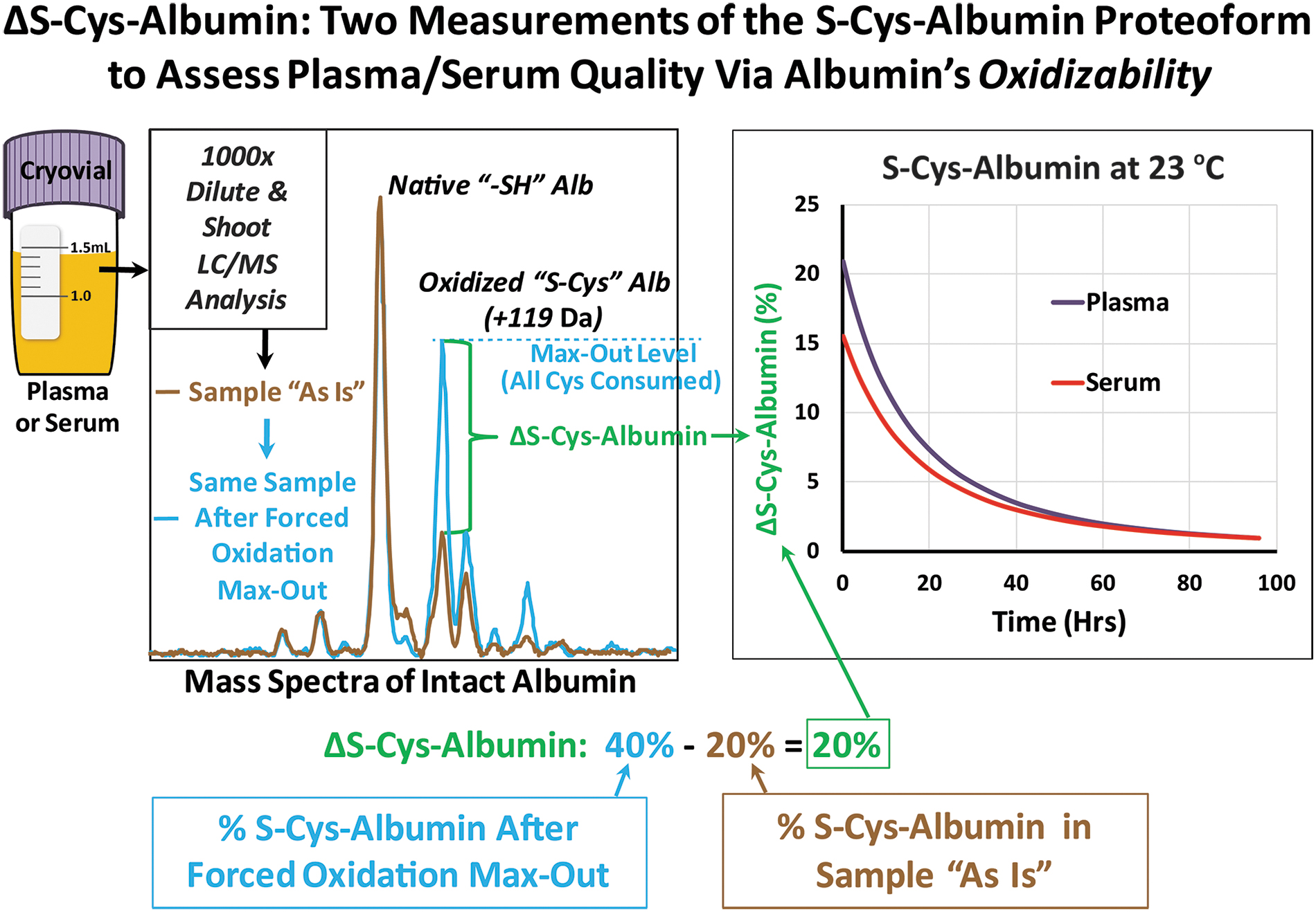
ΔS-Cys-Albumin concept: Half a microliter of P/S is diluted 1000-fold and injected onto a liquid chromatograph-mass spectrometer for analysis of intact albumin (brown spectrum) and determination of the percentage of albumin that is S-cysteinylated (oxidized). A 9.5-μL aliquot of the same sample is then intentionally incubated at 37°C for 18 hours to drive the percentage of albumin in the S-cysteinylated form to its maximum possible value—which is always less than 100% 29 —and the sample is measured once again (blue spectrum). The difference between the two measurements, known as “ΔS-Cys-Albumin,” is in the range of 12%–29% for fresh plasma samples and as low as zero for samples that have been exposed to thawed conditions (> −30°C for prolonged periods of time). 29 Since the multireaction rate law for formation of S-cysteinylated albumin at room temperature was also established, 29 measurements of ΔS-Cys-Albumin can be related to an estimated time of exposure to the equivalent of room temperature. This figure was originally published in Jeffs et al. 29 P/S, plasma or serum.
Based on these facts, our research group recently developed an assay known as “ΔS-Cys-Albumin” as a QC/QA marker of P/S integrity. 29 This assay measures the S-Cys-Albumin percent change (ΔS-Cys-Albumin) between the original P/S sample (presented with either a known or unknown storage history) and an aliquot of the same sample intentionally driven to its maximum ex vivo oxidation (S-cysteinylated) state through incubation at 37°C for 18 hours. This inexpensive and rapid assay requires only 10 μL of a P/S sample and employs a very simple dilute-and-shoot liquid chromatography-mass spectrometry (LC-MS)-based analysis. Since the associated rate law model for the formation of S-Cys-Albumin at room temperature was also established, 29 back calculation of the approximate time at which a given P/S sample must have been exposed to the equivalent of room temperature conditions was enabled—making it possible to place an approximate exposure “time stamp” on every sample.
As described above, we applied the ΔS-Cys-Albumin assay to a case study of nominally pristine serum samples collected under NCI sponsorship and discovered a previously undisclosed biospecimen integrity discrepancy. 29 To evaluate the potential utility of this assay to reveal variations in biospecimen handling and storage conditions, we have now applied it to 413 clinical plasma samples from the Women Epidemiology Lung Cancer (WELCA) study that were collected under a single SOP but from 12 different collection sites and then sent to a single repository for long-term storage at −80°C. In this study, we provide insights into the effects of initial P/S handling conditions on ΔS-Cys-Albumin and evaluate the meaning of these results with regard to their implications for the stability of important clinical analytes within P/S samples collected in a similar manner.
Materials and Methods
Materials
Trifluoroacetic acid (TFA, Cat. No. 299537) and formic acid (Cat. No. 06440) were obtained from Sigma-Aldrich (St. Louis, MO). LC/MS grade acetonitrile (Cat. No. A955-4) and water (Cat. No. W6-4) were acquired from Thermo Fisher Scientific (Waltham, MA). Protein captraps were from Optimize Technologies (Cat. No. 10-04816-TM).
Samples
General patient information
EDTA plasma samples from stage I to IV lung cancer patients and age and gender-matched controls from the Women Epidemiology Lung Cancer (WELCA) study were collected at 12 different collection centers in France. 46 This study was approved by Institutional Review Board of the French National Institute of Health and Medical Research and by the French data Protection Authority (IRB-INSERM, No. 3888 and CNIL No. C13-52). All specimens were collected in compliance with the Declaration of Helsinki principles. Once collected, they were coded and deidentified to protect patient identities. As part of the WELCA Study and for all samples analyzed here, all-female lung cancer patients were recruited between September 2014 and November 2016 in collection sites 2 to 14, and age-matched all-female controls were recruited between June 2015 and December 2016 at collection site 18. All women living in Paris and the lle de France area, newly diagnosed with lung cancer were considered as eligible cases. Age-matched controls were randomly sampled from women without a history of cancer living in the same area.
Inclusion/exclusion considerations
Primary inclusion and exclusion criteria for the WELCA study in general are provided elsewhere. 46 For the purposes of this study we considered that a wide array of clinical conditions can result in altered circulating albumin concentrations, including advanced age.47,48 With the exception of dehydration, most such conditions result in decreased albumin concentrations. 47 Abnormal changes to the ΔS-Cys-Albumin values observed in fresh P/S samples necessarily involve albumin as well as cysteine and cystine concentrations. 29
To make the ΔS-Cys-Albumin assay as widely applicable across as many pathological conditions as possible, our initial survey of the population reference range of ΔS-Cys-Albumin in nonacute cardiac patients excluded only patients with severely limited kidney function (i.e., eGFR <30 mL/min × 1.73 m2; which can cause extreme elevation of cysteine and cystine49–51 as well as albumin loss, 47 potentially resulting in extraordinarily high ΔS-Cys-Albumin measurements) and/or severe hemolysis (i.e., >250 mg/dL; which can also cause abnormal ΔS-Cys-Albumin measurements 29 ). The results of this survey indicated that the range of ΔS-Cys-Albumin in these nominally unhealthy patients was in line with (although slightly more compact and lower on average than) the theoretical ΔS-Cys-Albumin range of 11%–38% that was predicted based on known reference ranges for albumin, cysteine, and cystine. 29 Notably, ∼50% of the patients in this population were diabetic. Given our goal of making the ΔS-Cys-Albumin assay applicable to as wide a clinical patient population as possible, we only excluded severely hemolyzed specimens (>250 mg hemoglobin/dL) from this study. The ΔS-Cys-Albumin results from freshly processed and stored sample described below showed no indication of abnormally elevated ΔS-Cys-Albumin.
Specimen handling and storage
All peripheral blood samples were drawn and processed following a written standardized protocol. 46 For the lung cancer patients, each hospital had their own laboratory where the samples were processed. Control specimens were collected at the homes of individual donors by a nurse who deposited the whole blood samples at a designated laboratory. Alternatively, control specimen donors had the option of traveling to the laboratory themselves to donate their samples. For both the cases and controls, whole blood that had been collected in EDTA-containing tubes was placed on ice packs at ∼4°C and then transported to the laboratory. (While these instructions were provided to the hospitals and nurses involved in specimen collection and transport, it is possible that in some cases they may not have been followed exactly and therefore the whole blood may have been exposed to room temperature conditions before processing it into plasma.)
Once at the laboratory, blood samples were spun for 15 minutes at 3000 rpm and 4°C in a standard centrifuge. In each of the 12 collection centers, the collected plasma samples were aliquoted then temporarily kept at −80°C or −20°C while they awaited shipment to the central repository (see Results section for site-specific temporary storage details). Approximately every 3 months samples were transported on dry ice to the central repository for final storage at −80°C. No freeze/thaw cycles occurred before shipment to Arizona State University (Borges Lab) for analysis. Received plasma samples were aliquoted on ice and kept frozen at −80°C before analysis. Sample handling information, including collection site, precentrifugation delay (from blood drawing to centrifugation), postcentrifugation delay (from centrifugation to storage), total prestorage delay time (from blood drawing to storage), and initial storage temperature before shipment to the central repository were gathered and tabulated from the 413 corresponding sample collection information sheets. A freshly collected and processed 300-mL EDTA plasma sample from an individual, nominally healthy donor was obtained from BioIVT and served as a QC sample to ensure batch-to-batch quantitative reproducibility.
Experimental procedures
Sample preparation
Plasma samples, run in random order, were simply diluted 1000-fold before injection onto an LC-MS. Generally, a 10-μL aliquot of plasma was thawed at room temperature. Once completely thawed, 0.5 μL of plasma was added to 500 μL of 0.1% (v/v) TFA and mixed thoroughly by vortexing for 20 seconds. Then 10 μL of the freshly diluted plasma solution was injected onto the LC-MS instrument immediately. To intentionally drive the degree of albumin S-cysteinylation to its maximum value, the residual 9.5 μL plasma sample was incubated in a dry oven at 37°C for 18 hours in a 600-μL Eppendorf snap-cap test tube. Afterward, 0.5 μL of the sample (in fully oxidized state) was diluted 1000-fold in 0.1% (v/v) TFA and then injected onto the LC-MS, following the same steps described above. For incubated aliquots, however, batches of diluted samples were loaded onto an autosampler kept at 4°C and injected in an overnight run. We have previously verified the stability of S-Cys-Albumin measurements under these conditions. 40
LC-ESI-MS analysis
The separation and relative quantification of intact albumin proteoforms were performed on an Agilent 1260 Infinity II UHPLC connected to an Agilent 6530 Electrospray Ionization Quadrupole Time-of-Flight (Q-TOF) LC-ESI-MS instrument. A 10 μL of sample was loaded at 200 μL/min in 80% water containing 0.1% formic acid (Solvent A)/20% acetonitrile containing 0.1% formic acid (Solvent B) onto an Optimize Technologies protein captrap configured for bidirectional flow on a 6-port diverter valve. The same solvent composition was held for 3 minutes at 200 μL/min to rinse the protein captrap. Following were stepwise ramps of the flow composition to elute the trapped albumin at 200 μL/min: 3.0–3.1 minutes, ramped to 65/35 A/B and held until 4.5 minutes; 4.5–4.6 minutes, ramped to 55/45 A/B then held until 7.5 minutes; 7.5–7.6 minutes, ramped to 20/80 A/B and held until 8.6 minutes; at last, ramped back to 80/20 A/B by 8.7 minutes. This stepwise gradient results in near complete chromatographic separation of albumin from apolipoprotein A-I. A 4-minute postrun program was implemented at 200 μL/min of 80/20 A/B to re-equilibrate the system with the solvent composition of sample loading. The 6-port valve was initially set to the loading position and then switched to the inject position at 3 minutes, directing the eluate from the captrap to the mass spectrometer. The mass spectrometer was set to run in positive ion, TOF-only mode, and to collect spectra in the m/z 100 to 3200 range. ESI settings for the Dual AJS ESI capillary microflow nebulizer ion source were as follows: VCap 5700 V, Nozzle Voltage (Expt) 2000 V, Drying Gas nitrogen 7 L/min at 325°C, Nebulizer nitrogen 45 psig, Sheath Gas 11 L/min at 250°C. Data were acquired in profile mode at a rate of one spectra per second.
Data processing
Across the chromatographic peak apex of albumin ∼0.3 minute of recorded spectra were averaged. Then deconvolution by the MaxEnt algorithm was carried out on the electrospray ionization charge-state envelope with Agilent MassHunter Qualitative Analysis vB.07.00 software. The MaxEnt algorithm parameters were set as follows: mass range 60000.00–72000.00 daltons, mass step 1.0000 daltons, using limited m/z range 1000–2500 m/z, subtracting baseline with baseline factor 0.7, adduct as proton, peak height filter with peak signal-to-noise ratio ≥10.0, maximum number of peaks limited (by height) to the largest 100, calculating average mass using top 90% of peak height, minimum consecutive charge states 10, and minimum protein fit score 8. In the deconvoluted spectra, the peak heights of mass spectral peaks of interest were exported to a spreadsheet for further statistical analysis.
The percent abundance of S-cysteinylated albumin (oxidized form, S-Cys-Albumin) was calculated by dividing the height of the peak representing S-Cys-Albumin by the sum of the peak heights for native albumin and S-Cys-Albumin then multiplied by 100 (Fig. 1). The change in the percent S-Cys-Albumin (ΔS-Cys-Albumin) for each plasma sample was determined by subtracting the percent S-Cys-Albumin of the sample in its original state from the percent S-Cys-Albumin of the same sample in its fully oxidized state.
Statistical analysis was performed in GraphPad Prism 7: For the four groups with different initial handling conditions, outliers were removed by log-transformation and the ROUT method at Q = 1%. Outlier-removed data were then reverse-transformed by taking the anti-log of each value. To identify differences among groups, the Kruskal–Wallis test at 95% confidence level followed by the Benjamini–Hochberg false discovery correction procedure at a 5% false discovery rate was carried out. Correlation of ΔS-Cys-Albumin with various delays were evaluated by Spearman's rank correlation. Differences between lung cancer patients and controls were assessed by means of the Mann–Whitney test at a 95% confidence level. GraphPad Prism 7 was also used to plot all of the figures.
Results
For all 413 blood plasma samples obtained from the WELCA study, ΔS-Cys-Albumin was measured in 33 batches. Samples were analyzed in random order. A QC EDTA plasma sample was aliquoted and measured in each batch and indicated reasonable total interassay precision (%CV = 12.4%; at an S-Cys-Albumin value of 20% this corresponds to an absolute ΔS-Cys-Albumin variability of 2.5%).
Effect of initial handling conditions on ΔS-Cys-Albumin
The effects of initial storage temperature (before shipment to the central repository) and total prestorage delay on ΔS-Cys-Albumin were evaluated first. ΔS-Cys-Albumin data were divided into four groups based on the initial handling conditions of the corresponding plasma specimens: Group 1: samples stored at −80°C within 3 hours from the blood draw; Group 2: samples stored at −80°C but exposed to 4°C or higher temperature for longer than 3 but less than 13 hours before final storage at −80°C; Group 3: samples stored at −80°C but exposed to 4°C or higher temperature for longer than 17 hours before final storage at −80°C, and Group 4: samples stored at −20°C for 42 up to 456 days before final storage at −80°C at the central repository. (Notably, for Groups 1–3, no samples were exposed to 4°C or higher temperatures in the 13–17 hour time frame—hence the gap in the time frames.)
As might be expected based on the known stability of ΔS-Cys-Alb, 29 pronounced, statistically significant differences were observed in each group compared with one another (Fig. 2). Since ΔS-Cys-Albumin measures the difference in the percentage of S-Cys-Albumin between the original status and fully oxidized status of a sample, a smaller ΔS-Cys-Albumin indicates that more ex vivo oxidation had occurred in the sample before any analysis. The ΔS-Cys-Albumin values of samples stored temporarily at −20°C were found to be significantly lower than those stored initially at −80°C, indicating a definitively higher ex vivo oxidation level of albumin and compromised sample integrity. Moreover, for all the samples stored initially at −80°C, longer prestorage delay at 4°C also led to strikingly decreased ΔS-Cys-Albumin values, implying potentially jeopardized sample integrity.
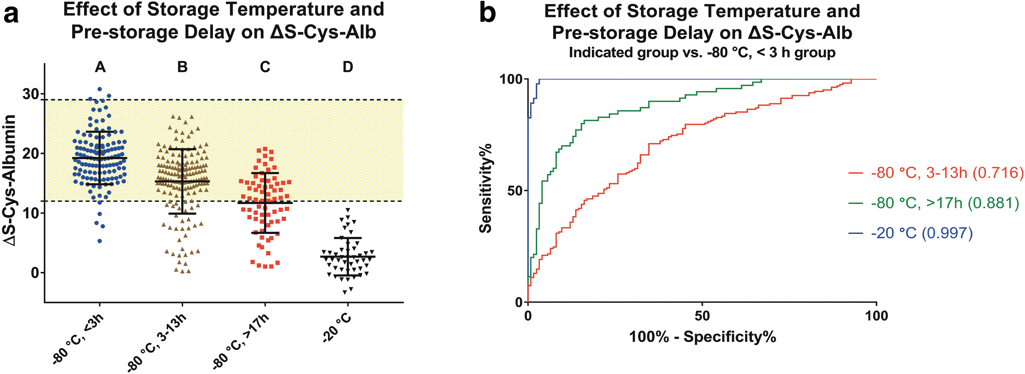
Effect of prestorage delay and storage temperature on ΔS-Cys-Albumin.
As reported in our previous work, 29 the range for ΔS-Cys-Albumin observed in freshly collected plasma from 97 nonacute cardiac patients was 12%–29% (yellow shaded area in Fig. 2). In total, 94% of the ideally handled plasma samples from the WELCA study—that is, those stored in −80°C within 3 hours from blood drawing—exhibited ΔS-Cys-Albumin between 12% and 29%, indicating that this reference range is likely to be valid with regard to its application to lung cancer patient plasma samples. Eighty percent, 54%, and 0% of the plasma samples with prestorage delays between 3 and 13 hours, over 17 hours, and temporarily stored at −20°C, respectively, fell in the 12%–29% range.
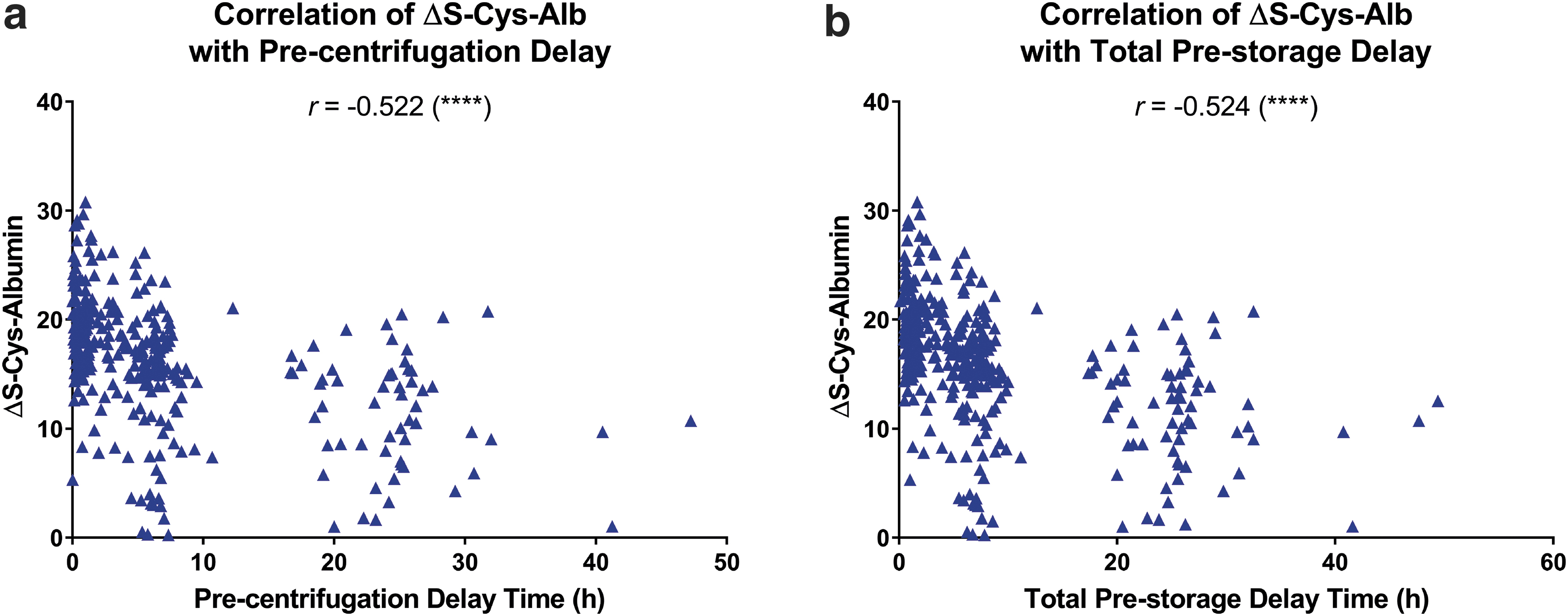
Next, the correlation of ΔS-Cys-Albumin to precentrifugation and total prestorage delay were assessed (Fig. 3). Cases and controls were combined for this analysis. For correlation of ΔS-Cys-Albumin to precentrifugation delay, samples with a postcentrifugation delay longer than 1.5 hours or that were temporarily stored at −20°C were excluded. Similarly, for correlation of ΔS-Cys-Albumin to total prestorage delay, samples that were temporarily stored at −20°C were excluded. These exclusions ensured that the variable of interest was the primary driver of the reported ΔS-Cys-Albumin value.
Correlation between ΔS-Cys-Albumin and
Spearman's rank correlation analysis demonstrated that ΔS-Cys-Albumin was significantly correlated with precentrifugation delay (Fig. 3a) and total prestorage delay (Fig. 3b), both with a moderate negative relationship (r = −0.522 and −0.524, respectively). For both precentrifugation delay (Fig. 3a) and total prestorage delay (Fig. 3b), samples processed within 13 hours had significantly higher ΔS-Cys-Albumin values than samples which took over 17 hours to process (p < 0.0001; two-tailed Mann–Whitney test). While it was already well established that postcentrifugation delay decreases ΔS-Cys-Albumin, 29 these data revealed that precentrifugation delay can also adversely affect the ΔS-Cys-Albumin values of the final processed plasma samples. This relationship had not yet been empirically established. 29
ΔS-Cys-Albumin at different collection sites
The distribution of ΔS-Cys-Albumin observed at different collection sites is depicted in Figure 4. Plasma samples of lung cancer patients were collected at sites 2 to 14, and those of controls were obtained only at site 18. Among all 12 sample collection sites, sites 5 and 11 were clearly distinct from the other sites, with markedly lower ΔS-Cys-Albumin values (Fig. 4a). These findings were aligned with the fact that plasma samples collected from site 11 were all temporarily stored at −20°C (from 43 to 271 days) before being sent to the central repository, while in site 5 all samples except one were stored temporarily at −20°C (for 42 to 456 days). No other sites temporarily stored plasma specimens at −20°C. ΔS-Cys-Albumin values from site 18 were also distinctive—exhibiting a slightly lower and more widely spread range of ΔS-Cys-Albumin values (Fig. 4a).

Univariate distributions of ΔS-Cys-Albumin at
At each collection site, collected and initially processed plasma samples were periodically (almost every 3 months) transported to the central repository biobank for final storage at −80°C. Yet, since the plasma samples of the 208 lung cancer patients were collected from 11 dispersed sites, <3 batches each of lung cancer samples were acquired from sites 2 to 14. However, for site 18, a total of eight batches of plasma samples were gathered and sent to the central repository, two of which (batches 6 and 7, received by the central repository in September and October 2016, respectively) demonstrated a statistically significant lower range of ΔS-Cys-Albumin (Fig. 4b).
Estimated time of exposure to the equivalent of room temperature
The actual thawed-state times and temperatures to which the specimens in this study were exposed varied considerably. However, to couch the severity of these exposures in consistent, easily understood terms, a mathematical model was employed to convert all exposure times to what they would have been if the only exposure temperature had been 23°C: Using the rate law-based mathematical model for S-Cys-Albumin formation in P/S ex vivo established by our research group, 29 the approximate time that a P/S specimen has been exposed to the equivalent of room temperature (23°C) can be back calculated from a measured ΔS-Cys-Albumin value. Based on the calculations underlying figure 10 of Jeffs et al., 29 where the rate law model-predicted ΔS-Cys-Albumin values for plasma were provided for up to 8 days of exposure to 23°C, the equivalent time of exposure to room temperature of each WELCA plasma sample was estimated and depicted in Figure 5.
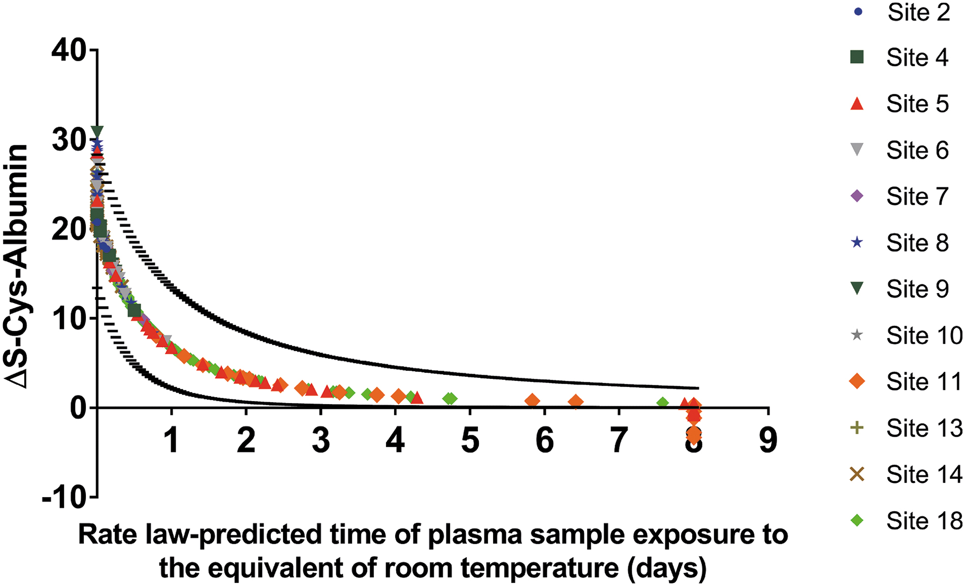
Estimate of plasma sample exposure times to the equivalent of 23°C based on the rate law for formation of S-Cys-Albumin. 29 The curved line traced out by the data points represents the rate law-predicted ΔS-Cys-Albumin value in a plasma sample with population-wide average initial concentrations of albumin, cystine, cysteine, and copper. The upper curved dashed line was calculated based on starting albumin and total copper concentrations of 2 SDs below the population mean but with a cystine concentration 2 SDs above the population mean; the lower curved dashed line was similarly calculated based on starting albumin and total copper concentrations of 2 SDs above the population mean but with a cystine concentration 2 SDs below the population mean. Thus, for each data point, the horizontal range between the two dashed black dashed lines represents the population-wide possible error for the calculated time of exposure to the equivalence of room temperature. Even though ΔS-Cys-Albumin should, theoretically, always be positive, some slightly negative ΔS-Cys-Albumin values were observed due to a modest degree of analytical error.
Univariate distribution of
The center line in Figure 5 depicts the trajectory assuming that each sample started with population average values of reduced and S-Cys-Albumin, cysteine, cystine, and copper; the dashed lines depict the ±2 SD (standard deviation) error around this estimate based on the known population distributions for these parameters. 29 Samples in Figure 5 were color coded by collection site. Most samples from site 5, 11, and some from site 18 stood out from the rest, particularly in the range where the equivalent time exposed to room temperature was longer than 1 day. Taken along with the site-specific sample handling conditions described above, these data reveal that samples stored at −20°C (for 42 to 456 days) exhibited the lowest ΔS-Cys-Albumin values and therefore the greatest exposure time to the equivalence of 23°C (as far as this particular marker is concerned).
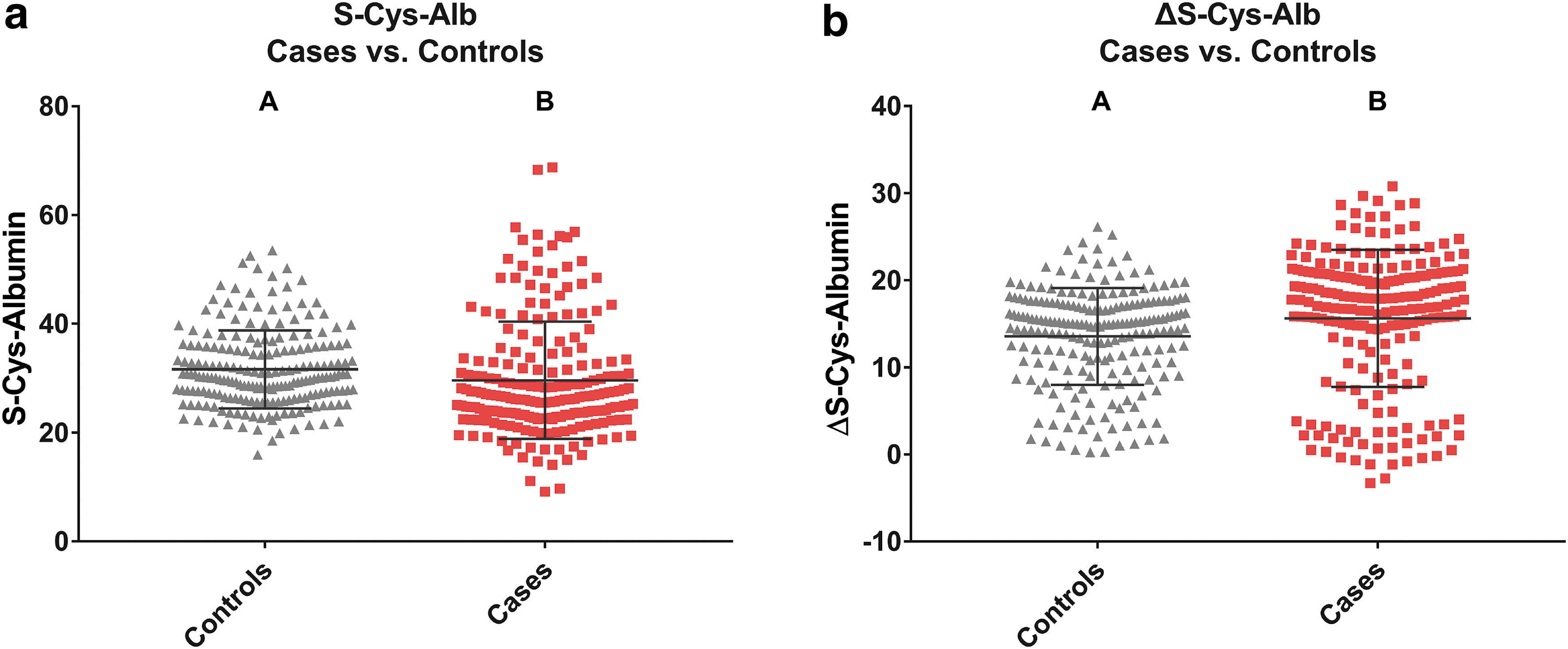
Comparison of ΔS-Cys-Albumin between lung cancer patients and controls
The oxidized form of P/S albumin has been proposed and employed as a marker for conditions related to oxidative stress,52–55 including chronic liver and kidney diseases and diabetes mellitus. 56 Given the data provided in this study and in previous publications describing the ex vivo oxidation (S-cysteinylation) of albumin,29,39,40 it is clear that neither S-Cys-Albumin or ΔS-Cys-Albumin should be put forth as biomarkers of disease in the absence of extremely rigorous control and documentation of preanalytical handling and storage conditions. Nevertheless, in a study like this one, we would be remiss to not compare S-Cys-Albumin and ΔS-Cys-Albumin in the control samples to that of the cancer patient samples (Fig. 6). Due to the large number of samples providing enormous statistical power, significant differences were detected for both S-Cys-Albumin and ΔS-Cys-Alb, but the distributions of the cancer patient population contained the entire control group population within itself and the mean ± SD values of the two patient groups were quite close (S-Cys-Alb: lung cancer cases 30 ± 11; controls 32 ± 7.2; ΔS-Cys-Alb: lung cancer cases 16 ± 7.9; controls 14 ± 5.6). As such, it is clear that the variations in ΔS-Cys-Albumin observed in this study were not caused by lung cancer and that ΔS-Cys-Albumin would not serve as an effective marker of lung cancer or any oxidative stress directly associated with it.
Discussion
The results presented above show that ΔS-Cys-Albumin in blood plasma is sensitive to common initial handling and storage conditions that leave samples in the thawed state—that is, at temperatures greater than −30°C.41,42 For reference, we have previously shown that S-Cys-Albumin is stable for at least 300 days when plasma is stored at −80°C. 40 Like many other analytes, the stability of S-Cys-Albumin is dramatically improved at −80°C relative to temperatures warmer than −30°C. 40 (For a summary of the stability of numerous clinical analytes in plasma stored for varying lengths of time at temperatures of −20°C and colder, see Hubel et al. 57 ) Nevertheless, storage of plasma at −80°C or colder does not guarantee the stability of all clinical analytes. 57 Our results also confirm that ΔS-Cys-Albumin is nonlinearly and inversely correlated with the time delay before storage—including delays before centrifugation. ΔS-Cys-Albumin in 94% of properly handled samples (i.e., those stored at −80°C within 3 hours from the blood draw) fell within the previously observed range for nonacute cardiac patients of 12%–29%. 29 (Further validation of this range in both cancer patients and healthy donors is presently underway.)
The thawed-state exposure times documented on paper and evidenced by the ΔS-Cys-Albumin assay in this study were categorized into four initial handling/storage conditions (as illustrated in Fig. 2a). Other investigators have reported clinical analytes that were unstable under each of these four conditions (Table 1). A total of at least 6, 18, 64, and 54 commonly measured clinical analytes have been found to be unstable under these four categories of handling/storage conditions, respectively. These unstable clinical analytes included metabolites (vitamins, fatty acids, etc.), peptides, and proteins (lipoproteins, prostate-specific antigens, etc.), hormones (insulin, leptin, etc.), cell-free DNA, and inorganic cations/anions (potassium, bicarbonate, etc.) (Table 1). (Undoubtedly, of course, there are certain to be hundreds of clinical analytes that remain stable under the conditions reported in this study. In fact, as part of our recent study of plasma protein glycan “nodes” in the WELCA sample set, we documented that glycan nodes remain stable under conditions more extreme than the conditions to which any of the WELCA samples were exposed. 58 )
Compilation of Studies Describing the Stabilities of Clinical Analytes in Blood Specimens Under the Initial Handling/Storage Conditions Documented in This Study
ACTH, adrenocorticotrophic hormone; APTT, activated partial thromboplastin time; FSH, follicle-stimulating hormone; HDL, high density lipoprotein; IDL, intermediate density lipoprotein; LDL, low density lipoprotein; PSA, prostate-specific antigen; PTH, parathyroid hormone; VLDL, very low density lipoprotein.
When viewed in line with the findings documented in Table 1, the results from this study showed that temporary storage of plasma at −20°C at satellite collection sites has a significant impact on the general integrity of samples treated in this manner. Although P/S samples appear to be frozen at −20°C, they are only actually fully frozen when the temperature is below −30°C41,42—although even storage at −80°C does not guarantee the stability of all potential analytes of interest. 57 Moreover, temporary storage at 4°C for more than 3 hours is generally ill advised, because many more analytes start to lose stability with delay times longer than 3 hours (Table 1). 11 We have provided elsewhere time course profiles at −20°C, 4°C, and 23°C for ΔS-Cys-Albumin in matched plasma and serum samples from donors starting at high, medium, and low initial ΔS-Cys-Albumin levels. 29
In light of the observations from this study, the ΔS-Cys-Albumin assay may serve as a valuable tool to assess P/S integrity—especially for P/S samples for which handling and storage paper trails do not exist and/or that may have been collected at multiple sites. Furthermore, it should be noted that pristine paper trails can be insufficient guarantees of sample integrity: As reported by Jeffs et al., 29 ΔS-Cys-Albumin measurements revealed an undisclosed integrity discrepancy between case and control samples in a set of serum samples for an early stage lung cancer study collected under NIH-sponsorship by seasoned investigators with a well-defined SOP. 29 As shown in Figure 4b, significant integrity discrepancies were observed among different batches of samples from the same site in this study (site 18). All samples from site 18 (the controls) were temporarily stored at −80°C in a private laboratory before being sent by batch to the central repository. Notably, samples from batches 6 and 7 were stored by the private laboratory during the course of a move to another location—that is, the samples may have been removed from −80°C for an unknown period of time. It is thought that a thawing incident may have occurred for these two batches during the move of the private laboratory.
ΔS-Cys-Albumin is a novel marker for P/S integrity. Because it requires LC/MS, many laboratories may not be equipped to run it. However, the easy dilute-and-shoot nature of the ΔS-Cys-Albumin assay along with its lack of requirement for a high-end LC-MS instrument means that nearly every MS facility and every major clinical reference laboratory should be readily capable of running it. Moreover, investigators should bear in mind that to make a credible evaluation of sample sets, it is not always necessary to analyze every single sample; rather, spot-checking a subset of each sample set that has been handled/stored together will, in many cases, suffice to empirically document P/S specimen integrity. Statistical power calculations to aid in this process were provided in our recent publication. 29
Conclusion
A simple dilute-and-shoot, intact-protein LC-MS assay known as ΔS-Cys-Albumin that quantifies cumulative exposure of P/S samples to their thawed conditions (> −30°C) was employed to evaluate the effect of various initial handling and storage conditions on plasma integrity in a clinical study involving multisite sample collection. ΔS-Cys-Albumin values were dramatically lowered under conditions of prolonged preprocessing/prestorage delay times at 4°C and at an elevated temporary storage temperature (−20°C) before shipment to the central repository where samples were permanently stored at −80°C. In accord with a previously established rate law for the ex vivo formation of S-Cys-Albumin in P/S, ΔS-Cys-Albumin values were found to be nonlinearly but predictably correlated with delay time before centrifugation as well as before storage. In consideration of the number of important clinical analytes that are unstable under the initial handling/storage conditions documented in this study, ΔS-Cys-Albumin will likely find utility as a tool to empirically evaluate P/S integrity.
Footnotes
Acknowledgments
The authors acknowledge resources and support from the Biodesign Institute core facilities at Arizona State University. The content is solely the responsibility of the authors and does not necessarily represent the official view of the National Institutes of Health.
Author Disclosure Statement
No conflicting financial interests exist.
Funding Information
The research reported here was supported in part by the National Cancer Institute of the National Institutes of Health under award number R33 CA217702 (to Chad R. Borges). The WELCA study was supported by the French “Institut National du Cancer” (Grant No. 2013-132), the “Fondation de France” (Grant No. 2015-60747), and the “Ligue Nationale Contre le Cancer” (Grant No. PRE2015.LNCC).