
Abstract
Anhydrous preservation is a promising approach for storage of living biomaterials at nonfreezing temperatures. Using the domestic cat model, the objectives of this study were to characterize changes in histology, DNA integrity, and viability of testicular tissues from adult versus prepubertal individuals during microwave-assisted drying. Testes from each age group were cut into small pieces before reversible membrane permeabilization, exposure to trehalose, and microwave-assisted drying during different time periods. In Experiment 1, water content was monitored for up to 40 minutes of drying. Tissues from adult or prepubertal cats experienced similar decreases of water content during the first 10 minutes. Desiccation progressed slowly between 10 and 20 minutes and then remained stable. In Experiment 2, structural properties were explored at 5, 10, and 20 minutes of desiccation. Percentages of normal seminiferous tubules were lower after 20 minutes drying in adult (43%) than in prepubertal tissues (61%). At the same time point, the proportion of cell degeneration was higher in adult (53%) than prepubertal tissues (28%). Percentages of intact DNA in tissues remained above 85% regardless of the microwave time in both age groups. Lastly, adult and prepubertal tissues only lost 33% of viability in both age groups. Collective results demonstrated for the first time that normal morphology, incidence of degeneration, DNA integrity, and viability of testicular tissues remained at acceptable levels during microwave-assisted drying for 20 minutes. Overall, prepubertal testicular tissues appeared to be more resilient to microwave-assisted desiccations than adult tissues. Importantly, water loss in the presence of trehalose after 20 minutes of desiccation already is compatible with long-term storage of testicular tissues at temperatures above −20°C, which is one step closer to future storage at supra-zero temperatures.
Introduction
Frozen biomaterial collections are invaluable tools in conservation biology and biomedical science.1,2 Specifically, long-term storage of frozen germplasms can be used to better understand fundamental cellular biology and reproduction. The untapped reserve of early germinal cells can also potentially be used to produce mature gametes and then embryos in vitro. 3 Therefore, the testicular tissue is a biomaterial of great value in male fertility preservation. 4 It contains early developmental stages of sperm cells that can possibly be grown in vivo (xeno-grafting) or in vitro to produce mature spermatozoa.
Understanding the resilience of living tissues to freezing temperatures is of paramount importance to ensure long-term storage of high-quality samples and to establish cryobanks. 5 However, relying on freezing temperatures involves specialized cryogenic equipment and facilities, a liquid nitrogen supply, high maintenance costs, and risks of contaminations.5–7 To tackle those issues, researchers have been inspired to use anhydrobiosis to develop simple and cost-effective storage methods at ambient temperatures,.8–11 Anhydrobiosis is a natural process used by a multitude of small organisms to resist dry conditions. It is based on the trehalose property to reach a high solute concentration without molecular mobility during dehydration, thus avoiding intra- and extracellular degradations.11–13
For several years, our laboratory has been studying dehydration in the presence of trehalose and storage at supra-zero temperatures for sperm cells, oocytes, and more recently for ovarian tissues.9,10,14 Among different methods to remove the water from the samples, desiccation provoked by exposure to microwaves is homogeneous in samples, which limits cellular damage.8,14 So far, resilience of living testicular tissues to dehydration has not been reported. Preservation of testicular tissue is complex due to the variety of cells, including spermatogonia and spermatocytes, which are primary cells leading to mature sperm cells during spermatogenesis. 15
In addition to preserving the histomorphology, DNA damage must be prevented while developing new preservation approaches. A loss in DNA integrity can lead to infertility in the parent as well as the future offspring.16,17 Cellular viability assessment also is an essential indicator that must be monitored. Cellular preservation techniques often involve physical and chemical agents that can affect cellular metabolism, causing cell toxicity leading to membrane destruction, changes in receptor mechanisms, inhibition of polydeoxynucleotide elongation, and enzymatic reactions.18,19 As we develop alternate solutions to preserve living tissues at nonfreezing temperatures, checking the above indicators in a systematic approach is a critical step.
To explore the influence of microwave-assisted desiccation on testicular tissues, the domestic cat is a good experimental model for other species as well as for humans.3–5 In human reproductive medicine and biomedical science, recovering and preserving tissues from prepubertal individuals is as important as for adults, to restore future fertility.20–23 It is therefore critical to study both age groups as we are evaluating a new fertility preservation technique.
Using the domestic cat model to develop future preservation at nonfreezing temperatures, the objectives of this first study were to characterize changes in histology, DNA integrity, and viability of testicular tissues from adult versus prepubertal individuals during microwave-assisted drying.
Materials and Methods
Collection and dissection of testes
Domestic cat testes were obtained after orchiectomy at local veterinary clinics. IRB approval was not needed. The Smithsonian Conservation Biology Institute's Animal Care and Use Committee granted a waiver of the animal care and use approval because samples were from routine neutering that otherwise would be discarded by veterinary clinics. Testes were transported in phosphate-buffered saline (PBS) at 4°C to the laboratory within 6 hours after surgery. Prepubertal animals (2–6 months of age) and adults (>2 years old) were collected (six testis pairs of each age category, one pair per day).
Testes were washed once with PBS and dissected from surrounding tissues. Samples were then placed in a medium composed of Hepes-Ham's F10 (No. 99168; Irvine Scientific) supplemented with 1 mM pyruvate, 2 mM
Testicular tissue preparation before microwave-assisted drying
Testicular pieces were threaded onto needles (30G × 1—0.3 × 25 mm) and immersed in Eppendorf tubes containing digitonin 1% (1 mg/mL in ultrapure water, Digitonin high purity, No. 300410; Merck KGaA, Darmstadt, Germany), for 3 minutes at room temperature (∼22°C) for permeabilization. Then, samples were washed three times in Eppendorf tubes containing the same medium at room temperature before being immersed in Eppendorf tubes containing 1 M trehalose solution in Tris-EDTA buffer (pH 8.00) for 10 minutes at room temperature (adapted from previous reports). 9
Microwave-assisted drying and rehydration
Samples were removed from needles and the excess of trehalose solution. After transferring the samples to filter paper over a polypropylene filter holder, they were placed onto a custom turntable inside the Microwave SAM 255 (CEM, Matthews, NC), setup at 20% power and maximum of 40°C,8,14 for different drying times. Samples were dehydrated in a microwave for up to 40 minutes, with water content evaluations every 5 minutes. After drying, tissue samples were rehydrated in Hepes-Ham's F10 medium supplemented for 30 minutes at room temperature (∼22°C) before evaluations.
Assessment of intratissue water level
To determine water content, control samples were sliced and immediately evaluated at time 0. Samples were also just exposed to preparation solution but not dried. Other evaluated samples were prepared and subjected to microwave drying for 5 minutes up to 40 minutes. Water content was measured by Karl Fischer titration, by placing the filter papers directly into a Mettler-Toledo V20 Volumetric Karl Fischer Titrator (detection limit 100 ppm). Methanol was used as the solvent and the titrant used was AquaStar CombiTitrant 5 (EMD Chemicals, Philadelphia, PA). All samples were weighed in a Mettler Toledo AX105 analytical balance to calibrate according each sample.8,9,14 All results were expressed as the percentage of water present in the sample.
Histomorphology evaluation
Testicular tissue samples were fixed in Bouin's solution in a refrigerator (4°C) overnight, washed five times in PBS, three times in 50%, 70%, 80%, 90%, and 100% ethanol for 10 minutes each in an orbit shaker. Then, samples were washed twice in xylene substitute and then embedded in paraffin. All paraffin blocks were sectioned in series (5 μm thickness), mounted on slides, and stained with Hematoxylin/Eosin. 4 Slides were analyzed using a microscope equipped for digital photomicrography (SPOT advanced software 5.0; Diagnostic Instruments, Inc., Sterling Heights, MI).
Seminiferous tubule and cell integrity were evaluated according to criteria established by Yildiz et al. 24 Intact tubules in general had no detachment of cells from the basement membrane, no rupture of stroma, no swelling of the fibroelastic capsule, normal junctions between cells, and no nucleus alterations, and were considered to have a normal structure. Tubules with changes in any of the previous criteria were classified as abnormal. 4 A total of 30 randomly selected seminiferous tubules for each animal in each experimental group were evaluated totaling 180 tubules for adults and 180 for prepubertal cats, and each tubule was classified as normal or abnormal. The percentage of normal seminiferous tubules was calculated relative to the total number of observed tubules, and percentage of basophilic nuclei cells (cells degeneration indicator) in each step.
DNA integrity evaluation by TUNEL assay
Levels of nuclear DNA fragmentation damage were assessed in both fresh and dehydrated samples using the TUNEL assay (Roche Diagnostics, Indianapolis, IN) adapted from Patrick et al. 10 Samples were fixed in 4% paraformaldehyde overnight. Samples were washed once in 50% and 70% ethanol, and next was the same procedure to prepare paraffin blocks for histomorphological evaluation.
All paraffin blocks were sectioned in series (5 μm thickness) and mounted on slides. Then, slides were washed in xylene substitute twice for 10 minutes, and washed in ethanol, for 3 minutes in xylene/ethanol (100%; 1:1) solution; and then for 3 minutes in ethanol (100%—twice, 95%, 80%, and 70%). Then the slides were washed three times in 1 μL Triton X/mL PBS for 5 minutes, and then were washed once in 5 μL Triton X/mL PBS for 30 minutes, all at room temperature. Then the slides were washed again once in 1 μL Triton X/mL PBS and positive controls were prepared and incubated with 100 μL RQ1 DNAse 10 × Reaction Buffer for 5 minutes at room temperature. In a dark room, the TUNEL assay was prepared by incubating in 9 μL Label Solution (fluorescein-dUTP)/μL Enzyme Solution (TdT), and for the negative control in 20 μL Label solution, all in a humidified darkened container at 37°C for 1 hour (Supplementary Fig. S1). Then, slides were washed thre times in 1 μL Triton X/mL PBS at room temperature, and the samples were incubated in Hoechst solution (5 μL Hoechst 33342 [1:100; Sigma-Aldrich, St Louis, MO] in 1 μL Triton X/mL) for 10 minutes. Excess liquid was removed and 30 mL of Vectashield/DAPI Fluoromount-G (Vector Laboratories, Burlingame, CA) was added to each slide. Samples were then covered with a coverslip and allowed to dry for 15 minutes at room temperature before observation with a microscope equipped with epifluorescence microscope (Olympus IX73; Olympus Corporation, Melville, NY) using SPOT software 5.0 (Diagnostic Instruments, Inc.).
A minimum of 1000 cells were analyzed per sample to determine the level of nuclear DNA fragmentation damage. The quantification of DNA fragmentation damage was based on the intensity of fluorescence (measured by analog to digital units) within each sperm head relative to the background fluorescence. 9 TUNEL-positive/negative cells were classified based on the presence (damaged)/absence (normal) of green fluorescence.
Cell viability evaluation
Calcein-AM and Ethidium homodimer-1 (EthD-1) from the LIVE/DEAD Viability/Cytotoxicity Kit (No. L3224; Invitrogen, Thermo Fisher Scientific, Waltham, MA) were used following the manufacturer's instructions. Cells present in tissue from all groups were extracted by slicing with a scalpel blade in Modified Ham's F-10 Basal Medium–HEPES (Irvine Scientific) supplemented. Sample tissues underwent a sequential enzymatic digestion in liberase (Liberase TL Research Grade—Thermolysin Low, No. 05401020001; Roche, Mannheim, Germany) solution (1:30—liberase/1 × Dulbecco's PBS [DPBS]) at 38°C for 30 minutes and mixed for 10 minutes each. Then, samples were added to a cold stop solution (1:9—1 × DPBS/FBS), and were centrifuged at 300 × g, for 3 minutes, supernatant removed, and pellets were resuspended in live/dead reagent (2.5 μL EthD-1/Calcein in 500 μL), at 38°C for 30 minutes in a dark room, 10 μL/mL Hoechst 33342 (1:100; Sigma-Aldrich) was added and incubated at 38°C for 10 minutes. Then, samples were transferred to slides and evaluated using epifluorescence microscope (Olympus IX73; Olympus Corporation). Cells stained with Calcein (green, Fig. 1A) were classified as live, and cells stained with EthD-1 (red, Fig. 1B) were classified as dead, and results were expressed as a percentage. 19

Representative pictures of viability evaluation in testicular cells.
To make a proper comparison between time points, proportional results of viability reduction (fx) were also expressed by the equation, fx = (XC − XD); XC = control value; XD = desiccation value.
Experimental design and statistical analysis
Experiment 1 was to determinate the water content after desiccation. Six pairs of testicles of adult males and 6 of prepubertal males (collected over several days; n = 6 replicates) were used to evaluate the water concentration. Testes were dissected and fragments were obtained, a part was selected to compose the control group (Control 1—fresh samples), and the other fragments were used for dehydration processing. Samples selected for dehydration were initially mixed in digitonin and later in trehalose. After this processing, samples were dried for 5, 10, 15, 20, 25, 30, 35, and 40 minutes before measuring water content.
Based on the results of Experiment 1, it was possible to select time intervals, where a significant reduction in tissue water content occurred. Thus, for Experiment 2, we selected the times for evaluation of cellular changes (5, 10, and 20 minutes in dehydration), in which we evaluated the histomorphology of the seminiferous tubules, as well as DNA damage and cell viability of the samples, after 30 minutes of sample rehydration. Twelve animals (six adults and six prepubertal) were collected over several days (n = 6 replicates).
Statistical analysis was performed using the R-project© statistical software version 3.3.2 (The R Foundation, Vienna, Austria). The experiment was conducted using a completely randomized design and the results expressed as mean ± standard deviation and verified for normality (Shapiro–Wilk test) and homoscedasticity (Levene's test). To evaluate the effect of the age of the animal, the data were submitted to Test T (p < 0.05) when parametric and Wilcoxon Mann–Whitney test (p < 0.05) when nonparametric (water content—10, 30, and 40 minutes). To assess the effect of time on each age group, data were subjected to analysis of variance followed by the Tukey's test (p < 0.05) when parametric, and the Kruskal–Wallis test followed by Dunn's test (p < 0.05) when not parametric (TUNEL).
Results
Water content progression during desiccation
In Experiment 1, no differences were observed at any time point between adult and prepubertal tissues (p > 0.05; Fig. 2). However, water content values were different (p < 0.05) between fresh samples (or after exposure to 1 M trehalose) at Time 0, 5 minutes of desiccation (leading to <40% of water content), or 10 minutes (water content below 20%). The water level then tended to decrease during the 10- and 20-minute intervals. After 20 minutes, water content appeared stable and this time point was chosen as the dehydration limit for further experiments.
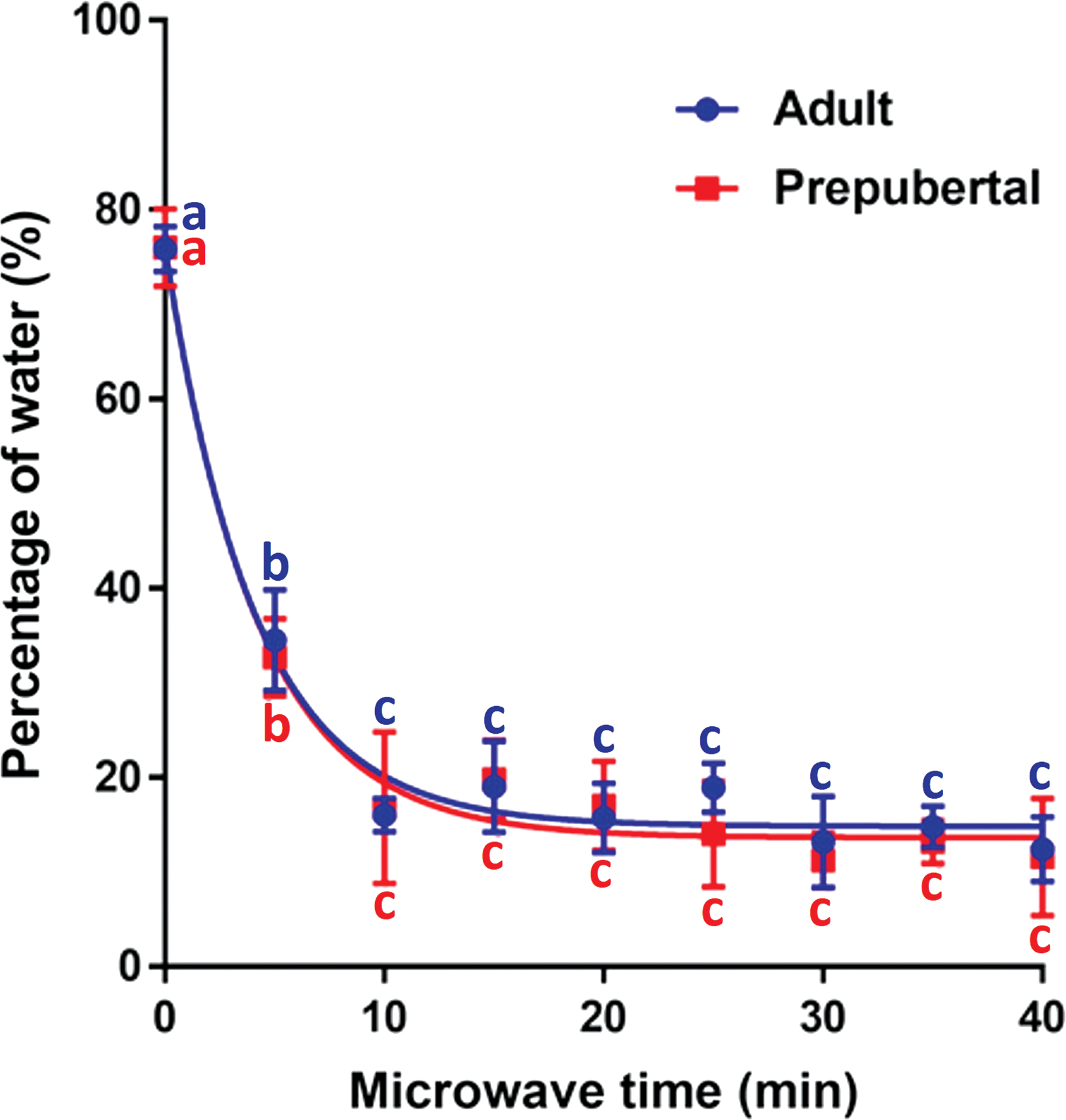
Percentage of water in testicular tissues from adult (n = 6 individuals) and prepubertal (n = 6 individuals) domestic cats following different times of microwave-assisted desiccation. Different lowercase letters represent differences across dehydration times (p < 0.05). Color images are available online.
Influence of microwave-assisted drying on histomorphology
In Experiment 2, percentages of seminiferous tubules with intact morphology in the control groups (almost 95%) were not different (p > 0.05) between adult and prepubertal tissues, except at 20 minutes of desiccation, where proportions in prepubertal tissues were higher (p < 0.05) than in adult tissues (Table 1). In adult tissues, a significant decrease of 18 percentage points was observed in morphological integrity after 5 minutes (p < 0.05) and continued to drop until 20 minutes (p < 0.05; Table 1). In prepubertal samples, a significant reduction only started after 10 minutes of desiccation compared with controls (p < 0.05; Table 1). In adult tissues, excessive dehydration changed the morphological structure of the tissue, causing the cells to detach from the basal membrane (Fig. 3D). Damage was uniformly distributed in the tissue sections from both age groups. Percentages of basophilic nucleus are reported in Table 2. The incidence of degeneration was higher in adults than prepubertal tissues after 10 and 20 minutes of dehydration (p < 0.05). It was possible to see a uniform increase in the number of cells with basophilic nucleus according to the dehydration time (Fig. 3A, E—control; 3B, F—5 minutes; 3C, G—10 minutes; 3D, H—20 minutes).
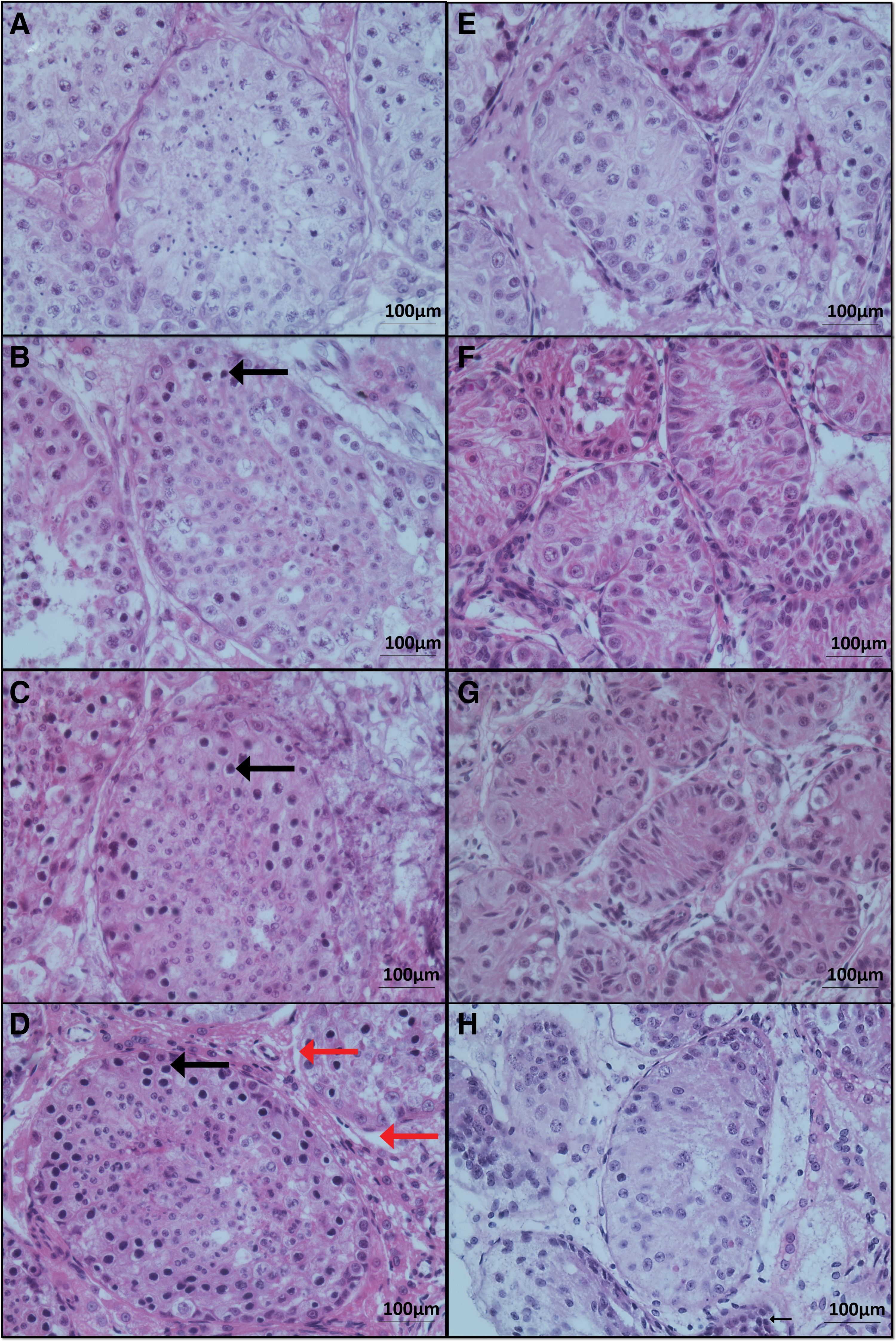
Histomorphological evaluation (Hematoxylin/eosin staining) of testicular tissues from adult (n = 6 individuals) and prepubertal (n = 6 individuals) domestic cats following different times of microwave-assisted desiccation.
Proportion of Normal Seminiferous Tubules (Histomorphology) in Testicular Tissue from Adult (n = 6) and Prepubertal (n = 6) Domestic Cats Following Microwave-Assisted Desiccation for 5, 10, or 20 Minutes
Values are expressed as mean ± SD. Different superscript lowercase letters in the same column means that there was a statistical difference among time evaluations (p < 0.05). Different superscript uppercase letters in the same row means that there was a statistical difference between adult and prepubertal cats (p < 0.05).
SD, standard deviation.
Proportion of Basophilic Nuclei in Adult (n = 6 Individuals) and Prepubertal (n = 6 Individuals) Testicular Tissue Following Microwave-Assisted Desiccation for 5, 10, or 20 Minutes
Values are expressed as mean ± SD. Different superscript lowercase letters in the same column means that there was a statistical difference among time evaluations (p < 0.05). Different superscript uppercase letters in the same row means that there was a statistical difference between adult and prepubertal cats (p < 0.05).
Influence of microwave-assisted drying on DNA integrity
Percentages of germ cells with intact DNA were not different between adult and prepubertal individuals at any time point (p > 0.05; Table 3). In both tissue types, percentages of cells with intact DNA steadily decreased over time, but the majority (about 85%) remained intact after 20 minutes of dehydration. Figures 4 and 5 show representative images of the TUNEL assay in testicular tissue sections from adult and prepubertal cats at different dehydration time points. There were no preferential localizations of cells with DNA damage within both tissue types.

DNA integrity in testicular tissues (TUNEL assay) from adult domestic cats following different times of microwave-assisted desiccation. First column represents cells with damaged DNA, second column all the cells (stained with DAPI).
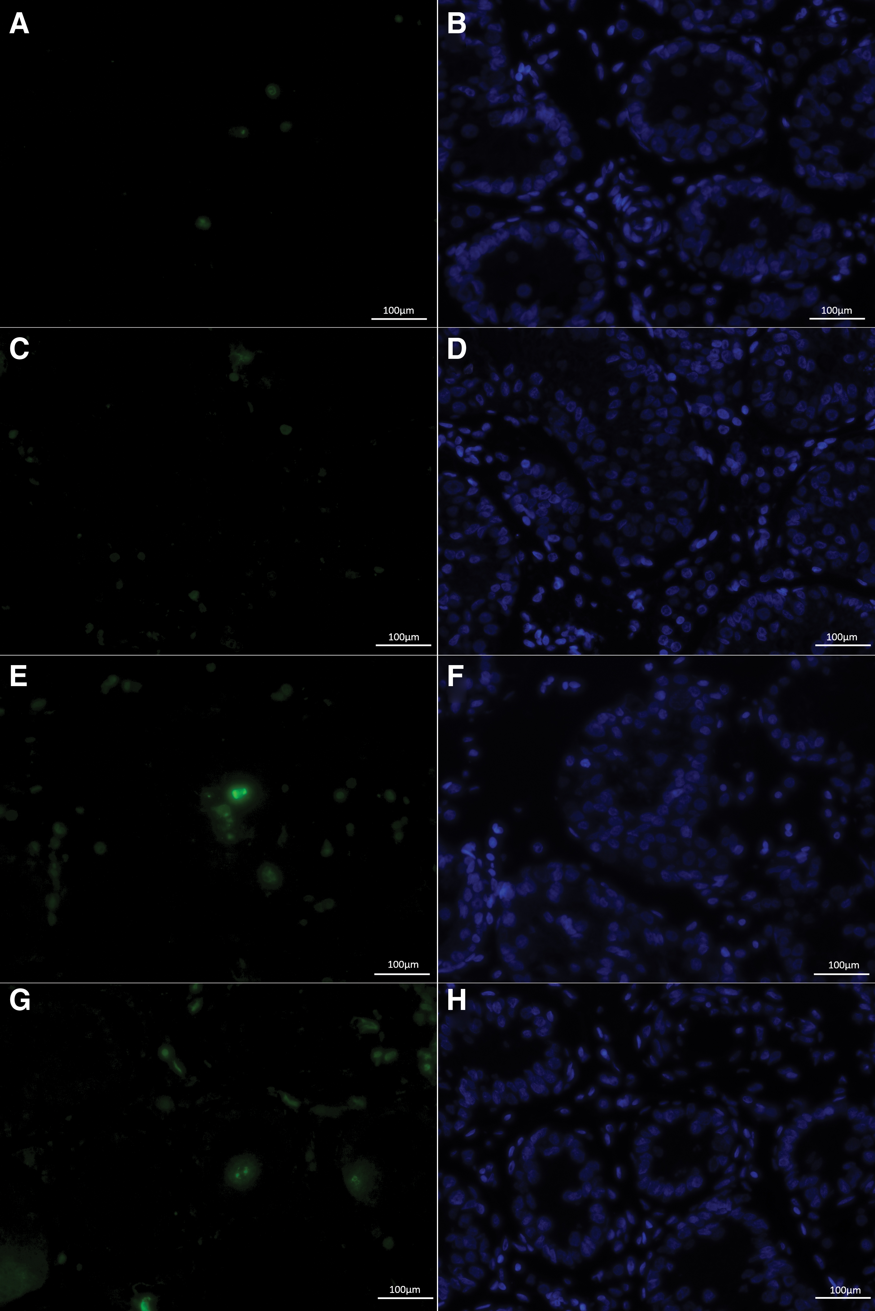
DNA integrity in testicular tissues (TUNEL assay) from prepubertal domestic cats following different times of microwave-assisted desiccation. First column represents with damaged DNA, second column all the cells (stained with DAPI).
Proportion of Cells with Intact DNA (Tunel Assay) in Testicular Tissues from Adult (n = 6 Individuals) and Prepubertal (n = 6 Individuals) Domestic Cats After Microwave-Assisted Desiccation for 5, 10, or 20 Minutes
Values are expressed as mean ± SD. Different superscript lowercase letters in the same column means that there was a statistical difference among time evaluations (p < 0.05). Different superscript uppercase letters in the same row means that there was a statistical difference between adult and prepubertal cats (p < 0.05).
Influence of microwave-assisted drying on tissue viability
Percentages of viable cells in prepubertal tissues were higher than in adult tissues in the control group (p < 0.05; Table 4). From 5 to 20 minutes, percentages of viability reduction did not differ between groups (p > 0.05). For each tissue type, the proportion of viable cells started to decrease after 10 minutes drying (p < 0.05). In adult tissues, there was no more decrease between 10 and 20 minutes (p > 0.05). However, in prepubertal individuals, percentages of viability were lower at 20 minutes while remaining at better levels than the adult tissues (p < 0.05). Overall, 33% of viability was lost over the 20 minutes of desciccation in both age groups (Table 4).
Proportion of Viability in Desiccated Adult (n = 6 Individuals) and Prepubertal (n = 6 Individuals) Testicular Tissues Following Microwave-Assisted Desiccation for 5, 10, or 20 Minutes
Values are expressed mean ± SD. In parentheses, percentage of viability reduction cell after processing (fx), fx = (XC − XD); XC = Control Value; XD = Desiccation Value. Different superscript lowercase letters in the same column means that there was a statistical difference among time evaluations (p < 0.05).
Different superscript uppercase letters in the same row means that there was a statistical difference between adult and prepubertal cats (p < 0.05).
Discussion
This is the first study describing the influence of microwave-assisted drying in the presence of trehalose on testicular tissues in the domestic cat model. After 20 minutes of microwave exposures, more than 50% of the histomorphometry was uniformly preserved, 85% of DNA was intact, and only 33% of viability was lost. Overall, testis samples from prepubertal individuals were more resilient to the preservation method than the adult ones.
Similar approaches have already been used for the preservation of structure and function of mature sperm cells, 10 but multicellular tissues are much more complex to dehydrate.
Dehydration time varies according to the initial water content present in the sample and the concentration of trehalose during preliminary exposure. 8 Using a concentration of 1 M trehalose, the water content in samples was monitored until 40 minutes of dehydration. Similar to recent reports in ovarian tissues, water content dropped faster during the first 10 minutes than for the rest of the dehydration process. 14 The water content present in the sample may influence the quality of tissue preservation, where it has been shown that high concentrations of remaining water can cause early tissue degeneration . 8 Based on our recent report in ovarian tissues, 14 we expect that trehalose penetrated the samples and were incorporated in the different cells composing the testicular tissue. Similarly, water removal was homogeneous in the tissue biopsy. This was confirmed by the incidence of histomorphometric or DNA damages that were uniformly distributed in the tissue sections. Importantly, water loss in the presence of trehalose after 20 minutes of desiccation is compatible with long-term storage of tissues at temperatures above −20°C. 14
Evaluations of testicular tissue histomorphology showed that an acceptable proportion of seminiferous tubules was preserved. Adult individual samples lost a little more than 50% of tubule integrity after 20 minutes of drying compared with prepubertal individuals. This was consistent with observations by Mota et al. 25 in cryopreservation studies of testicular tissues from immature individuals. This difference might be due to the tissue composition being more compact and homogeneous in prepubertal individuals than in adults. 26
Even after the longest dehydration time (20 minutes), structural integrity in young testes was similar to observations by Lima et al., 4 in the testicular cryopreservation of prepubertal cats after warming at 37°C. A frequent change was the spacing between the cells and the basement membrane becoming larger over time during dehydration. This revealed a loss of gap junctions, which are essential for the passage of several molecules, ions, and electrical impulses between the cellular cytoplasm.27–29
Incidence of basophilic nuclei also increased with the dehydration level. Basophilia is characterized by the deposition of dense granules in cell structures and may also be a parameter to determine cell disorder, or cells in degeneration stages. 30 Excessive dehydration times may have intensified the degeneration of tissue cells.
In both tissue types, percentages of cells with intact DNA steadily decreased over time, but the majority (85%) remained intact after 20 minutes of dehydration and was uniformly distributed. Alterations in chromatin can affect spermatogenesis, consequently altering the germ line, because most somatic histones are replaced by DNA packaging proteins. 31
The importance of the integrity of the morphological structure after processing is directly linked to the resumption of spermatogenesis. 4 No differences in DNA integrity were found among age categories, but significant differences were found across processing times. Previous reports showed that germ cells, even in the early stages of differentiation, are more sensitive and predisposed to DNA damage than sperm cells. These changes are also observed in other cell types such as sperm 10 or other preservation techniques like freeze drying. 32 Hydrogen bonds between the bases of the DNA strands can be easily broken due to nonphysiological temperatures, thus altering their conformation and being one of the factors that can compromise the viability of the cell.33,34 Compared with vitrification of testicular tissue, our results were close to those described by Lima et al. 4 that reported at least 60% cells with intact DNA postwarming.
The majority of tissue viability was preserved. However, total loss in viability in adult individual samples after 20 minutes of drying was more important than in immature individuals. This proved again the greater resistance of tissues from young individuals. Tissue processing likely influenced viability results since control samples did not contain 100% of viable cells. This factor is common in all tissue processing in which the substances used for evaluations have deleterious effects on the sample, so it was necessary to adjust the results, making a relative comparison to determine the real value of the results expressed. 18
Reduction of viability during the drying stages using the microwave was inevitable.9,14,35 However, promising results were obtained, since the percentage of viable cells were close to those obtained in bulls (58%), 36 mouse (25%–95%), 37 and cats (60%–80%), 4 using conventional techniques such as cryopreservation of testicular tissue.
This is the first evaluation of testicular tissue processing by microwave-assisted drying. Despite the expected changes in structural integrity and viability, the results are quite promising. To reach the vitreous/stable state it will be necessary to reduce the internal moisture or decrease the temperature.38,39 Interestingly, water loss in the presence of trehalose after 20 minutes of desiccation already is compatible with long-term storage of tissues at temperatures above −20°C, 14 which is one step closer to anhydrobiosis and storage at supra-zero temperatures. Future storage studies at different temperatures are warranted. This technique may be applied in the future fertility preservation strategies, reducing costs and stimulating the development of germplasm biobanks.40,41
Footnotes
Acknowledgments
The authors would like to thank Dr. Brent Whitaker (Animal Rescue, Inc.) and Dr. Keiko Antoku, and their staff for providing domestic cat testes and the Smithsonian Institution for providing all the necessary structure for the development of this work. L.D.M.S. and A.R.S. are National Council for Scientific and Technological Development (Conselho Nacional de Desenvolvimento Científico e Tecnológico—CNPq) investigators.
Author Disclosure Statement
The authors have stated that there are no competing interests. None of the authors has financial or personal relationships that may influence or distort the content of the article.
Funding Information
We would like to thank these funding agencies for financial support: CAPES (Coordenação de Aperfeiçoamento de Pessoal de Nível Superior—Brazilian Federal Agency for the Support and Evaluation of Graduate Education), for PDSE (Programa de Doutorado Sanduíche no Exterior—internship program PhD abroad).
References
Supplementary Material
Please find the following supplemental material available below.
For Open Access articles published under a Creative Commons License, all supplemental material carries the same license as the article it is associated with.
For non-Open Access articles published, all supplemental material carries a non-exclusive license, and permission requests for re-use of supplemental material or any part of supplemental material shall be sent directly to the copyright owner as specified in the copyright notice associated with the article.