
Abstract
Due to the induced oxidative stress that exists in sperm freezing/thawing procedures and handling media, the use of exogenous antioxidant agents seems necessary. Drug delivery by nanocarriers has been designed to overcome the limitations of antioxidants, such as high-dose toxicity and short biological half-life. In this study, we tried to investigate the effects of tretinoin-loaded solid lipid core nanocapsules (TTN-SLN) added to freezing/thawing and handling media (in three experimental groups) on sperm motility (total/progressive), viability, DNA fragmentation, and extracellular reactive oxygen species (ROS) levels. Sperm samples from at least 30 adult male NMRI mice were evaluated in this study. The results of experiments 1 and 2 showed that the addition of 0.5 μM TTN-SLN in freezing and thawing medium significantly increased sperm viability and total/progressive motility and decreased DNA fragmentation and extracellular ROS levels (p < 0.05). Adding 0.25 and 0.5 μM of TTN-SLN to the handling medium (experiment 3), increased sperm parameters and decreased DNA fragmentation and extracellular ROS levels significantly (p < 0.05) compared with the control group. Briefly, our results indicate that SLN can deliver the lowest concentrations of tretinoin in a controlled release mechanism into the intracellular space of sperm. Also, high-dose TTN-SLN is safe during freezing/thawing and handling processes of mouse sperm.
Color images are available online.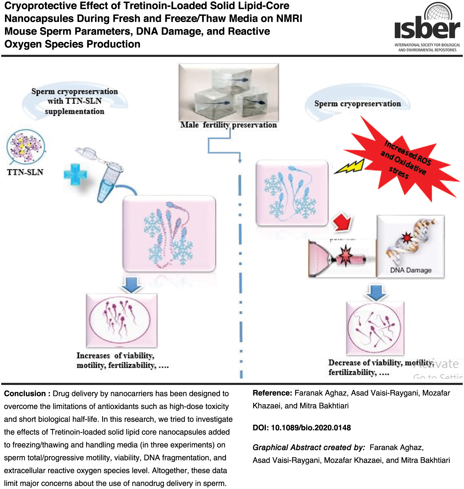
Introduction
Sperm cryopreservation has become a mainstay of the assisted reproduction technology (ART) laboratory and research centers, and increasingly sperm banking is offered to patients at risk of early menopause, and underpins fertility preservation for patients with cancer and other conditions.1,2 However, manipulation of sperm during in vitro or freezing/thawing processes involves the exposure of sperm to high levels of reactive oxygen species (ROS), which is proposed as the major contributing factors in cryodamage to spermatozoa.3,4 This cryodamage is still a great concern and could dramatically reduce sperm fertilization efficiency in ART centers.5,6 Therefore, a reasonable strategy to improve the cryopreserved sperm quality is the addition of various substances such as antioxidants to the freezing/thawing and handling media.
Antioxidants are molecules with the potential ability to prevent or reduce oxidative processes in other molecules by scavenging the produced free radicals. 7 Previous works focused on the use of applications of sericin, 8 vitamin E, 9 resveratrol,10,11 ascorbic acid, 10 genistein, 12 and melatonin13–16 as antioxidant supplementations on the improvement of sperm motility, protection of DNA, and reduction of ROS levels. 17
Tretinoin (all-trans retinoic acid, TTN), an active metabolite product of vitamin A, is widely used as a retinoid with potent antioxidant or free radical scavenging. 18 Experimental evidence confirmed TTN's beneficial effects, such as protection against various kinds of oxidative stress (OS) on follicular growth, oocyte development, fertilization, earlier embryonic growth,19,20 and spermatozoa capacitation.21,22 Also, it has been found that the inhibition of peroxidation processes in membranes, which could be similar to that of β-carotenes or α-tocopherol, may also be an important process for gamete and embryo development. Timm et al. 23 have shown that nondifferentiated HL-60 cells can be cryopreserved/thawed, and start cell proliferation after being treated with TTN. These findings suggested that TTN could be used as an antioxidant supplement for sperm cryopreservation.
In spite of these benefits, TTN has some drawbacks such as poor hydrosolubility, low bioavailability, and short biological half-life. 24 One of the most prevalent methods to overcome these challenges and intragamete delivery of insufficient amounts of TTN is administration of a nanoencapsulation system.25–27 To date, a variety of nanocapsule platforms have been employed for encapsulation of antioxidants such as liposomes, micelles, polymeric nanocapsules, and solid lipid core nanocapsules (SLNs). SLNs are colloidal carrier systems containing blending lipids as a solid core coated in surfactants. 28 SLNs have emerged as an alternative to other new approaches due to various benefits, such as the possibility of encapsulation of lipophilic drugs, improved physical stability, low cost compared with liposomes, and ease of scale-up and manufacturing. 29
Beneficial effects of hydrophobic drug-loaded SLNs in mouse gametes/embryos culture were explored previously (not published) by our research team. Yet, the encapsulation of the TTN into SLN during the freezing/thawing and handling media in mice has not been elucidated. As the first applications of SLN in studies involving gametes/embryo cryopreservation, this investigation was designed to determine whether supplementing the freezing/thawing and handling media with tretinoin-loaded solid lipid core nanocapsules (TTN-SLN) can protect mouse sperm against cryodamage or OS.
Materials and Methods
Materials
In this study, sperm samples of 30 adult male NMRI mice were used. Male NMRI mice were purchased from the Pasteur Institute, Tehran, Iran and housed under standard conditions. All applicable institutional guidelines for the care and use of animals were carried out in complete accordance with the regulatory guidelines of Animal Ethics and Welfare Committee (AEWC) of Kermanshah University of Medical Sciences. All protocols used in the study were approved by the Ethics Committee of Institutional Animal Care at the Kermanshah University of Medical Science, Kermanshah, Iran (IR.KUMS.REC.1399.1009), and Iran National Science Foundation (INSF, 97008908). All chemicals and reagents were purchased from Sigma-Aldrich Chemical Co. (St. Louis, MO, USA) unless otherwise stated.
Ethics statement
This research was carried out in compliance with the rules laid down by the Faculty of Medicine Ethics Committee of the Kermanshah University of Medical Sciences.
Preparation of TTN-SLN
TTN-SLN were prepared by the general self-assembly method. 30 Briefly, the aqueous phase (containing 0.25 mg of the Tween 80) was added to the lipid phase (containing 1 mg/mL of stearic acid dissolved in CH2CL2) under a stirrer. Then, 3 mg of TTN (dissolved in CH2CL2 in separate vials) was added to the previous solution drop by drop under ultrasonication (for 15 minutes at 80 W). Then the vial was dialyzed against distilled water for 12 hours, and purified by a 0.2 μm filter membrane to remove the unloaded TTN. Unloaded SLN were prepared using the same procedure. In addition to the physical appearance, ease of reconstitution, and stability of storage, the nanocapsules were checked for variations in desired quality attributes when added to the freezing/thawing and handling media. The TTN-SLN was freeze dried and stored at −20°C in a dark environment.
Characterization of prepared nanocapsules
Fourier-transformed infrared spectroscopy
The chemical composition of the synthesized nanocapsules was determined using Fourier-transformed infrared (FT-IR) spectroscopy (model FTIR Prestige-21; Shimadzu Co., Japan). The sample spectra were measured over a 200–4000/cm scanning range, with a 4/cm spectral resolution.
Basic physicochemical properties
A Zetasizer instrument (model 3600; Malvern Instruments Ltd., Worcestershire, United Kingdom) was used to determine the average particle diameter (size), size distribution and polydispersity index (PDI), and zeta potential (ZP) of nanocapsules (TTN-SLN). Morphology and surface characteristics were assessed with scanning electron microscope (SEM) microscopy (KYKY-EM3200).
Drug loading and encapsulation efficiency
Our research team used Ultraviolet-visible (UV-Vis) spectrophotometry (Philips PU 8620, USA) to determine the entrapment efficiency (EE%) or drug loading content (DL%) as defined previously 31 versus a calibration curve.
EE% and DL% of TTN were calculated through the following equation:

In vitro release profile
In vitro release profiles of nanocapsules (TTN-SLN) and TTN solutions were investigated by the dialysis bag method. 30 Briefly, the TTN-SLN and TTN (comparable to 1 mg of each one) were redispersed in 80 mL of release medium, that is, phosphate-buffered saline (PBS; pH 7.4) for the 55 hours at 37°C ± 0.5°C in dialysis bags. At different time intervals (0.5, 1, 2, 3, 4, 5, 6, 24, 48, and 55 hours), 1 mL of the receptor phase was taken out and replaced with the same volume of fresh fluid. The quantity of TTN in each sample release medium was evaluated by a UV spectrophotometer set (Philips PU 8620, USA) at 360 nm.
Collection and processing of semen
In three experiments, the specific sperm parameters (motility, morphology, and vitality) were assessed using light microscopy in compliance with the guidelines of the World Health Organization. 32 After basic sperm analysis, the sperm samples were freshly analyzed for DNA fragmentation by the sperm chromatin dispersion (SCD) test.
Sperm freezing and thawing
Sperm cryopreservation was performed as described previously. 8 The cauda epididymis and vasa deferentia were collected from male NMRI mice (6–8 weeks) in a sperm collection dish (Falcon, Franklin Lakes, New Jersey, USA). For cryopreservation, five aliquots (500 μL) of each sample were diluted (1:0.7), drop by drop, with a freezing medium (SpermFreeze, a commercial cryoprotectant consisting of 15% glycerol in HEPES buffer). After 10 minutes at 37°C, the cryovials were placed onto a polystyrene raft floating in liquid nitrogen vapors for 15 minutes (slowly frozen) and then immersed in liquid nitrogen (−196°C liquid N2) for storage.
Following a minimum time of 48 hours of cryostorage, the samples were thawed for 4–5 minutes in a tap water container with a temperature of 35°C. After that, the sperm samples with 50.50 ± 22.90 × 10⁶ concentration were resuspended in the Hamsef.10 medium supplemented with 10% human serum albumin. Following 30 minutes of incubation, they were centrifuged at 300 g for 7 minutes. Subsequently, they were immediately analyzed for sperm motility, viability (detected by Eosin 0.5% staining), and DNA fragmentation (detected by SCD staining).
Sperm motility and viability analyses
Sperm motility analysis was done as described previously. 8 Briefly, 10 μL of sperm suspension was dropped onto a slide and covered with a coverslip. The sperm motility percentage was measured by optical microscopy at 400 × magnification and categorized into three grades according to the WHO criteria (WHO 2010): progressive, nonprogressive, and immotile. 32 At least 200 sperms per sample were counted, which was repeated three times. Based on the methodology used by Banihani et al., 33 the sperm viability of each treatment group was evaluated by 0.5% Eosin staining, under 400 × magnification. Finally, the percentages of unstained (live) sperms and partial or complete red-colored (dead) sperms were calculated.
Assessment of sperm DNA fragmentation
In all three experiments, the newly updated and improved version of the SCD method was used to determine DNA fragmentation. 34 In brief, an aliquot of each semen sample was diluted in PBS medium to a concentration of 10–15 million spermatozoa/mL. In the meantime, an Eppendorf tube containing agarose was put in a water bath at 95°C–100°C for 5 minutes to melt the agarose. Then, it was moved to a 37°C water bath. Sixty microliters of diluted semen was transferred to the Eppendorf tube after temperature equilibration and was gently mixed following combination with the fused agarose. Subsequently, 20 μL of this suspension was pipeted into precoated slides with agarose and protected with a 22 × 22 mm coverslip. To solidify the agarose, the slides were put on a cold plate in the refrigerator (4°C).
After 5 minutes, the slides were taken out of the refridgerator, and the coverslips were gently removed. The slides were then immediately immersed horizontally in an acid solution, previously prepared by mixing 80 μL of HCl from an Eppendorf tube in a kit containing 10 mL of distilled water, and incubated at room temperature (22°C) for 7 minutes. The slides were then completely immersed horizontally in the lysing solution in 10 mL for 25 minutes. After 5 minutes of washing in a tray with plenty of distilled water, the slide was dehydrated in ethanol baths of increasing concentration, including 70%, 90%, and 100% for 2 minutes each and then were air dried. Finally, the slides were allowed to air dry before staining. For bright field microscopy, the slides were horizontally covered with a mix of Wright's staining solution (Merck, Darmstadt, Germany) and PBS (Merck) (1:1) for 5–10 minutes. Slides were washed in tap water for a short time and allowed to dry. Eventually, the samples were analyzed using light microscopy (Olympus BX-40; Olympus U-RFL-T, Tokyo, Japan) at 400 × magnification (minimum of 300 spermatozoa per semen sample was counted). Spermatozoa, with a broad and spotty halo around it, contained intact DNA in the compact nuclear core, and spermatozoa, displaying a small or absent halo around the nucleus corresponding to those with fragmentation. 35
Measurement of extracellular ROS levels
Following each experiment, the sperm samples were collected for extracellular ROS measurements with a chemiluminescence assay as previously reported. 36 First, Luminol (5-amino-2, 3-dihydro-1, 4-phthalazinedione; Sigma, St. Louis, MO, USA) as a detector was used, and 100 mmoL/L stock solution of luminol was formulated in dimethyl sulfoxide. Then 10 mL of working solution (5 mM) was added to the 300 mL of each sperm sample. Chemiluminescence was measured for 15 minutes using a luminometer, and results were expressed in relative light units (RLU)/s/106 sperm. This procedure was performed five times for each experiment.
Experimental design
Experiment 1: Cryopreservation of sperm samples in freezing medium supplemented with various concentrations of TTN-SLN
Sperm samples from 10 adult male NMRI mice were obtained in experiment 1. After a basic analysis, the samples were frozen in the presence of basic freezing medium plus TTN-SLN with various concentrations (0 [control group], 0.25, 0.5, 1, and 5 μM), for a minimum period of 48 hours. The thawing cycle proceeded, as was mentioned above. After 7 minutes of centrifugation at 300 g, the sperm motility, viability (detected by staining with Eosin 0.5%), and DNA fragmentation were analyzed immediately.
Experiment 2: Thawing of frozen sperm samples in thawing medium supplemented with various concentrations of TTN-SLN
In the second experiment, sperm samples were collected from 10 adult male NMRI mice. As mentioned above, following swimming and centrifugation, each sample was cryopreserved through a freezing procedure. For the thawing process, each sample was divided into five cryovials containing thawing medium supplemented with TTN-SLN with various concentrations (0 [control group], 0.25, 0.5, 1, and 5 μM). After 7 minutes of centrifugation at 300 rpm, the cryovials were incubated for 1 hour and were immediately analyzed for sperm motility, viability (detected by 0.5% eosin staining), and DNA fragmentation.
Experiment 3: Handling of samples in medium supplemented with various concentrations of TTN-SLN
In the third experiment, sperm samples were collected from 10 adult male NMRI mice. Each sample was divided into five cryovials containing handling medium supplemented with TTN-SLN with various concentrations (0 [control group], 0.25, 0.5, 1, and 5 μM). After 7 minutes of centrifugation at 300 rpm, the cryovials were incubated for 1 hour and were immediately analyzed for sperm motility, viability (detected by 0.5% eosin staining), and DNA fragmentation.
Statistical analyses
Each experiment was performed five times, and all data were expressed as mean ± standard deviation. First, data were tested for normality analysis of parameters with the Kolmogorov–Smirnov test. Then, statistical comparisons between the experimental groups were analyzed using a factorial ANOVA followed by posthoc analysis for multiple comparisons (Tukey's HSD test). All statistical analyses were conducted using the SPSS software for Windows (version 23.0). p-Value of ≤0.05 was considered as statistically significant.
Results
Physicochemical characterization of nanocapsules
The analysis of size, PDI, ZP, EE%, and DL% of TTN-SLN are shown in Figure 1 and Table 1. The size-average diameters of B-SLN (basic SLN or SLN without tretinoin) and TTN-SLN were 145 ± 6.1 and 185 ± 7.2 nm, respectively. The ZP and PDI of TTN-SLN were 16 mV and 0.16, respectively.
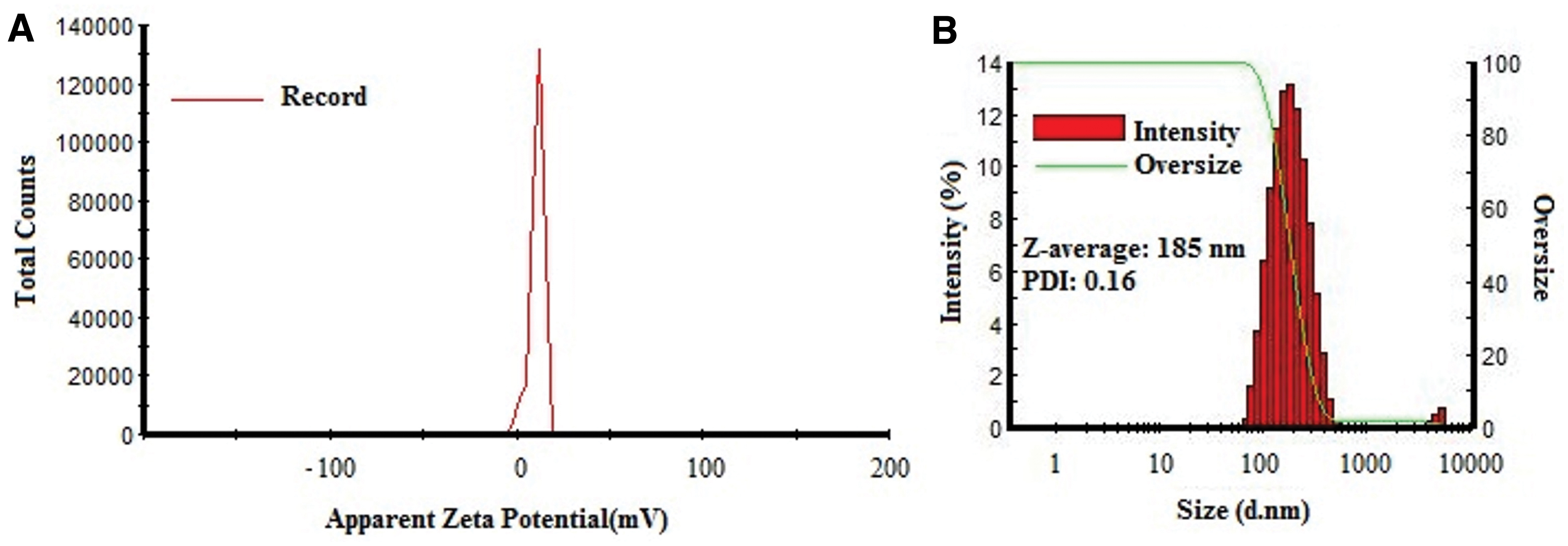
Physicochemical characterization of TTN-SLN by Zetasizer instrument;
Physicochemical Features of the Different Nanocapsules
Data are presented as absolute values (mean ± SD). The experiment was repeated three times.
SLN, Solid Lipid nanocapsules; blank, nanocapsules without drug; B-SLN, basic SLN; DL%, drug loading capacity; EE%, eapsulation efficiency %; SD, standard deviation; TTN-SLN, tretinoin-loaded solid lipid core nanocapsules.
To evaluate the EE% and DL% of the drug into the SLN, the calibration curve of TTN was conducted by the molarity of solutions between 0.5 and 2 μg/mL and samples of TTN-SLNs were prepared and analyzed by UV–vis spectrophotometry (Philips PU 8620, USA). As indicated, high EE% was achieved with values of 92% and DL% of 21% for SLN (Table 1).
The morphological study of TTN-SLN by SEM microscopy is illustrated in Figure 2. SEM results demonstrated that TTN-SLN particles are spherical and regularly distributed between the prepared nanocarriers. Consistent with the findings of Zetasizer, the SEM also confirmed that TTN-SLN particles were in nanosize range with no aggregation and adhesion.
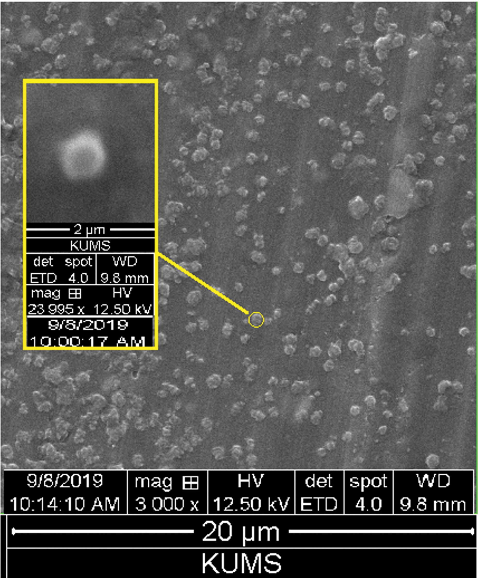
SEM analysis of the optimized TTN-SLN. Yellow outlined indicates nanoparticles with higher magnification. SEM, scanning electron microscope. Color images are available online.
In vitro release profile
The in vitro release of TTN from TTN-SLN studies was carried out under physiological pH (7.4), up to 55 hours (Fig. 3). We observed that the TTN from pure drug is released 100% over 10 hours. In comparison, the TTN was released from their corresponding nanocapsule (TTN-SLN) at a second-order rate with ∼25% of drugs released over 10 hours followed by ∼38% after 24 hours and finally 60% of the drug was released from nanocapsule over 55 hours of incubation time.
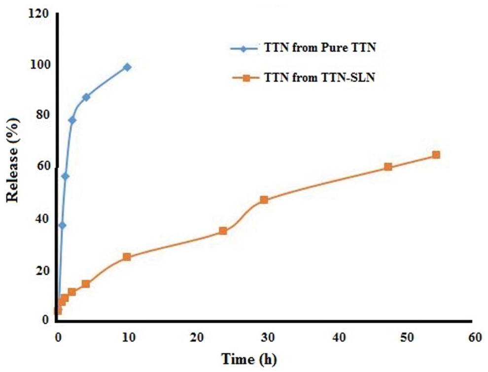
In vitro drug release profiles of TTN from the pure TTN and TTN-SLN with the dialysis bag system in the PBS medium pH 7.3 at 37°C for, 55 hours. PBS, phosphate-buffered saline. Color images are available online.
FT-IR analysis
Figure 4 shows the FT-IR spectra of the pure TTN, B-SLN, and TTN-SLN. The TTN spectrum (Fig. 4) showed the peaks at 3429, 3043, 2927, 1685, 1604, and 1088/cm. The SLN spectrum showed the peaks at 3437, 3012, 2920, 2854, 1693, 1639, 1562, 1415, 1246, and 1103/cm. 37 The TTN-SLN spectrum (Fig. 4) showed the peaks at 3415, 3043, 2918, 2850, 1690, 1606, 1577, and 1083/cm experiment, as reported by our research team previously (accepted for publication).
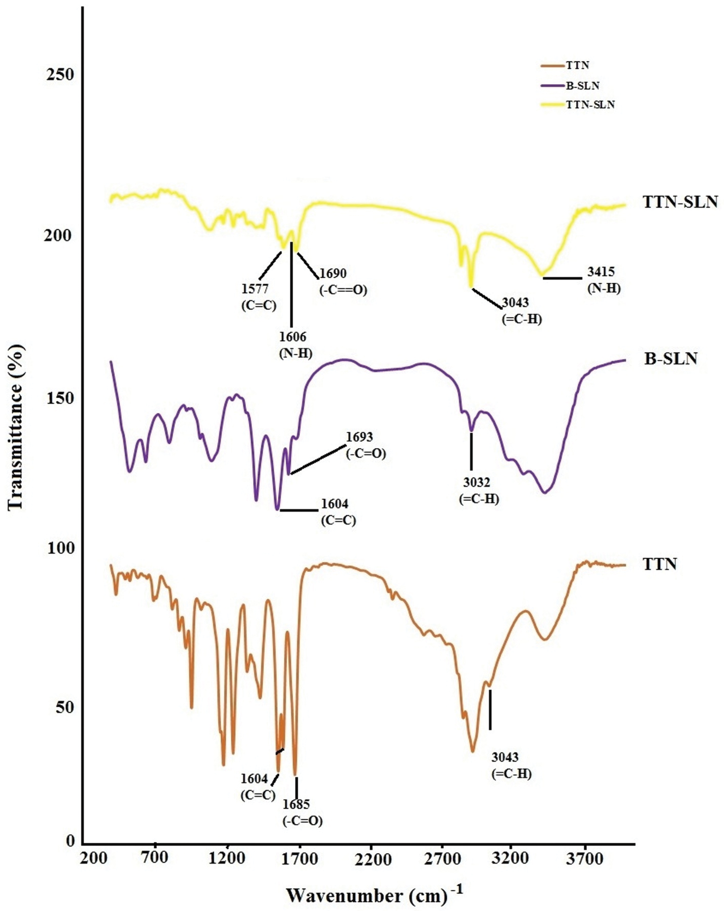
FT-IR diagrams of the TTN, B-SLN, and TTN-SLN. B-SLN, basic SLN; FT-IR, Fourier-transformed infrared. Color images are available online.
Experiment 1: Effect of TTN-SLN with various concentrations added to freezing medium on sperm parameters and DNA fragmentation
As shown in Table 2, significant differences were found in total and progress motility, viability, and DNA fragmentation index (DFI) among the groups. Our findings suggested that the total motility and progress motility were highest value in the treated group with 0.5 and 1 μM TTN-SLN compared to the control and other groups (Table 2; p < 0.05). No substantial differences were found between the maximum dose of TTN-SLN or B-SLN (5 μM) groups and the control group when comparing total and progressive motility. The results achieved from Eosin staining showed that the viability significantly higher in the treated group with 0.5 μM TTN-SLN (58.78 ± 1.87) compared to the control and other groups (Table 2; p < 0.05). No significant differences were detected between the 0.25 and 1 μM of TTN-SLN groups, whereas both of them were significantly higher than the control group. The DFI was significantly lower in the treated groups with 0.5 and 1 μM TTN-SLN (24.73 ± 1.98) than the control (37.40 ± 1.34). However, the differences between the highest doses of TTN-SLN or B-SLN groups (5 μM) and the control group were not significant. The results summarized in the last column of Table 2 show that there was a beneficial significant difference in ROS level of 0.5 TTN-SLN in comparison with the control group, which was similar to the findings found in the sperm parameters and DFI analysis.
Effect of Tretinoin-Loaded Solid Lipid Core Nanocapsules' Various Concentrations (0 [Control Group], 0.25, 0.5, 1, and 5 μM) Added to Freezing Media on Motility, Morphology, Viability, DNA Fragmentation Index, and Reactive Oxygen Species Level
Basic medium: freezing medium without sperm and TTN-SLN; Control medium: freezing medium that contain sperm but without TTN-SLN; The experiment was repeated five times.
Data are presented as absolute values (percentages ± SD), and different superscript letters (a, b, c) indicate significant differences among experimental groups (p ≤ 0.05).
DFI, DNA fragmentation index; ROS, reactive oxygen species.
Experiment 2: Effect of TTN-SLN with various concentrations added to thawing medium on sperm parameters and DNA fragmentation
Table 3 shows the effect of TTN-SLN with various concentrations added to the thawing medium on total/progressive motility, viability, and DFI indices. Total motility and viability of sperm were significantly increased in the supplemented group with 0.5 μM TTN-SLN compared with other groups (Table 3; p ≤ 0.05). The rates of progressive motility and DFI in 0.25 and 1 μM TTN-SLN showed no significant differences (Table 3). The rates of total/progressive motility, viability, and DFI in 5 μM B-SLN, 1 or 5 μΜ TTN-SLN, and the control group also showed no considerable differences. Our results indicated that the ROS level in 0.5 μM TTN-SLN group was significantly lower than other groups (Table 3, the last column).
Effect of Tretinoin-Loaded Solid Lipid Core Nanocapsules' Various Concentrations (0 [Control Group], 0.25, 0.5, 1, and 5 μM) Added to Thawing Media on Motility, Morphology, Viability, DNA Fragmentation Index, and Reactive Oxygen Species Level
Basic medium: thawing medium without sperm and TTN-SLN; Control medium: thawing medium that contain sperm but without TTN-SLN; The experiment was repeated five times.
Data are presented as absolute values (percentages ± SD), and different superscript letters (a, b, c) indicate significant differences among experimental groups (p ≤ 0.05).
Experiment 3: Effect of TTN-SLN with various concentrations added to handling medium on sperm parameters and DNA fragmentation
As shown in Table 4, our findings suggested that the values of total motility and viability were at the highest value and DFI% was in the lowest value in the treated group with 0.25 and 0.5 μM TTN-SLN compared with other groups (Table 4; p < 0.05). Progressive motility in the 0.25 μM TTN-SLN group was significantly lower than the 0.5 μM TTN-SLN group. Similar to the findings found in Experiments 1 and 2, the differences between the highest doses of TTN-SLN or B-SLN groups (1 and 5 μM) and the control group was not significant. Also, ROS levels in the 0.25 and 0.5 μM TTN-SLN groups were significantly lower than the control group (Table 4, the last column).
Effect of Tretinoin-Loaded Solid Lipid Core Nanocapsules' Various Concentrations (0 [Control Group], 0.25, 0.5, 1, and 5 μM) Added to Handling Media on Motility, Morphology, Viability, DNA Fragmentation Index, and Reactive Oxygen Species Level
Basic medium: handling medium without sperm and TTN-SLN; Control medium: handling medium that contain sperm but without TTN-SLN; The experiment was repeated five times.
Data are presented as absolute values (percentages ± SD), and different superscript letters (a, b, c) indicate significant differences among experimental groups (p ≤ 0.05).
Discussion
Sperm cryopreservation and thawing procedures, as two of the most important procedures in reproductive medicine, minimized sperm motility and viability and raised the DFI due to the occurrenct of OS.38–40 This cryodamage ultimately impairs sperm content and decreases fertilizing capacity. For these reasons, the management of elevated OS by adding optimal doses of exogenous antioxidants with available formulations of freezing/thawing media is conceived as an essential region in the cryobiology research field. But the addition of antioxidant supplements has some limitations, such as low solubility in water, low bioavailability, short biological half-life, and high-dose toxicity. To overcome these issues, the current studies have suggested nanoencapsulation as a new nanodrug delivery strategy. Therefore, this work represents the first investigation on nanoderived TTN into the sperm cells by SLN, in an attempt to reduce the cryodamage during freezing/thawing procedure.
The results obtained from experiment 1, for the first time, showed that the lowest concentrations of TTN-SLN (0.5 and 1 μM) preserved the highest viability (according to Fig. 5, live and dead sperm are counted), total motility, and progress motility rates and reduced the levels of DFI following cryopreservation (Fig. 6). Although the percentages of total and progressive motility, viability, and DFI were significantly different between the low concentrations of TTN-SLN treatments, these parameters in the 0.5 μM TTN-SLN group were significantly higher than the other groups. These findings confirmed that the lowest concentrations of TTN-SLN (0.5 and 1 μM) could suppress cryodamage during sperm cryopreservation.
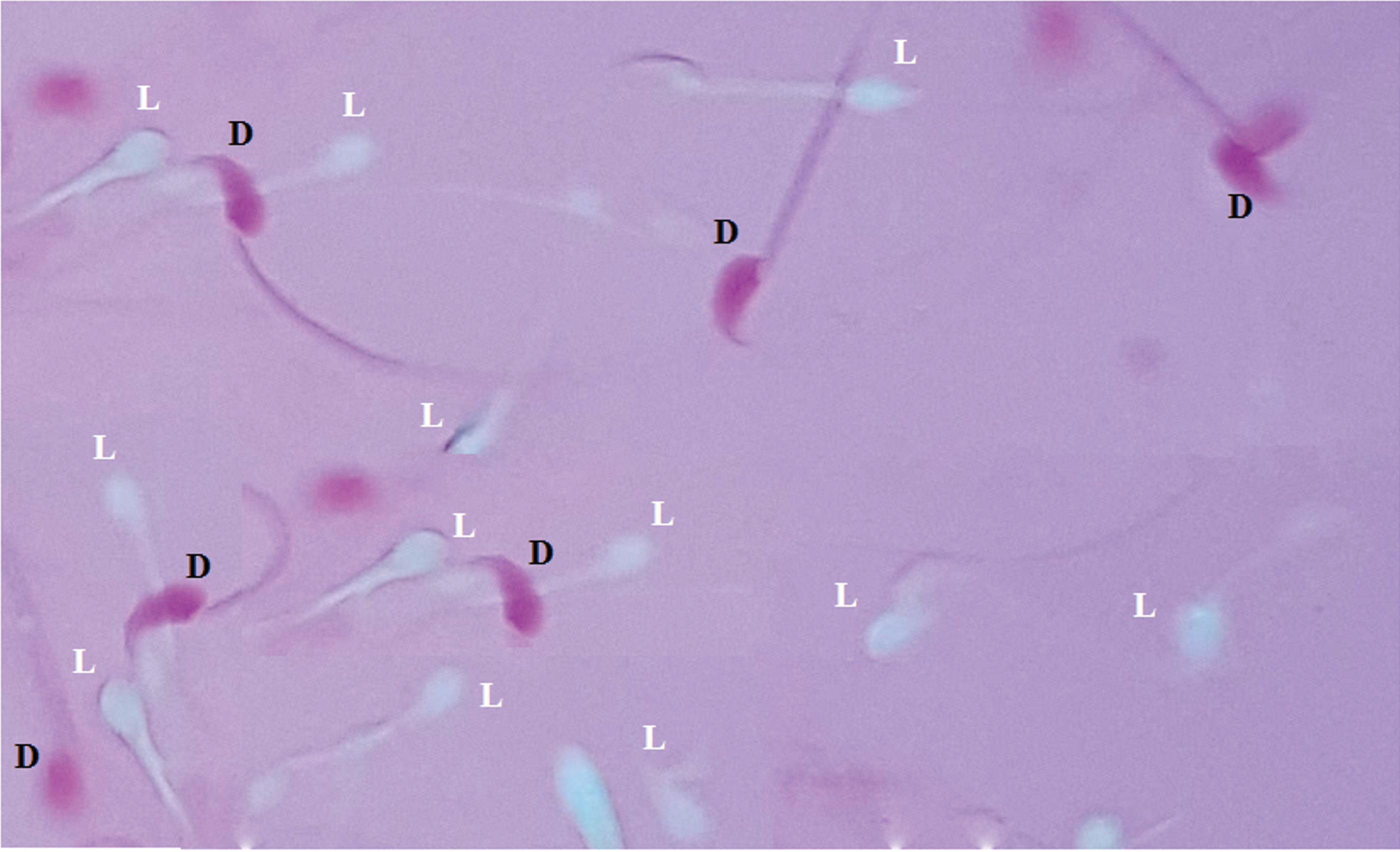
Bright field morphological evaluation image of stained sperm with Eozin and their classification into (D) dead sperm; (L) live sperm. Color images are available online.

Bright field morphological evaluation image of stained sperm with SCD and their classification into (U) unfragment sperm; (F) fragment sperm. SCD, sperm chromatin dispersion. Color images are available online.
Previously, it was indicated that sperm damage could also occur during the thawing process. For example, Gosálvez et al. 41 reported that sperm DNA damage had increased sequentially throughout a 4-hour period after thawing. Thus, in experiment 2, we investigated the effects of various TTN-SLN concentrations added to the thawing medium on sperm total motility and progressive motility, viability, and DNA fragmentation of mouse spermatozoa. The results of experiment 2 showed that the supplementation with the lowest concentrations of TTN-SLN (0.5 μM) could reduce the DFI percentage in frozen/thawed mice sperm, 2 hours after thawing. Also, sperm motility and viability with 0.5 μM of TTN-SLN were enhanced compared with the control group. It was also found that the lowest concentration of TTN-SLN (0.25 or 1 μM) during the thawing media exerted no significant inhibitory effects on DFI% in vitro.
In experiment 3, our findings suggested that the total motility and viability were in the highest value, and DFI% was in the lowest value in the treated group with TTN-SLN at both concentrations of 0.25 and 0.5 μM compared with other groups (Table 4; p < 0.05). It was also concluded that the lowest concentration of TTN-SLN (0.25 μM) during handling media showed no significant effects on progressive motility similar to the TTN-SLN 0.5 μM group, but both of them reduced extracellular ROS levels compared with other groups.
This improved sperm quality after the freezing/thawing and handling procedures may be contributing to the intracellular-controlled delivery of TTN by SLN. Therefore, all of the nanocapsule's basic physicochemical properties are considered as more probable reasons for its higher cellular uptake of TTN-SLN compared with the pure antioxidants. This is due to the importance of particle mean diameter (size), size distribution, PDI, and ZP for increased cellular uptake of nanoparticles. In the present study, the results of zetasizer and SEM demonstrated that TTN-SLN particles possess spherical shapes and regular distribution between prepared nanocarriers, and also have a smooth surface without aggregation, indicating an excellent capping strength of TTN with the size ≤200 nm (Figs. 1 and 2a).
This size has the capability to escape from circulation and successfully accumulate in the intracellular matrix. Also, for all formulations, the PDI was <0.16, which demonstrates narrow size distributions of all preparations. Thus, the smaller size of TTN-SLN may have made it easier for passage through the biological barriers, providing better integration and delivery of TTN to the sperm subcellular compartments, such as mitochondria and nuclei.
Another important physicochemical property regarding cellular uptake is the surface charge or ZP, which corresponds to the electric potential of the nanoparticles influenced by the composition of the nanocapsules and the medium. 42 Light positively charged carriers seem to increase the cell surface affinity and uptake of nanocarriers more extensively compared with negatively charged nanocarriers. 43 In our results, the ZP of TTN-SLNs was 13.3 mV, which is considered to be the main reason for higher cellular uptake of TTN-SLNs compared with pure TTN. These findings confirmed that the 0.5 μM of TTN-SLN can be replaced with high doses pure of TTN for inhibition of cryodamage.
The extracellular ROS levels could be used as an indicator of low sperm quality and high DFI. 44 Our chemiluminescent measurements revealed that the TTN-SLN is more efficient to scavenge the excessive production of extracellular ROS in freezing/thawing and handling media, which provided another explanation for improved sperm quality and DFI after cryopreservation.
In addition, TTN-SLN or B-SLN groups are safe, up to 5 μM tested, and have not found any cytotoxicity associated with its supplementation in the freezing/thawing and handling media in three experiments. This is probably correlated with in vitro controlled TTN release owing to the fit of amphiphilic balance in TTN-SLN nanoscopic structures. Only less than 60% of TTN cumulatively was released from TTN-SLN complex in 55 hours, whereas ∼100% was released from pure drugs over 10 hours. Therefore, SLN has a significant influence on the release profile of TTN due to the high stability of TTN-SLN with controlled leakage of antioxidants after an intracellular operation. The findings also confirmed that fabricated TTN-SLN and sustained drug-releasing profiles have been controlled and followed by a slow-release pattern. Because the surface-attached drugs may separate and float in the release solution in initial hours; later, the drug entrapped in the middle of nanoparticles may gradually be released.
Some limitations of the current study, which should be investigated by future studies, are the testing of internalization mechanisms of nanocapsules in sperm, what happens to this material after fertilization and during embryo development, and what are the consequences of nanomaterial accumulation on fertility, embryo development, and health of the offspring.
Conclusion
For the first time, the results of this investigation revealed that the application of the lowest concentrations of TTN-SLN (0.5 μM) supplementation for three procedures of sperm freezing/thawing and handling media could increase the sperm parameters, including enhancement of sperm total/progressive motility and vitality, protection of sperm DNA fragmentation, effective suppression of intra/extracellular ROS level, and decreasing oxidative damage in sperm freezing/thawing and handling media. Since our findings are a starting point for the utilization of nanotechnology in reproductive medicine, further independent studies seem necessary to confirm the findings. These results are of great importance for improving the efficiency of sperm cryopreservation to help fertile/subfertile men.
Footnotes
Acknowledgment
The authors want to thank the coworkers who helped with the tests and the collection of data
Authors' Contributions
Processed data, evaluated data, and writing of the article: F.A. and M.B.; Performed experimental research and contributed to the study of the results: F.A. and M.K.; Engaged in experimental work and analyzed data: F.A. and A.V.; Engaged in experimental work and updated article: F.A. and M.B.; Selected suitable animal, rated embryos, and gathered research data: F.A.; Planned tests, analyzed data, and revised the article: F.A, M.B., A.V., and M.K.
Confirmation Statement
Our research is supported by Kermanshah University of Medical Sciences that is primarily involved in education or research.
Author Disclosure Statement
No conflicting financial interests exist.
Funding Information
This study was funded by the Iran National Science Foundation (INSF, 97008908).