




Abstract
Introduction:
Even with the significant advancements in sperm cryopreservation, the addition of intracellular or extracellular antioxidants in preparation and freezing media remains an understudied topic.
Objective:
We examined the effects of hypotaurine and melatonin on the routine and functional tests of sperm and the expression of HspA2 and Caspase9 during the human sperm rapid freezing process.
Methods:
Following the collection of 34 normospermia semen samples, each sample was split into four experimental groups: fresh (F), freezing control (C) (human tubal fluid medium and 0.5M sucrose), and two freezing groups with the inclusion of 2 mM melatonin (MEL) and 50 mM hypotaurine (HYP). A straw held 100 μL of the sample, which was then cryopreserved in liquid nitrogen to accomplish rapid freezing. Before and after rapid freezing-thawing, the sperm classical parameters and the expression levels of HspA2 and Caspase9 were assessed.
Results:
The HYP group exhibited higher normal morphology (p < 0.001), viability (p < 0.001), and higher acrosome integrity (p < 0.001) and lower DNA fragmentation index (DFI) (p < 0.001) than the C and MEL groups. No significant difference was observed in the total and progressive motility percentage among the antioxidant and frozen control groups. The MEL group had a significantly higher level of HspA2 mRNA compared with F and C groups (p < 0.05). The expression of Caspase9 was unaffected by including MEL and HYP in all experimental groups.
Conclusion:
Hypotaurine, as an extracellular antioxidant, is more effective than melatonin as an intracellular antioxidant in reducing deleterious cryoinjuries on morphology, viability, acrosome reaction, and DFI.
Introduction
Sperm cryopreservation has evolved as an important strategy for male fertility preservation during assisted reproduction techniques. The common methods of sperm cryopreservation in clinics include rapid freezing and vitrification. In rapid freezing, sperm is frozen by liquid nitrogen vapor cooling followed by plunging into liquid nitrogen, while in vitrification, sperm is plunged directly into liquid nitrogen. 1 Spermatozoa are especially susceptible to cryoinjury due to the lipid composition of their plasma membranes and their restricted ability for active repair. 2 In this setting, it is evident that cryopreservation causes various disruptions to sperm cells, such as osmotic and oxidative stress, along with physical harm resulting from the formation of ice crystals. Considering the significance of these pathophysiological mechanisms, various efforts have been undertaken to enhance cryopreservation results by improving different permeable and nonpermeable cryoprotectant agents (CPA) to mitigate these stress-related effects. 3 Permeable CPAs such as glycerol have been found to cause harmful osmotic and cytotoxic effects on sperm during cryopreservation. 4 Therefore, the focus of research on human sperm cryopreservation has shifted toward rapid freezing and vitrification methods that utilize nonpermeable CPAs such as sucrose and trehalose. 5 Rapid freezing and vitrification are not only cost-effective, more rapid, and easier but also surpass slow freezing in preserving the motility and DNA integrity of human sperm. 6 A previous study has shown rapid freezing is more effective than vitrification. Nevertheless, they concluded that a sucrose concentration of 0.25 mol/L is suitable for human sperm rapid freezing and vitrification. 7
In addition, cryopreservation may induce diverse deleterious effects on sperm, particularly regarding molecular and subcellular damage that impacts different cellular components. 8 Reactive oxygen species (ROS) can diminish sperm functional parameters and cause DNA damage by affecting its structure, which in severe cases triggers apoptosis and cell death, by inducing caspase factors. 9 Moreover, this cellular stress leads to the destruction of intracellular proteins, which is faced with heat shock proteins (HSPs). 10
Antioxidants such as melatonin, “an intracellular antioxidant” and hypotaurine, “an extracellular antioxidant,” serve as the primary defense against the generation of ROS and the oxidation of proteins and lipids during sperm cryopreservation processes. Melatonin, also known as N-acetyl-5-methoxytryptamine, is a molecule that has both hydrophilic and lipophilic properties. It is found in both the watery cytosol and lipid-rich membranes of cells, and it can interact with ROS in the mitochondria. 11 Furthermore, melatonin can preserve the structure and function of mitochondria, which, in turn, enhances the probability of cell survival following cryo-osmotic stress. 12 Furthermore, hypotaurine serves as a precursor to taurine, functioning as a nonenzymatic scavenger that possesses membrane-stabilizing properties and can inhibit enzymes that produce ROS. 13 The viability and motility of human sperm during slow freezing were significantly increased when post-thaw sperm were selected and frozen in the presence of hypotaurine. However, hypotaurine does not protect the sperm from changes in the gene expression of HspA2. 14 While both antioxidants have been extensively studied as protective agents in sperm cryopreservation, there is, to our knowledge, no research comparing the effects of melatonin and hypotaurine together on sperm during this process. In previous studies, we have attempted to optimize sperm cryopreservation methods for a limited number of sperm initially through procedural modifications and subsequently by using hypotaurine.14,15 Therefore, in this study, our objective was to compare the effects of melatonin, an intracellular antioxidant, and hypotaurine, an extracellular antioxidant, during sperm cryopreservation with nonpermeable CPAs to evaluate their effectiveness on classical sperm parameters and HspA2 expression.
Materials and Methods
Human sample collection and analysis
The study methodologies conformed to the standards set by the 1964 Helsinki Declaration. Samples and demographic data were collected from Royan Institute, Tehran, Iran, after obtaining permission from Royan Institute Ethics Committee, Tehran, Iran (IR.ACECR.ROYAN.REC.1401.106). Before beginning the study, all participants were informed about the nature and purpose of the study, and written informed consent was obtained from each patient who provided a semen sample. Signed informed consent was obtained from all subjects.
Following approval of all procedures, 34 normospermic semen samples were collected from infertile couples who had gone to the Royan Institute for seminal fluid analysis. Semen samples were collected by masturbation into sterile plastic containers after 2–4 days of sexual abstinence. After liquefaction, sperm concentration, and motility were assessed according to the World Health Organization’s (2021) guidelines using a Computer Assisted Sperm Analyzing system (CASA; Microptic Co., Spain) with 10× magnification. Normal sperm morphology was evaluated by Papanicolaou staining and then examined under an oil immersion light microscope (Olympus™, CX21, Japan) at 1000× magnification for sperm normal morphology, as well as head and tail deformities. A minimum of 200 sperm were counted on each slide to determine the morphology. 16
Study design and experimental groups
Rapid freezing and thawing
The semen samples underwent centrifugation utilizing 45% and 90% gradient centrifugation (ALLGrad, LifeGlobal, USA) and were subsequently processed with human tubal fluid (Sigma, USA) supplemented with human serum albumin 10% (Vitrolife, Sweden) (HTF-HSA 10%). The processed samples were then subjected to centrifugation at 300 g for 15 minutes. After collecting the 90% layer and washing it with 2 mL HTF (300 g, 5 minutes), the resultant pellets were then re-suspended in 1 mL of HTF. Following CASA analysis of the sperm parameters, each sample was split into four groups: fresh (F), freezing control (C) (which included HTF medium and 0.5M sucrose), and two freezing groups with inclusion of 2 mM melatonin 17 (MEL) (Sigma) and 50 mM hypotaurine 14 (HYP) (Aldrich, USA). To achieve rapid freezing, the sample was diluted with HTF until the sperm concentration reached 15–20 × 106/mL for each group. 15 For the control vitrified group, 0.5 mol/L sucrose (Sigma) was combined (1:1) with the suspended sperm in HTF-HSA 0.5%. The samples were incubated at room temperature for 10 minutes before freezing. Four experimental groups included F, C, the 50 mM HYP, and 2 mM MEL. Hypotaurine and melatonin were added to the suspended sperm and left at room temperature for 10 minutes and next mixed with the mentioned freezing solution and kept in a room for sperm equilibration with sucrose (10 minutes). Finally, the sperm samples were loaded into 0.5 mL standard cryostraws (IMV, Aigle, France) (100 µL/straw). After equilibration at room temperature for 10 minutes, all loaded straws for comparison in the experiment were placed horizontally at 5 cm above the liquid nitrogen surface (–130°C) for 10 minutes followed by plunging into liquid nitrogen. After 1 week, samples were thawed by submerging a cryostraw in a 37°C water bath for 2 minutes before the heat seals at both ends were cut off and the sperm suspension was expelled into 2 mL pre-warmed HTF medium for dilution and evaluation. 18
Sperm viability and morphology.
The morphology of sperm was assessed by Papanicolaou (PAP) staining. PAP staining is one of the most significant and well-established sperm procedures extensively used in andrology laboratories and reproductive clinics. The smears were then examined under an oil immersion light microscope (Olympus, CX21) at 1000× magnification for sperm normal morphology, as well as head and tail deformities. Eosin–nigrosin staining was used to assess the viability of human sperm. Dead sperm with damaged membranes showed red, while live sperm appeared white. A minimum of 200 sperm were counted on each slide to determine viability, and the proportion of living sperm was expressed as a percentage. 19
Acrosome reaction
The acrosome integrity was assessed using the fluorescein isothiocyanate-labeled conjugated Pisum sativum agglutinin (FITC-PSA, Sigma) staining protocol. The specimen was smeared on the microscope slide, dried, and fixed in ethanol at 20°C for 30 minutes. The smear was stained with FITC-PSA and incubated at 4°C for 60 minutes. The number of 200 cells was evaluated in each replicate using fluorescence microscopy at 100× magnification at 450–490 nm excitation. 20
DNA fragmentation
A halo sperm kit (SDFA kit; Ideh Varzan Farda, Iran) was used to assess the sperm chromatin condition. This method is not complicated and time-consuming, although it is economical. In brief, 20 μL of low-melting agarose was combined with 50 μL of semen samples. Then, 20 μL of the mixture was placed on the pre-coated glass slide. A coverslip was placed on the drop to spread evenly over the slide. The slide was placed in the refrigerator at 4°C for 5 minutes. Next, the coverslip was slowly removed from the slide and the denaturing solution (A) was added. The slides were kept in a dark room at room temperature for 7 minutes. The lysing solution (B) was added to the slide and placed at room temperature for 15 minutes. The slide was washed with distilled water for 5 minutes. For dehydration, the slides were immersed in 70%, 90%, and 100% ethanol solutions for 2 minutes, respectively, and dried for 2 minutes at room temperature. First, the slides were stained with solution C for 75 seconds, then solution D for 3 minutes, and finally solution E for 2 minutes. After that, the slides were washed with distilled water, and 200 sperm cells were examined with bright-field microscopy. The DNA fragmentation rate was evaluated according to halo formation around the sperm head. A medium or large halo around the sperm head indicated DNA without fragmentation, and the absence of a halo represented fragmented DNA. The percentage of sperm with fragmented DNA (no halo) was reported. 21
RNA extraction and cDNA synthesis of sperm cells
Total RNA from the sperm was isolated using the TRIzol reagent (Kiazist-Iran) with the manufacturer’s instructions. Spectrophotometry was used to assess the quantity and quality of extracted RNA (NanoDrop 1000; Thermo Fisher Scientific, USA). All total RNA samples had spectrophotometric optical density ratios of 260/280 > 1.8 and 260/230 > 2. 22
Quantitative real-time polymerase chain reaction analysis
The process of reverse transcription was utilized to synthesize cDNA, using the PrimeScript RT Reagent Kit from Takara, Japan. The resulting cDNA was then subjected to quantitative real-time polymerase chain reaction (PCR), using the primers listed in Table 1. The normalization of samples was performed using β-actin as the internal control for gene expression. The experimental procedures were conducted employing 96-well plates manufactured by Applied Biosystems. The reaction mixtures used in the experiment were composed of 10 mL volume, comprising 2.5 mL of SYBR Green PCR (manufactured by Applied Biosystems), 1 mL of both forward and reverse primers, 2 mL of cDNA, and approximately 3.5 mL of bi-distilled water, which was added to achieve the final volume of 10 mL. Simultaneous quantitative PCR assays were achieved for fresh and cryopreserved samples from each group. Dissociation curves were generated to validate the specificity of the products. The amplification parameters were a temperature of 60°C for 1 minute. The melting curve was analyzed to verify the presence of a single peak per reaction, which was subsequently validated by conducting PCR product electrophoresis on a 2% agarose gel. The Ct (threshold cycle) obtained for each sample was first normalized with the Ct of the reference gene, which is considered to be the β-actin gene, and then the final data analysis was performed with the comparative Ct method. Data analysis was performed by the comparative 2-(ΔΔCT) method. 22
Sequences of the Primers Used for the Quantification of the Housekeeping Gene (β-actin) and Target Genes
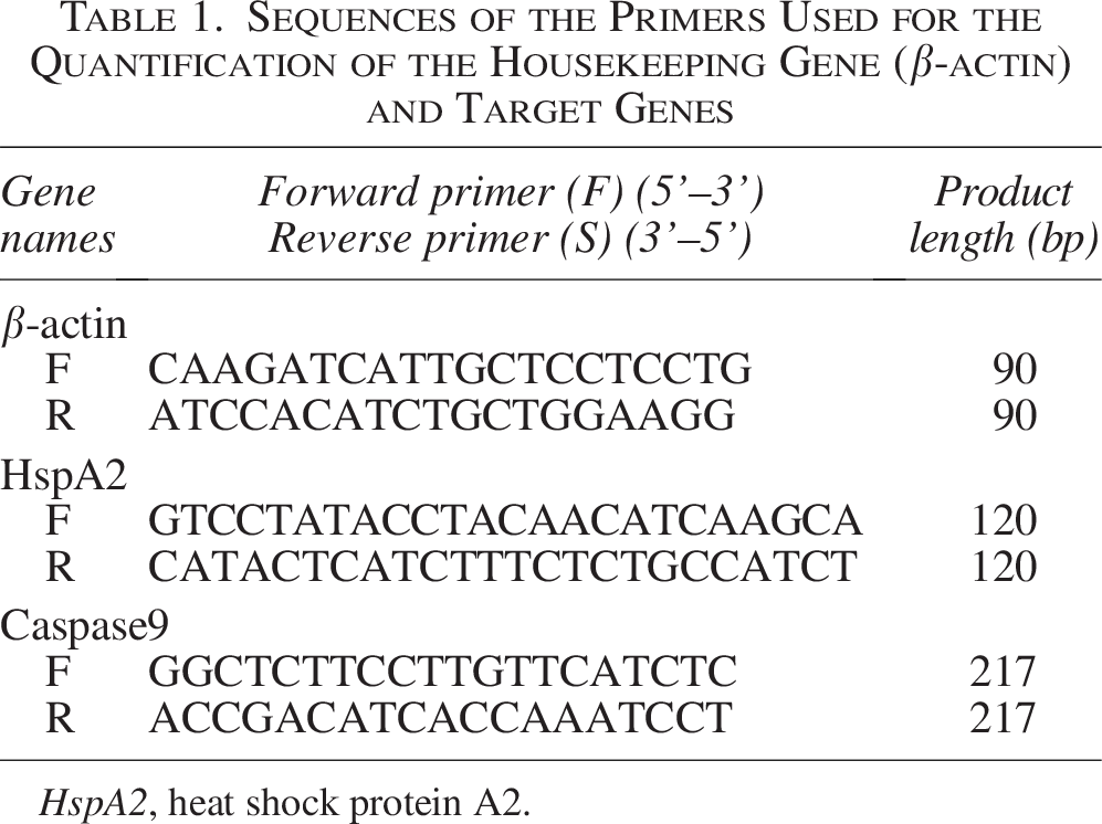
HspA2, heat shock protein A2.
Statistical analysis
The data were analyzed after checking for normality by Kolmogorov–Smirnov test and by one-way analysis of variance and Tukey’s post hoc test. The data were analyzed by SPSS version 20. The data were presented as means ± standard error of the means (SEM), and the significance of the results was considered at the level (p < 0.05).
Results
Sperm parameters in the fresh and freezing groups
The mean ± SEM of sperm classical parameters, DNA fragmentation index (DFI), and acrosome integrity are shown in Table 2. Fresh samples exhibited significantly higher mean for progressive motility, total motility, morphology, viability, and acrosome integrity than all freezing groups (Fig. 1). In contrast, the F group had the significantly lowest percentage of DFI (Fig. 2) (p < 0.05).

Comparison of sperm classical parameters among fresh and frozen samples (n = 34). While using antioxidants did not positively affect sperm motility, hypotaurine preserved morphology and viability in contrast to melatonin. All data are expressed as the mean ± SEM; p < 0.05. The uppercase and lowercase letters indicate significant differences among groups. SEM, standard error of the mean.

Comparison of acrosome integrity and DNA fragmentation among fresh and frozen samples (n = 34). Hypotaurine exhibited higher integrity of acrosome and DNA compared with melatonin. All data are expressed as the mean ± SEM; p < 0.05. The uppercase and lowercase letters indicate significant differences among groups.
Sperm Parameters and Characteristics in Normozoospermic Men (n = 34)
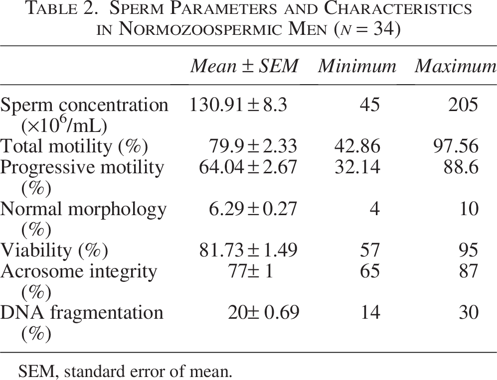
SEM, standard error of mean.
Following the procedure of sperm cryopreservation, there was no significant difference in the percentage of progressive and total motility among the freezing groups. In contrast, the HYP group exhibited a significantly higher normal morphology (1.51 ± 0.19%) compared with the MEL and C groups (0.90 ± 0.8% and 1.96 ± 0.36%, respectively, p < 0.001). Similarly, treatments altered viability but were not significant (44.96 ± 2.79%) (Fig. 1).In addition, the mean of acrosome integrity was found to be significantly higher in the HYP group (43.01 ± 1.78%) compared with the C (28.63 ± 0.99%) and MEL groups (32.51 ± 1.05%) (p < 0.001) (Fig. 2).
As depicted in Figure 2, the level of DNA fragmentation in the HYP group (31.39 ± 0.93%) was significantly (p < 0.001) lower than that of the C (41.30 ± 0.89%) and MEL (40.21 ± 1.05%) groups. The levels of DNA fragmentation were similar among the MEL and C groups.
The expression level of HspA2 and Caspase9 in the fresh and frozen groups
In terms of HspA2, melatonin had a significant (p < 0.05) effect on overexpression of HspA2 (2.19 ± 0.23) compared with F (1.33 ± 0.22) and C (1.24 ± 0.16, p < 0.01) groups. The relative expression of HspA2 mRNA did not differ between MEL and HYP groups. There was no significant difference in sperm Caspase9 mRNA levels between F and frozen groups (Fig. 3).

Comparison of the expression level of HspA2 and caspase 9. While relative expression of HspA2 mRNA was higher in the 2 mM melatonin (MEL) group than in fresh and freezing control groups, relative expression of Caspase9 mRNA did not differ between the MEL and 50 mM hypotaurine groups. Relative expression of Caspase9 mRNA did not differ between the four groups (p > 0.05). The (**) indicate significant differences among groups.
Discussion
Using the hypotaurine antioxidant during the rapid freezing approach improved the sperm’s classical criteria, such as viability and morphology, but had no obvious effect on total motility or progressive motility. Similarly, Seify et al. stated that hypotaurine along with vitrification improved viability and morphology. 14 Thus, the data suggest that the extracellular antioxidant (hypotaurine) is more effective than the intracellular antioxidant (melatonin) during human sperm rapid freezing to reduce damage to viability and morphology during cryopreservation.
Increased acrosome integrity by hypotaurine was consistent with DFI decreased in this group. It seems that the main effect of hypotaurine was to maintain acrosome integrity, which was supported by previous studies in rams, 23 dogs, 24 and bulls. 25 However, melatonin’s role in rapid freezing is controversial, and some studies stated that melatonin has effects on sperm acrosome integrity and DNA damage reduction in rooster, 26 horse, 27 and human sperm. 28 The discrepancy between present findings and previous studies regarding melatonin may be attributed to variations in melatonin concentration and freezing techniques.
The HYP group exhibited higher percentages of acrosome integrity and a reduced DFI compared with the MEL and C groups. Hypotaurine’s inhibition of superoxide dismutase inactivation and taurine’s osmolyte protection against hypertonic stress confirm their importance in sperm freezing.29,30 In addition, the neutralization of hydroxyl radicals by hypotaurine and its role as a nonenzymatic scavenger help prevent DNA damage by protecting against guanine oxidation. 31
As putative mechanisms, taurine and hypotaurine contribute to cold preservation by reducing oxidative stress-induced cold damage through osmotic regulation, reducing lipid peroxidation, and increasing membrane stabilization, including Ca2+ regulation. Altogether, our data in human sperm for hypotaurine support the theory 32 that taurine and hypotaurine may play a role in indirectly safeguarding against cryoinjuries that are a result of oxidative stress, by engaging in osmoregulation, providing antioxidant benefits, stabilizing membranes, and managing the conjugation and regulation of Ca2+. 14
By examining HspA2 to assess the damage caused by freezing cryopreservation, our results revealed an increase in this gene within the MEL antioxidant group. To our knowledge, there has been a lack of research exploring the relationship between melatonin and HspA2 in human sperm. By the vitrification method, Seifi et al. found HspA2 gene expression was reduced in all vitrified groups compared with the F group. 14 It is possible to assume that the higher levels of HspA2 in the MEL group indicate a protective mechanism against cellular damage. 33 Indeed HSPs may play a role in cellular stress response in creating cell resistance by (1) preventing the accumulation of misfolded proteins in the cell and returning them to a functional state and (2) restoring the plasma membrane. 34 In this regard, reducing or disrupting the expression of HspA2, seen in infertile individuals, is associated with increased frequency of chromosomal aneuploidy, DNA damage, ROS, apoptosis, and abnormal morphology.35–37 Also, HspA2 forms a complex with sperm adhesion molecule 1 and arylsulfatase A in sperm adhesion to the zona pellucida (ZP), and decreased HspA2 levels are associated with defects in ZP adhesion. 38 However, further studies are needed to fully elucidate the functional importance of HSPs in the “freezing” process, particularly when using intracellular and extracellular antioxidants. Effects of hypotaurine on apoptotic gene expression in previous studies on sperm were not reported. We investigated the apoptotic cascades, which due to uncertainty and significance in the expression of Caspase3. It seems that the expression of the Caspase9 gene follows the apoptotic signals caused by freezing and osmotic shock, leading to the release of cytochrome c from mitochondria and activation of apaf-1 (apoptosome), leading to procaspase conversion. Caspase9 becomes an active dimer, which eventually causes apoptosis by affecting Caspase3. 39 As reported by Najafi et al., melatonin protects human sperm against oxidative damage by acting on the MT1 receptor and kinase activity and the decrease of Caspase3. 40 Nevertheless, our results indicate no substantial variation in the mRNA abundance of Caspase9 between the MEL and HYP groups.
Conclusion
In conclusion, understanding the effects of intracellular and extracellular antioxidants, one of the major effective parameters and one of the molecular mechanisms involved in the reduction of cryoinjuries during rapid freezing, becomes increasingly important, given the diminished cryodamages. This study provides the first analysis of the comparison between extracellular antioxidants (hypotaurine) and intracellular antioxidants (melatonin) through the human rapid freezing method. Hypotaurine (50 mM) is more effective than melatonin (2 mM) as an intracellular antioxidant in reducing deleterious cryoinjuries on morphology, viability, acrosome reaction, and DFI. Further studies that include protein expression and functional tests may provide additional clues to the involvement of factors in, and the mechanisms underlying, the effective roles of intracellular and extracellular antioxidants to reduce cryodamages during rapid freezing in human sperm.
Authors’ Contributions
A.Z.: Main contribution toward the project, conceptualization, data collection and analysis, investigation, writing—original draft, and writing—review and editing. L.R.G.: Study design, conceptualization, data curation, formal analysis, and writing—original draft. V.E.: Methodology and writing—original draft. A.A.: Conceptualization, investigation, visualization, writing—original draft, and writing—review and editing. M.R.V. and A.S.: Projects supervision, study design, methodology, validation, and writing—review and editing. All authors have read and approved the final article.
Footnotes
Acknowledgments
The authors thank all participants who were involved in the present study. They would also like to extend their appreciation to the Royan Institute laboratory staff, particularly Mrs. Elham Abedheydari and Dr. Mina Sharbatoghli for their sincere contributions to all laboratory procedures, and the Andrology laboratory staff in the Royan Clinic for samples.
Author Disclosure Statement
The authors declare that they have no known competing financial interests or personal relationships that could have appeared to influence the work reported in this article.
Funding Information
This research was financially supported by the University of Science and Culture, Tehran, Iran, and Royan Institute, Tehran, Iran.
Confirmation Statement
The Royan Institute, Tehran, Iran, and the University of Science and Culture, Tehran, Iran, established as public, nongovernmental centers in 1991 and 1993, respectively, are recognized for their commitment to multidisciplinary integration and collaboration among scientific, academic, and medical personnel. They focus on understanding male and female infertility, embryo development, stem cell biology, and biotechnology. All authors listed on this article confirm that they are faculty members or students at the Royan Institute and the University of Science and Culture. These institutions are acknowledged as centers of excellence in research and technology and are leaders in various fields of biological sciences. Each author confirms that their research is supported by an institution that is primarily involved in education or research.
Data Availability
All data and material used and analyzed during the current study are available from the corresponding author upon reasonable request.