
Abstract
Background:
Electrostatic complexes of poly (l-Arginine) (pArg) and hyaluronic acid (HA) have been investigated for their functional applications to supply free or polymeric form of l-Arginine (Arg) to target cells. As a vital amino acid, Arg plays significant role in multitude of pathophysiological processes ranging from wound healing to cancer. However, serum arginase expression and toxicity of Arg at cellular level renders exogenous delivery of this amino acid a challenging task. We showed that polyarginine-hyaluronic acid ionic nanocomplexes (pArg-HA iNCs) could be an effective way to deliver Arg to target cell populations.
Materials and Methods:
These electrostatic complexes were prepared by mixing HA (average m.w. of 200 kDa) with pArg (m.w. 5–15 kDa; Sigma) in aqueous solutions and purifying over glycerol. Nanocomplexes were characterized for their particle size, surface charge, capacity to release l-Arg, and intracellular uptake of complexes.
Results:
Synthesized nanocomplexes showed hydrodynamic diameter ranging from 140–306 nm depending on the content of pArg or HA within the formulation. With surface charge (ζ-potential) of −29 mV, the nanocomplexes showed pH-dependent release of Arg. At pH 7.4, pArg-HA iNCs released 30% of the total Arg-content, while at pH 5.0, 60% of Arg was released after 24 h. These electrostatically stabilized complexes were found to promote growth of human dermal fibroblasts (HDF) in wound-healing assay and increased nitric oxide (NO) activity in these cells in a time-dependent manner. Nanocomplexes also showed cellular uptake and enhanced dose-dependent toxicity against two pancreatic cancer cell lines, i.e. MIA PaCa-2 and Panc-1. Interestingly, the cytotoxic effect was synergized upon pre-treatment of the cells with a frontline chemotherapeutic agent, gemcitabine (GEM), and was not observed when the cells were treated with Arg alone.
Conclusion:
As such, this communication shows the prospect of pArg-HA iNC electrostatic nanocomplexes to interact and interfere with intracellular Arg metabolic machinery conducive to rescuing different pathological conditions.
Introduction
Arginine (Arg), a dibasic amino acid, is involved in a number of metabolic pathways through activation of inducible nitric oxide synthase (iNOS) that produces nitric oxide (NO) as an active metabolite.1,2 In case of wound healing, exogenous delivery of Arg was found to accelerate proliferation and remodeling phase of wound healing. 3 Supplemental Arg feeding demonstrated beneficial effects in wound repair.4,5 Occurrence of an injury is known to activate arginase action around the injury site and an overexpression of the enzyme is likely to impair endogenous Arg supply and diminish iNOS. Arginase metabolizes L-Arg to L-Ornithine and urea.6,7
In proliferative cancer cells, on the other hand, exogenous administration of free Arg was found to have broad-spectrum, dose-dependent effect on cellular fate. Due to decreased expression of argininosuccinate synthetase and/or ornithine transcarbamylase, several types of tumor were found to be auxotrophic for Arg. Deprivation of Arg thus leads to significant vulnerability of these tumor cells, leading to cell death. However, peptides with polyarginine (pArg) motifs also trigger cellular cytotoxicity because of their ionic binding interactions with vital biologically charged molecules. 8 Being cationic in nature, pArg also possess cell membrane translocation activity.9,10
To gain therapeutic advantage from Arg in their free or polyvalent form, and to bypass Arg-mediated toxicity or cellular dependence, we investigated the prospect of intracellular delivery of pArg, mediated through electrostatic complexes with hyaluronic acid (HA). Due to the presence of complementary charge distribution, pArg and HA usually form polyelectrolyte complexes.
We hypothesize that, such electrostatic complexes will be able to release bioavailable quantity of Arg, either in their polyvalent or monovalent form, within the proliferating cells intracellularly (Fig. 1). We prove our hypothesis by investigating the effects on pArg/HA-based ionic complexes (abbreviate in this work as pArg-HA iNCs) on fibroblast and on pancreatic cancer cells. Earlier work had shown that it is possible to generate nanocomplexes with pArg and HA at the size range of 120–146 nm depending on the feed ratio of HA and pArg, which also governed the zeta potential of such ionic complexes.
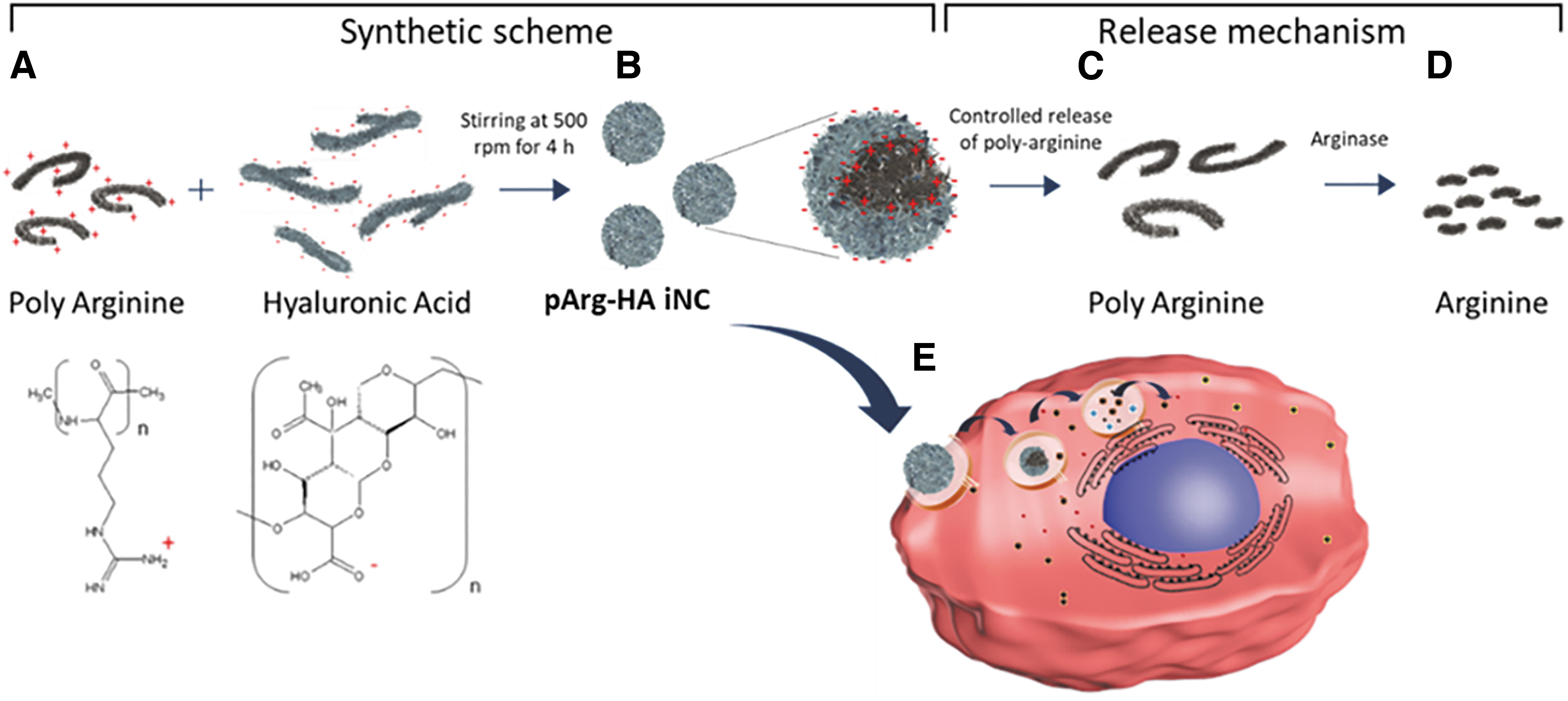
Schematic representation of
HA is an anionic, naturally occurring, biodegradable polysaccharide with mucoadhesive properties and no reported toxicity. 11 HA is a suitable component and has found extensive applications as delivery vehicle for CD-44 overexpressed receptors; as such these receptors act as endogenous ligand for HA.12,13 Arg's putative role as supplement for wound healing14,15 and the effect of pArg on cancer cell targeting and destruction have inspired us to identify a safer delivery strategy for this biologically active polypeptide utilizing its complex-forming capacity with HA. It has been noted that continued dose of Arg infusion at an injury site improved the wound healing process, pertaining to the sustained NO availability. 16 Decrease in the activity of iNOS demonstrated reduced collagen synthesis and impaired wound healing. 17
It has also been established that iNOS inhibition in mice decreased collagen concentration in the wound fluid, whereas supplemental Arg or ornithine feeding increased appropriate collagen production in wounds. 18 Therefore, we set out to investigate if HA-pArg nanocomplexes could act as a stable nanoplatform to inhibit the release and degradation of pArg in serum conditions and liberate active Arg through decomplexation from the nanoparticle intracellularly inside target cells.
In this article, we have designed and synthesized pArg-HA iNC, which can deliver sufficient Arg concentration intracellularly for interfering with cellular metabolism. We identified intracellular NO expression in fibroblast in response to pArg-HA iNC treatment. We also investigated the effect of such complexes on pancreatic cancer cells to identify the prospective therapeutic gain that is achievable when these complexes are sequentially administered with a clinically relevant anticancer drug. For wound healing conditions, we conducted qualitative and quantitative analysis of cellular migration and wound healing activity in human dermal fibroblast (HDF) cells, and for cancer setting, we used actively proliferating pancreatic cancer cell lines, such as MIA PaCa-2 or Panc1.
Materials and Methods
Synthesis of pArg-HA iNc
iNCs were prepared by mixing HA (average m.w. of 200 kDa within the range of 151–300 K; Lifecore Biomedical) and pArg (m.w. 5–15 kDa; Sigma) aqueous solutions. Briefly, 4.5 mL of an aqueous solution containing different concentrations of HA (0.44–2.67 mg/mL) was added to 4.5 mL of a solution containing pArg (0.53 mg/mL) with constant stirring at room temperature. For isolation, 1 mL of the nanoparticles was transferred to Eppendorf tubes and centrifuged (16,000g, 30 min, 25°C) in 20 μL of a glycerol bed. Supernatants were discarded, and pArg-HA iNCs were resuspended in water by vigorous shaking. Arg quantification was carried out using L-Arginine/Urea/Ammonia Assay Kit from Megazyme, following manufacturer's protocol.
Characterization of ionic nanocomplexes
Morphological examination of the nanocomplexes was carried out by transmission electron microscopy (TEM) (CM12 Philips, Eindhoven, Netherlands). The samples were stained with 1% (w/v) phosphotungstic acid for 10 s, immobilized on copper grids with Formvar, and dried overnight before TEM analysis. Differential light scattering (DLS) measurements was conducted in deionized water using a Malvern instrument (Malvern ZS 90).
In vitro release studies
The synthesized pArg-HA iNCs were stirred at 100 rpm separately in solutions maintained at pH conditions (pH 3.0, pH 5.0, and pH 7.4) for defined time intervals (1, 4, 8, 24 h), centrifuged, and 100 μL of clear aliquots of each were taken to estimate the amount of Arg released by L-Arginine/Urea/Ammonia assay method from Megazyme.
Wound healing assay
HDF cell suspension was prepared following standard protocols. 103 cells were seeded into each well of the culture inserts. Cells were incubated at 37°C and 5% CO2 for 24 h. A confluent cell layer was confirmed before removal of culture insert. Fresh medium is added after the removal of the culture insert. We observed that gap obtained between confluent cells captured bright field image. For further experiment, media supplemented with 100 nM pArg-HA iNC and equivalent concentration of Arg were then added to evaluate cell behavior. Post 24 h, cells were fixed using 4% paraformaldehyde and stained with Phalloidin. Wound area was observed under the microscope, using fluorescein isothiocyanate (FITC) filter and 10 × objective lens.
Fluorescence microscopic detection of intracellular NO
The microscopic images were captured using a fluorescence microscope (Axio Observer.A1; Zeiss, India), as well as confocal microscopy (Confocal Leica TCS SP5). Media was removed from the wells and was discarded. The wells were washed gently two times with 250 μL Dulbecco's phosphate-buffered saline (DPBS) (containing magnesium and calcium). One hundred microliter of 1 × Nitric Oxide Fluorometric Probe was added to each tested well. The plate wells were covered to protect the reaction from light and incubated at 37°C for 30–120 min. Cells were observed with a fluorescence microscope using FITC filter set.
Cancer cell lines and cell culture maintenance
The adherent pancreatic cancer cell lines Panc 1 and MiaPaca2 were collected from American Type Culture Collection (ATCC; Manassas, VA) and then maintained in high glucose Dulbecco's Modified Eagle Medium (DMEM) (supplemented with 10% fetal bovine serum, 50 IU/mL penicillin G, and 50 μg/mL streptomycin) in a humidified incubator under 5% CO2 environment. Subculturing was executed when the cells reached 90% confluence.
In vitro cytotoxicity assay using Alamar Blue
The cytotoxicity of pArg-HA iNCs on pancreatic cancer cell lines Panc 1 and MiaPaca2 was performed by Alamar Blue assay. Briefly, Panc 1 and MiaPaca2 cells (1.25–2.5 × 10⁴) were separately seeded in 96-well plates followed by incubation with supplemented DMEM media overnight at 37°C under 5% CO2 environment. Thereafter the media was discarded, and the cells were fed with fresh media containing pArg-HA iNC 2 formulation (total volume 200 μL in each well) with different iNC concentration ranges. After 48 h, the cells were treated with the half maximal inhibitory concentration (IC50) concentration of gemcitabine (GEM). The control group was not treated with any drug. At the end of 72 h of iNC treatment period, and 24 h of GEM treatment period, 20 μL of Alamar Blue solution was added in each well and incubated for 3–4 h at 37°C, CO2 incubator. The solution in 96-well plates was mixed in a shaker for 2–3 min. The plate was then analyzed in an ELISA reader. Cell viability % was plotted against the different concentrations of iNCs, and IC50 value was determined. Bliss calculation was carried out by Combenefit software. 19 Arg
Preparation of dye loaded Arg-HA iNC systems for confocal microscopy
Dye-loaded iNC systems were developed by the addition of 2 mL, Alexa fluor 647 labelled 1 mg/mL concentration of HA into 5 mL pArg (5 mg/mL) under stirring condition at room temperature (RT). Alexa fluor 647 labelled HA was added dropwise by an automated syringe with a speed of 0.25 mL/min. These complexes were then transferred into filter bedded Eppendorf for centrifugation at 16,000 rpm for 30 min at RT. The precipitate was collected followed by being treated in Mia PaCa 2 cell lines for 6 and 12 h for confocal imaging.Arg
In vitro cellular uptake study
Cellular uptake of pArg-HA iNC 2 system was monitored in vitro on pancreatic cancer cell, MiaPaca2, by confocal laser microscopy. MiaPaCa-2 cells (1 × 10⁵ cells) were seeded in ibidi® glass bottom dish (35 mm) overnight. The cells were then targeted with fluorescent (Alexa Fluor 647) tagged nanoparticles (at a concentration of 0.5 mg/mL) at 37°C for 6 and 12 h followed by washing with cold phosphate-buffered saline (3 × ), fixed with 4% paraformaldehyde, and costained with 4′,6-Diamidino-2-phenylindole. For confocal laser microscopy, around 104 cells were seeded on a cover slip and placed in a 35-mm tissue culture dish with media and incubated at 37°C overnight. The confocal images were captured using Zeiss Axio Observer Z1 microscope equipped with LSM700 laser scanning module (Zeiss, Thornwood, NY) at 40 × magnification with 40 × /1.3 Plan-Apochromat lens.Arg
Results and Discussion
Synthesis of pArg-HA iNC
pArg-HA iNCs were synthesized through polyelectrolyte complexation under aqueous condition. The provident advantage of this method is tunable electrostatic characteristic obtained simply by varying the ratio between oppositely charged polyions such as pArg and HA. 20 We prepared these nanocomplexes by incorporation of variable concentration of HA to a fixed concentration of pArg solution, accompanied by constant stirring and capturing of the nanoparticles on a glycerol bed. These iNCs were formed by electrostatic interactions between the positively charged amino group of the guanidine moiety on the PArg and the negatively charged carboxylate groups with the HA. Complexes were prepared, with a concentration ratio of HA to PArg varying from 0.8:1 to 5:1 as shown in the Table 1.
Formulations of pArg-HA iNCs with Varying Ratio of HA Added to a Fixed Concentration of pArg
pArg-HA iNCs, polyarginine-hyaluronic acid ionic nanocomplexes.
Characterization of pArg-HA iNCs
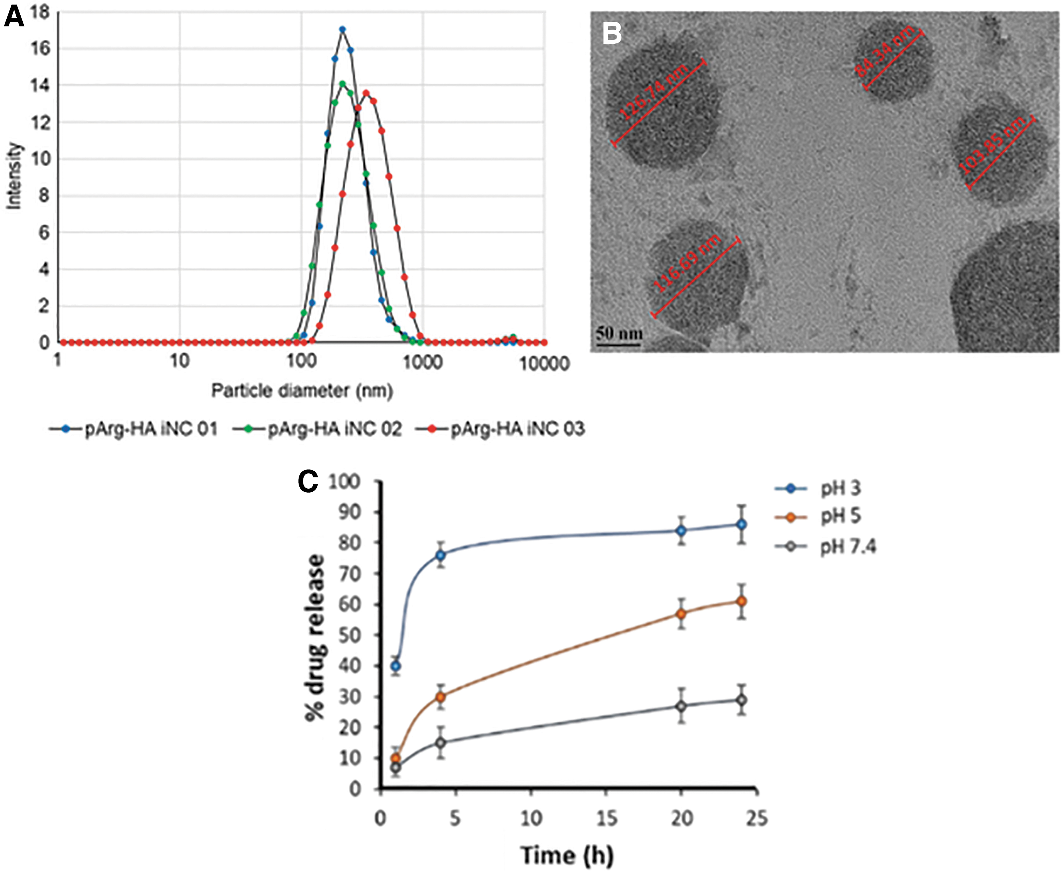
pArg-HA iNCs were characterized by DLS for particle size analysis, TEM, and zeta potential analysis. We observed that increasing HA content within the electrostatic complexes increases the particle size of the complexes. Depending on the ratio of pArg to HA, particle size distribution for different formulations was found to be within 140–306 nm (Fig. 2A). HA is a swellable polymer, and thus, the increment of particle size with increased amount of HA was expected. For further analysis, sample containing HA to pArg ratio 1.6:1 was used. DLS showed particle size for this sample to be around 150 nm, whereas TEM images revealed size in the range of 83–126 nm with spherical morphology (Fig. 2B). Drying effect on TEM grid might be responsible for size differences observed compared with DLS results. Zeta potential analysis showed −29 mV charge on the surface of iNCs, which indicates that HA is mostly interspersed at the surface of iNCs. Total Arg loading was quantified by Megazyme® assay method, where iNCs were treated with excess hyaluronidase and arginase. Total loading content of Arg was found as 71% in this formulation, where pArg to HA ratio was maintained at 1.6:1 (i.e., pArg-HA iNC 2). In this assay, Arg hydrolyzed to urea, which is further hydrolyzed to ammonia. In presence of ammonia, NADPH forms NADP+. NADPH consumption is measured by the decrease in absorbance at 340 nm.
In vitro release studies
Arg release from pArg-HA iNC 2 formulation was studied at pH7.4, pH5.0, and pH3.0. Amount of Arg released from iNCs was quantitated by Megazyme assay method as mentioned above. At each time point, released Arg separated from iNCs by centrifugation method, and only free Arg was quantified. pArg-HA iNC showed 85% Arg release in 24 h at pH 3.0, whereas less than 30% Arg was released in 24 h at pH 7.4 from these iNCs (Fig. 2C). At pH 5.0, which is endosomal pH, iNCs showed sustained release with 60% Arg released in 24 h. The Arg release pattern observed from this experiment showed a pH-dependent release behavior. Such pH-dependent release suggested that pArg-HA iNCs can trigger Arg release in acidic compartments after cellular uptake, namely the endosomal (pH 5.0–6.5) and the lysosomal compartment (pH 4.5–5.0).
Wound healing assay
To investigate how effectively Arg affects cellular migration at injury site, we conducted wound healing assay. Fibroblast cells play a major role in wound healing and show arginase expression in vitro as well. We used primary dermal fibroblast (HDFa) cells to conduct wound healing assay. A constant 11.1 × 105 μm2 wound area was created in ibidi 2-well culture insert for Arg, pArg-HA iNC 2 (with pArg to HA ratio of 1.6:1), and control analysis. Figure 3A–C shows HDFa cell migration into wound area in Arg, pArg-HA iNC treated, and in control well. We found a most dense, regenerated cellular network in pArg-HA iNC treated cells compared to Arg treated or untreated cells. Furthermore, we quantified the migrated cell area using ImageJ software. We found that 79% wound area was covered by pArg-HA iNC treated HDFa cells in 24 h, whereas 68% wound area was covered by Arg treated HDFa cells and only 35% wound area was covered by untreated HDFa cells. Homopolymers containing a high percentage of cationic amino acids have been shown to have a unique ability to pass through the plasma membrane of cells and, consequently, have been used to enhance the uptake of different biopolymers and small molecules. 8 In the proliferative phase, NO affects arginase to enhance collagen production and cell proliferation. 21 Both supplemental Arg and ornithine appear to be beneficial in wound healing. 18 The products of these pathways give feedback to regulate both the pathways, affecting activity at the different stages of wound healing. Our observation is most likely related to NO-mediated effect, which increased the cellular availability of Arg from pArg-HA iNCs.
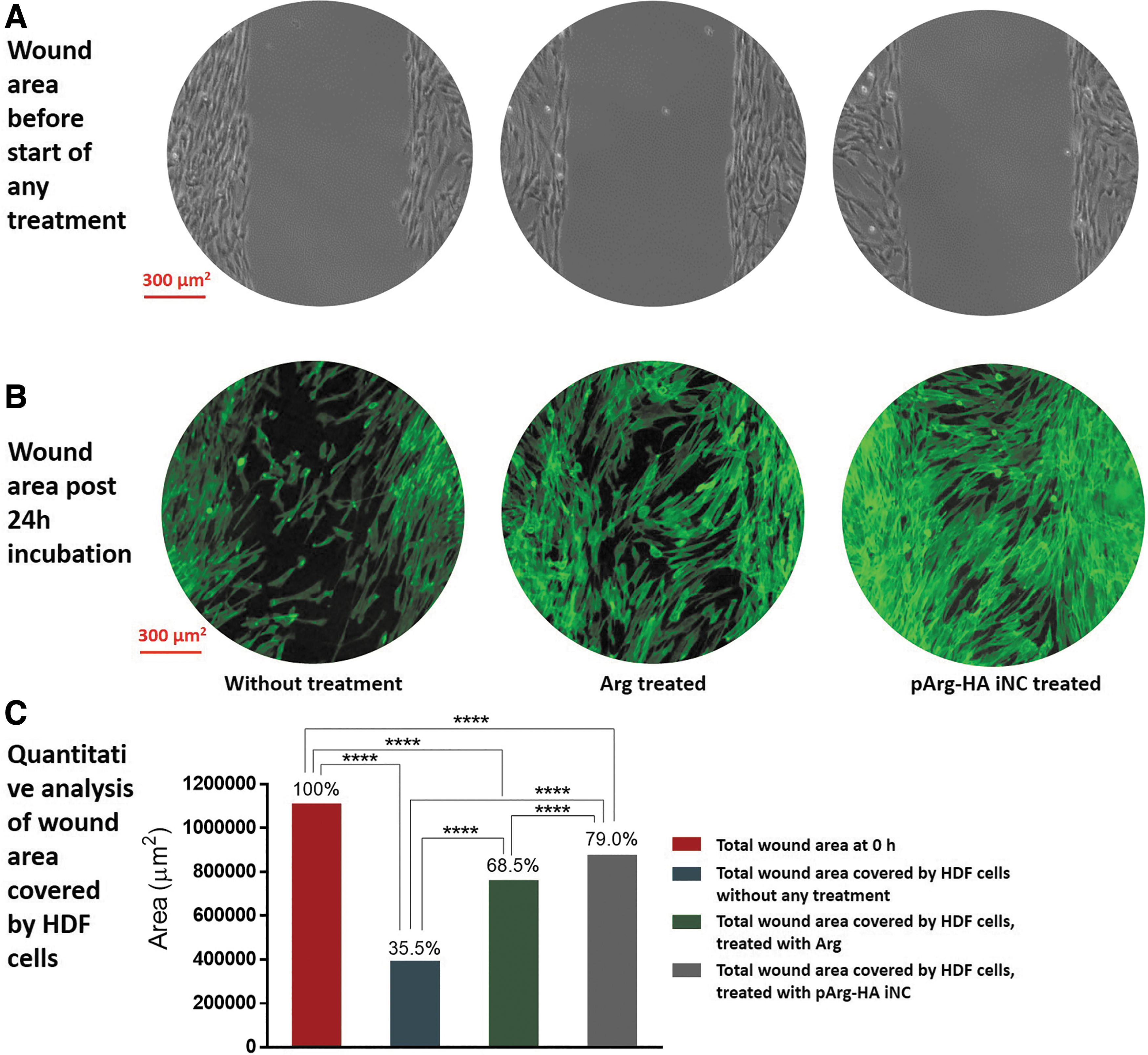
Wound healing assay.
Intracellular NO assay
To identify if the ionic complexes of Arg is responsible for enhanced, in vitro wound healing capacity, we evaluated the effect of Arg delivered by the complexes on intracellular NO. Generally, Arg is known to activate NO synthetase pathways and expected to enhance intracellular NO. To evaluate intracellular NO, we imaged pArg-HA iNC 2 formulation (with pArg to HA ratio of 1.6:1) treated HDFa cells using NO Fluorometric Probe. The chemical principle of this assay hinges on the fact that NO probe passively diffuses into cells and is deacetylated by cellular esterases to a nonfluorescent intermediate, which is further oxidized to a highly fluorescent, triazolo-fluorescein analog in presence of intracellular NO. This NO probe is cell permeant and stable at cellular pH conditions. iNOS derived NO may be produced by fibroblasts, when activated by cytokines, inflammatory mediators, growth factors, and hypoxic conditions. 22 However, NO production by these cells may be limited by cofactors and the availability of Arg itself. We analyzed HDF cells post 24 h treatment with pArg-HA iNC. In confocal microscopy-based experiments, we found prominent signal of NO fluorometric probe at cellular cytoplasm, which revealed activation of Arg mediated NO synthetase pathway. Fluorescence intensity showed 6.7-fold increase in green fluorescence, which is directly proportional to intracellular NO (Fig. 4A–C). The effects of NO on wound repair are diverse, involving angiogenesis, inflammation, cell proliferation, matrix deposition, and remodeling, as well as mediation of apoptosis. These effects contribute to the increased activity of the iNOS. The products of the iNOS production (NO and citrulline) decrease the arginase activity.23,24 Thus we envision that the iNCs of pArg-HA systems most likely interfere with intracellular iNOS production pathways by liberating free Arg inside target cells. Currently we are investigating more detailed cellular pathway for this observation.
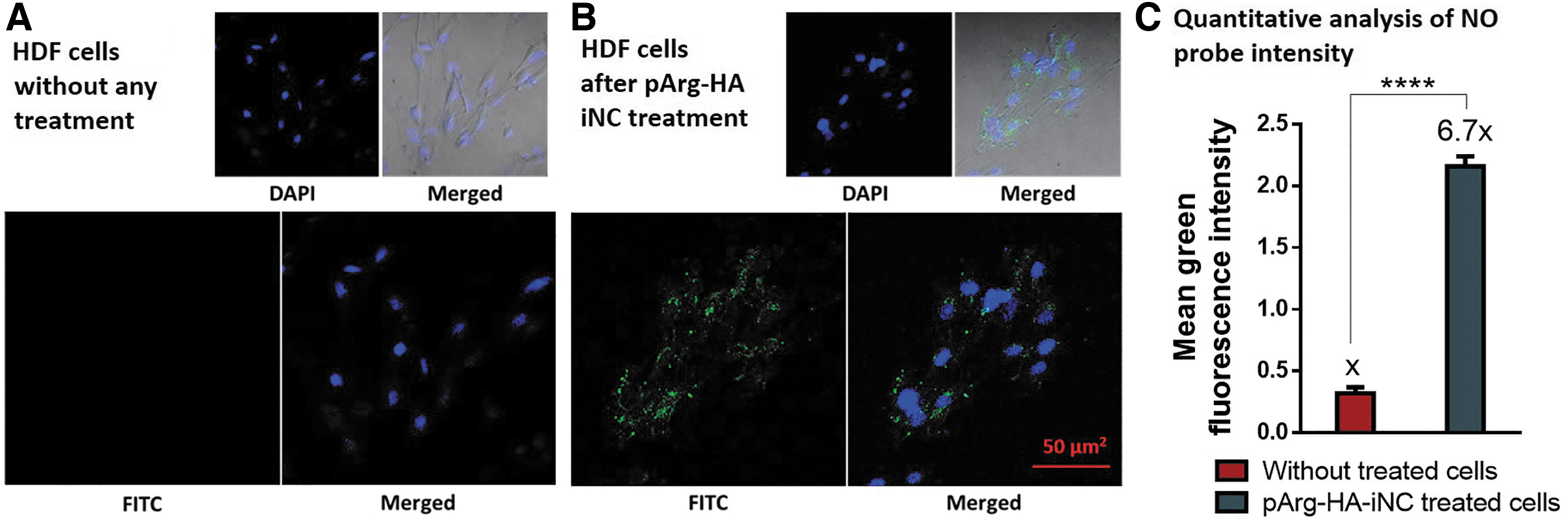
Intracellular NO analysis of HDF cells using NO specific fluorescent probe.
Effect of pArg-HA iNCs on cancer cells
The polycationic nature of pArg has facilitated the use of this polypeptide for cell targeting and membrane translocation of biomolecular cargo. Moreover, a dependence between cell permeability and the increase in the number of Arg residue has been established, where 6–12 Arg residues within pArg sequences elicit optimal translocation activity. Arg-rich peptides oftentimes trigger cellular cytotoxicity because of their polarity and electrostatic binding capacity with critical biologically charged molecules. Therefore, although Arg-rich peptides are highly efficient in mediating in vitro cellular uptake, in vivo translation of these peptides has been limited by poor specificity and off-target toxicity. Therefore, we set forward to investigate if pArg, delivered through ionic complexes with HA, can be a viable option to deliver pArg to affect cancer cells. Our hypothesis is that pArg when delivered through pArg-HA iNCs will be uptaken by cancer cells, and upon disintegration of complexes, liberated pArg can impart cytotoxic effect or chemosensitize a frontline chemotherapy.
We first evaluated the cellular uptake of pArg-HA iNCs in pancreatic cancer cell lines, such as MIA PaCa-2. Electrostatic complexes were developed by the addition of 2 mL, Alexa fluor 647 labelled 1 mg/mL concentration of HA into 5 mL Poly Arg (5 mg/mL) under stirring condition at RT. In subsequent stages, Mia PaCa 2 cell lines were treated for 6 and 12 h with these complexes and were observed for confocal imaging postincubation. As demonstrated in Figure 5, time dependent internalization of nanocomplexes was evident in MIA PaCa-2 cells after 6 and 12 h. We observed the accumulation of red fluorescence from Alexa Fluor 647 throughout the cytoplasm that indicated successful internalization of the complexes into the cells. The nanoparticles were also found to be accumulated around the nucleus. Due to strong electrostatic nature, we do not expect decomplexation of the nanocomplexes in cell culture media.

We also observed that these iNCs were able to impact cellular viability. Compatibility of HA has been well established, and thus, we envision that this cytotoxic onslaught is mostly mediated by pArg or monomeric Arg. The cytotoxicity of ionic complexes was evaluated on two types of pancreatic cancer cell lines, namely Mia PaCa 2 and Panc1. These two cell lines exhibit RAS mutation, which is evident in >95% of human pancreatic cancer. The nanocomplexes have shown significant, yet identical cytotoxic efficacy on both cell lines with IC50 value 30.6 and 32.01 nM for MIA PaCa-2 and Panc-1, respectively. Apart from the antiproliferative activity of poly Arg, we also studied the cytotoxic effect of free Arg on two pancreatic cancer cell lines Mia PaCa 2 and Panc1 and did not find any significant effect of free Arg on these cell lines (Fig. 5C). Furthermore, we were interested to investigate the effect of most widely accepted antimetabolite chemotherapeutic drug for pancreatic cancer, GEM, at its IC50 concentration, when the cells were preincubated with pArg-HA iNCs. Therefore, we pretreated pancreatic cancer cell lines with ionic complexes (pArg-HA iNC 2, pArg:HA ratio of 1.6:1), before treating the cell lines with GEM at a concentration equivalent to the IC50 of the drug against MIA PaCa-2 and Panc-1 cell lines. We observed that the cytotoxicity of the ionic complexes was effectively enhanced by post-treatment of the cancer cells with GEM. Further reduction of the IC50 value of ICs to 18.33 and 19.69 nM in Mia PaCa 2 and Panc1 cell lines post treatment with GEM may open a new direction of combinational chemotherapy toward pancreatic cancer treatment.
We next examined the nature of the observed enhancement of interaction between pArg-HA iNC 2 and GEM on MIA PaCa-2 and Panc-1 cell lines. We executed widely used Bliss analysis using Combenefit software to analyze such interaction (Fig. 6). 19 Interestingly, highly significant synergy has been detected using Bliss synergy model, for the combinations of pArg-HA iNCs along with GEM on Panc1 cell line, but not in MIA PaCa-2 cell lines. Such observation may not be entirely unexpected, since pancreatic cancer cells show widely differing metabolic profile and demand for biological macromolecules. MIA PaCa-2 is strongly glycolytic compared to Panc-1 cells and, therefore, might exhibit such differential metabolic vulnerabilities. 25
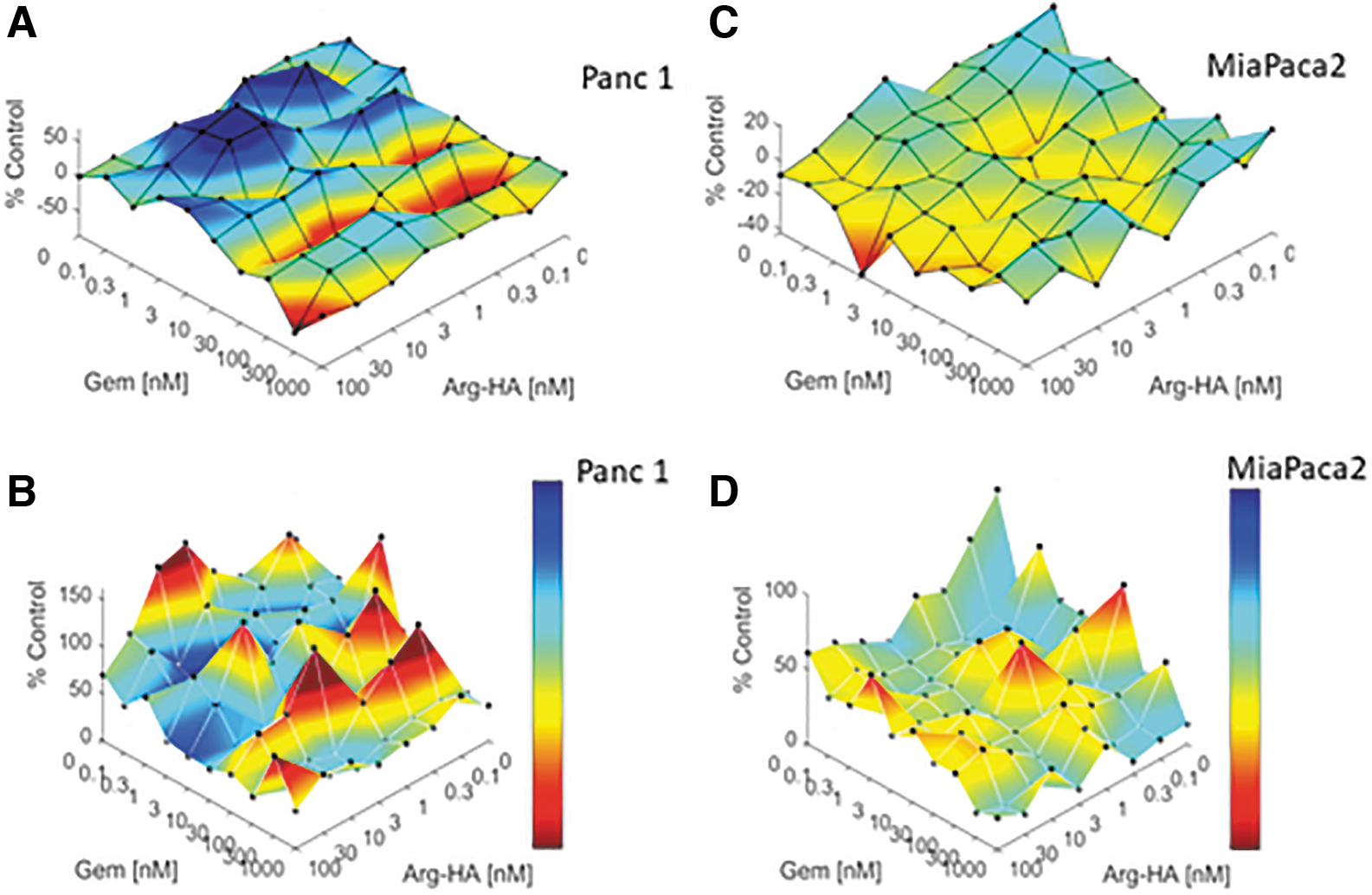
Synergistic effect of pArg-HA iNC 2 in combination with GEM on Panc1
Conclusions
pArg-HA iNCs demonstrated a potential delivery vehicle for Arg delivery in wound healing application. We successfully synthesized stable pArg-HA iNC for sustained Arg delivery. These pArg-HA iNCs showed enhanced cellular migration to cover wound area in vitro and internalization and suppressive properties in cancer cells. In addition, pArg-HA iNCs demonstrated a significant synergistic effect in combination with GEM on Panc1 cell line that may establish a new direction in future therapeutic strategy. So far poly(ionic) species are involved; several electrostatic factors, that is, ionic strength, ionic species, and osmolarity show significant effects on cellular biology. 26 We are currently deconvoluting the mechanistic pathways through which these electrostatic complexes of pArg interact with cellular metabolic machinery.
Authors' Contributions
N.R.K. and D.D. conducted experiment and contributed equally. S.M. and M.Q. designed and supervised research; M.Q. conceived the idea. All coauthors participated in article preparation before submission. The article has been submitted solely to this journal and is not published, in press, or submitted elsewhere.
Disclaimer
Its contents are solely the responsibility of the authors and do not necessarily represent the official views of the NIH. Any opinions, findings, and conclusions or recommendations expressed are those of the author(s) and do not necessarily reflect the views of the National Science Foundation.
Footnotes
Author Disclosure Statement
No competing financial interests exist.
Funding Information
Synthetic part of this research was supported by NSF grant no. IIA-1355466 from the North Dakota Established Program to Stimulate Competitive Research (EPSCoR) through the Center for Sustainable Materials Science. Partial support for the work was received from NIH grant number 1 P20 GM109024 (to M.Q.) from the National Institute of General Medicine (NIGMS). TEM material is based upon work supported by the NSF under grant no. 0923354.