
Abstract
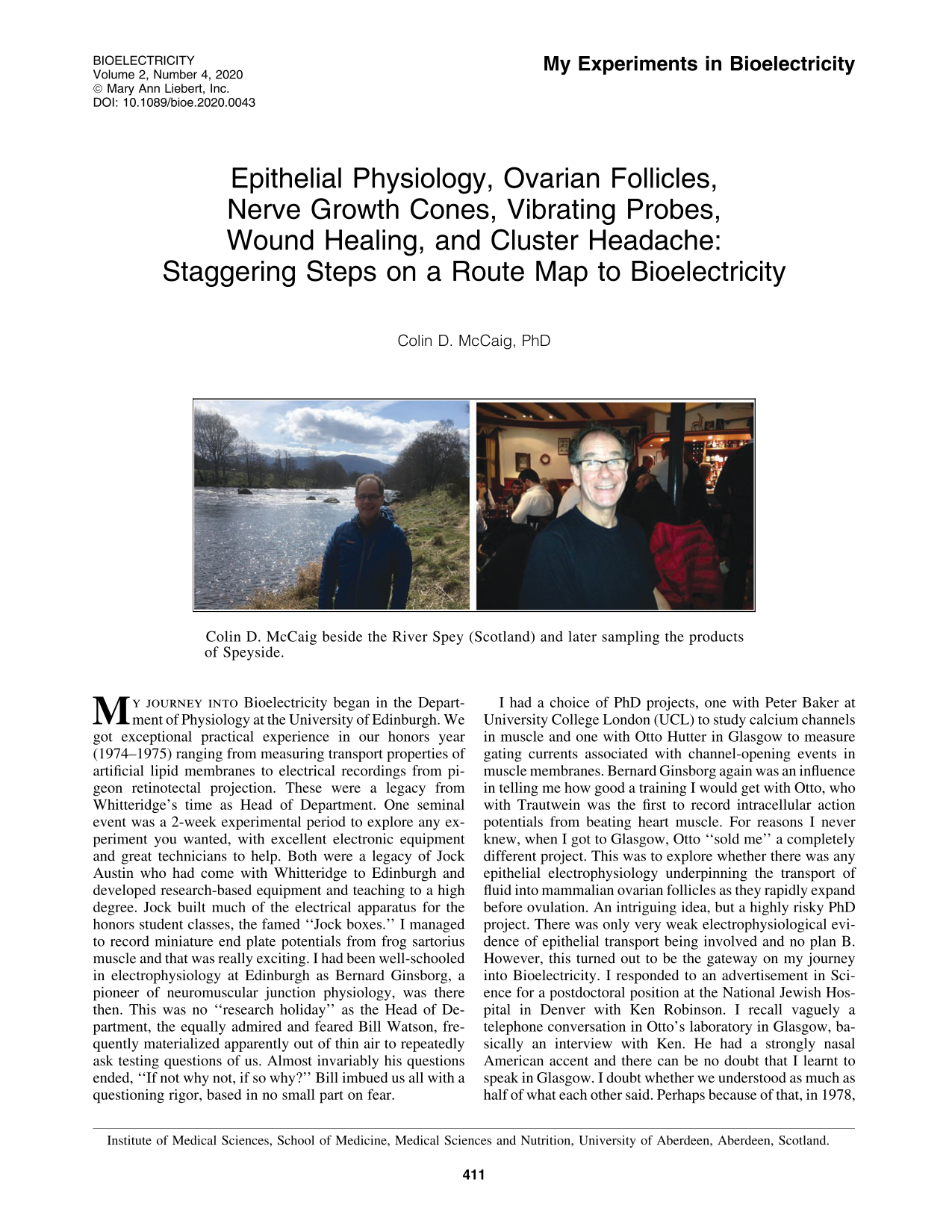
Colin D. McCaig beside the River Spey (Scotland) and later sampling the products of Speyside.
My journey into Bioelectricity began in the Department of Physiology at the University of Edinburgh. We got exceptional practical experience in our honors year (1974–1975) ranging from measuring transport properties of artificial lipid membranes to electrical recordings from pigeon retinotectal projection. These were a legacy from Whitteridge's time as Head of Department. One seminal event was a 2-week experimental period to explore any experiment you wanted, with excellent electronic equipment and great technicians to help. Both were a legacy of Jock Austin who had come with Whitteridge to Edinburgh and developed research-based equipment and teaching to a high degree. Jock built much of the electrical apparatus for the honors student classes, the famed “Jock boxes.” I managed to record miniature end plate potentials from frog sartorius muscle and that was really exciting. I had been well-schooled in electrophysiology at Edinburgh as Bernard Ginsborg, a pioneer of neuromuscular junction physiology, was there then. This was no “research holiday” as the Head of Department, the equally admired and feared Bill Watson, frequently materialized apparently out of thin air to repeatedly ask testing questions of us. Almost invariably his questions ended, “If not why not, if so why?” Bill imbued us all with a questioning rigor, based in no small part on fear.
I had a choice of PhD projects, one with Peter Baker at University College London (UCL) to study calcium channels in muscle and one with Otto Hutter in Glasgow to measure gating currents associated with channel-opening events in muscle membranes. Bernard Ginsborg again was an influence in telling me how good a training I would get with Otto, who with Trautwein was the first to record intracellular action potentials from beating heart muscle. For reasons I never knew, when I got to Glasgow, Otto “sold me” a completely different project. This was to explore whether there was any epithelial electrophysiology underpinning the transport of fluid into mammalian ovarian follicles as they rapidly expand before ovulation. An intriguing idea, but a highly risky PhD project. There was only very weak electrophysiological evidence of epithelial transport being involved and no plan B. However, this turned out to be the gateway on my journey into Bioelectricity. I responded to an advertisement in Science for a postdoctoral position at the National Jewish Hospital in Denver with Ken Robinson. I recall vaguely a telephone conversation in Otto's laboratory in Glasgow, basically an interview with Ken. He had a strongly nasal American accent and there can be no doubt that I learnt to speak in Glasgow. I doubt whether we understood as much as half of what each other said. Perhaps because of that, in 1978, I became Ken's first postdoc, interestingly without being a “doc” (a story for another time and place)! Ken introduced me to his mentor, the wonderful Lionel Jaffe, and his work on ion currents in development, especially in eggs and plants. Lionel was a polymath who co-invented the vibrating probe with Rich Nuccitelli, and he and Ken led me to the American school of the early 1900's that pioneered the study of electrical events in developmental biology, most notably Child and Lund.
Ken was a physicist-turned-biologist who had worked in the Peace Corps in the Philippines and we started every morning with fine doughnuts, strong coffee, politics, physics, and physiology, usually in that order. It was a happy mix. My main project was to determine whether developing neurites really responded with directed growth to an applied direct current electrical field (DC EF) of physiological magnitude. This had been shown by Ingvar 1 in 1920 in Ross Harrison's laboratory at Yale soon after tissue culture was invented, but his one-page article had never been followed up. Indeed, Ingvar was the first to confirm experimentally the phenomenon of galvanotaxis. Our culture of choice was the disaggregated neural tube from early Xenopus embryos just before motor nerve outgrowth begins. Most neurons therefore would be cholinergic motoneurons. The culture method had been set up by a summer graduate student, Laura Hinkle, but she had to finish before she gathered much data. She had a few pictures of growing neurites that looked like they had followed the EF vector. Over the next year, I amassed the data that appeared in our Journal of Physiology article in 1981. 2 This required making little maps of the locations in the culture chambers of about 10–15 nerve cells per dish just as they sprouted neurites, hoping that they would continue to grow and that I would find them again as they changed shape, switching on the EF and photographing each cell sometimes every hour, for 18–20 h. My apartment was close to the laboratory and so I regularly was back after dinner for more photography. We wound our own spools of black and white film and so spent endless hours developing and printing every frame in the dark room and further endless hours hand measuring each timed sequence of growing neurites from the prints. No computers or advanced imaging technologies in those days, but the upside was an intimacy with how your cells were behaving that is lost from more automated experiments. These were thrilling experiments as neurites showed stunning directed growth behaviors and depending on your name, you could almost find your own initials spelt out in the cultures (Fig. 1). Chemotropic turning assays, where your molecule of choice was released from a micropipette to form a gradient closeby a nerve growth cone, were novel and popular in the early 1980's. None then, or since elicited anything like as robust a turning response as I watched unfold in real time with small DC EFs. Some neurites would turn through 180° or more to redirect and grow cathodally. One thing frequently overlooked about this work, even by me, was that it was the first to document the curious observation that myoblasts differentiated to became bipolar and did so perpendicular to the EF. Indeed, in co-cultures, nerves and muscles aligned themselves in a DC EF as they do in a developing embryo (Fig. 1). I remember showing Lionel Jaffe these early striking turning and orientation responses and him saying without a hint of hyperbole, “someone will win a Nobel Prize from all of this.” In those days, Journal of Physiology used alphabetical authorship, but would allow a statement that the authorship did not reflect the contribution of each author. I chose not to do this and sometimes regretted that, since I learnt that many who saw that article referred to as Hinkle et al., did not realize that all the data, all the figures, and all the words were mine. An early lesson maybe, not really damaging, just mildly annoying. We had submitted our story without success to Nature and to Science and those delays almost got us scooped. However, Mu-ming Poo's similar Journal of Neuroscience article appeared a few months after ours in 1982. 3 It took me some years to learn that since both the Nature and Science reviews were very good, we should have fought our corner and not accepted rejection. Another early lesson. Ken's laboratory and I moved to Connecticut in 1979 and it was here that I first met the sadly departed Richard Borgens, another of Jaffe and Vanable's students from Purdue University, Indiana. Rich was the first to use applied DC EF stimulation successfully, if controversially, in animal models of spinal cord injury, first in rodents and subsequently dogs. 4 He also pioneered applied DC EF stimulation in human spinal cord-injured patients in a Phase 1 clinical trial in collaboration with Shapiro et al. 5 Some sensory functions, but no motor functions, improved. I still hope to see this reinvestigated with more advanced microelectronics techniques.

The direction of growth of differentiating neurons and myoblasts from frog embryos in an applied electric field. Top two panels—nerves have grown strikingly toward the cathode (scale bars = 100 μm). Initially, nerve cells were spherical and undifferentiated. Around 18 h later, neurites have sprouted and grown preferentially toward the cathode (shown at left) in the presence of a steady DC EF. Lower left: myoblasts have differentiated from spherical cells to elongate perpendicular to the EF; lower right: a nerve and a myoblast have differentiated from spherical cells in a steady EF for around 18 h (cathode at left). The myoblast is innervated and their respective orientation mimics their anatomical relationship in the developing neural tube and somites of the frog embryo. EF = 120 mV/mm (top left) and 170 mV/mm in other panels. DC EF, direct current electrical field.
I moved to John Harris's laboratory in Dunedin, New Zealand (1980), where I had relatives. John had worked with Bernard Ginsborg's colleagues Katz and Miledi and with Geoffrey Burnstock at UCL, and together we were first to show that motoneuron cell death was a natural developmental event in mammalian spinal cord in vivo. 6 John provided great training in developmental neuroscience as he hosted evening laboratory meetings to discuss a clutch of seminal articles from Nicole Le Douarin through Rita Levi-Montalcini and Victor Hamburger to Dale Purves and Lynn Landmesser.
After almost 3 years, I was back in Edinburgh with a prestigious Beit Memorial Fellowship that allowed me to follow up the effects of DC EFs on nerve growth in my own laboratory. This was my independent entry to Bioelectricity in the Physiology Department hosted by my original mentor Bill Watson (1983). Local politics and the ousting of Watson as Department head, which reached the national press no less, meant I would need to take my fellowship elsewhere if I wanted to secure a tenured lectureship. Watson had decided that most of his academic colleagues were incompetent and that he alone would teach the entire Physiology syllabus, which he did. I took my Beit Fellowship to an empty laboratory in Physiology in Aberdeen (1986) hosted by Cecil Kidd and was supported into a permanent position by winning a Wellcome Trust University Lectureship award in 1988.
Soon after my return from New Zealand, I had met Dennis Bray at British Developmental Biology Society meetings and he was intrigued by how robustly growth cones responded to the EF. That encouraged me to embark on a series of pharmacological studies that explored the underpinning mechanisms. I was among the first to use this strategy. These were productive years for me in growth cone physiology and nerve guidance in a DC EF. I remain pleased with my eleven single-author articles between 1986 and 1990. Most importantly, I had outlined roles for specific Ca channels and for asymmetric growth cone filopodia and cytoskeleton in the cathodal turning response. I submitted this to Nature, got excellent reviews, and got rejected. A few months later, I was invited by Stan Kater to a meeting at the Cajal Institute in Madrid to celebrate “One Hundred Years of the Nerve Growth Cone.” I was somewhat surprised when I was asked by a colleague (a referee of the article, no names no pack drill) when my single-author Nature article was coming out. He had assumed it would be accepted and I (again) had failed to fight hard enough for it. Those articles appeared back to back in Journal of Cell Science.7,8
Two further highlights whose significance has yet to be exploited come from this period. One was the discovery that DC EFs also stimulated robust back branching of neurites from along the shaft and that this too was directed cathodally. 9 I also showed that repeatedly reversing the EF with around a 30 min cycle allowed forward growth of neurites to continue in opposing directions. This was because anodal-directed retraction was slower than cathodal-stimulated forward growth and so oppositely directed neurites could still benefit from their respective cathodally oriented cycle. 10 Borgens and colleagues used this practically to address the issue of ascending and descending axons receiving DC EF stimulation in spinal cord-injured animals and in their human studies.4,5
I was able to begin recruiting around the end of the 1980's and Ann Rajnicek (a Bioelectricty Editor) joined me from Ken Robinson's laboratory. Ann did some very nice work on the mechanisms underpinning contact guidance in directing nerve growth cones using nanogrooved surfaces that we obtained from the sadly departed Adam Curtis in Glasgow.11,12 Adam had set up one of the earliest Cell Biology Departments in the United Kingdom. Ann also elucidated more of the mechanistic detail that drives electrical guidance of neuronal growth cones in two fine articles.13,14
I was fortunate to get an intercalated medical student Lynda Erskine and she became intrigued enough by developmental neuroscience to abandon medicine for science and do a PhD. She showed that the neurotransmitter acetylcholine, which is released by migrating Xenopus growth cones, may be part of a feedback guidance mechanism that transduces the EF into directed growth through asymmetrically distributed Ach receptors. Blocking growth cone neuronal Ach receptors with
Despite all this, convincing neuroscientists that EFs are physiological and that they influence nerve growth and guidance profoundly remained a challenge. I decided that it might be helpful if we diversified and were able to show that bioelectricity had profound effects across many other biological systems. I had been intrigued by Vanable and Jaffe's work on wound healing 17 and began to collaborate with an ophthalmologist in Aberdeen John Forrester, who had been trained in cell biology by Adam Curtis. We embarked on corneal epithelial studies, back to my epithelial bioelectricity PhD roots.
We had some nice cultures showing strongly directed cell migration, then Min Zhao joined us and he drove that project forward with great vigor. Initially, Min had only 12–18 months of funding, so he took a risk moving from UCL where he had worked with Geoffrey Burnstock. In another of these scientific coincidences, my Dunedin mentor John Harris also worked with Burnstock: small world! Among other things, my rich and hugely rewarding collaboration with Min established roles for growth factors in transducing the EF signal, in particular, epidermal growth factor (EGF) and for EF-induced asymmetric rearrangements of receptors, second messengers, and cytoskeleton, which underpinned directed cell migration (Fig. 2a).18–20

One intriguing discovery the significance of which remains elusive was that DC EFs directed the axis of cell division (Fig. 2b). I remember Min showing me some black and white images of migrating cells and I saw a few had been taken during cell divisions and that the daughter cells lay along the EF vector. I asked him to follow this up and he found remarkably that the EF stimulated corneal epithelial cells to divide more, and that it oriented the mitotic spindle along the EF vector. 21 There are a number of stem cell situations in brain and gut for example, where the axis of cell division is critical for the choice of cell differentiation pathway and I wonder whether some of them are influenced by local EFs in vivo.
We recruited Bing Song, now a Professor in Cardiff, and his beautiful experiments importantly took our EF work in vivo. Together with Min, he established rat models of corneal wounds and we had a panoply of pharmacological agents, which we knew from epithelial electrophysiology would influence the wound-induced DC EF in specific ways. Thank you Otto Hutter for giving me the curved ball PhD—it did come in useful, eventually. Bing even bought a camp bed from our Wellcome Trust grant so that he could stay overnight to deliver regular eye drops to his corneal wounded rats to suppress or enhance their wound-induced EFs. This work produced two excellent articles, which established not only that electrical cues regulate the orientation and frequency of cell division and the rate of wound healing in vivo but also that nerves are guided and nerve sprouting is stimulated by a naturally occurring EF in vivo.22,23 Bing made the front cover of Journal of Cell Science 117: 4681–4690 and a news and views article covered his great work (Figs. 3, 4).

The naturally occurring wound-induced DC EF that arises in vivo immediately after wounding directs nerves to the wound edge and reorients nerve sprouts to turn cathodally toward the wound edge.
23

Wound-induced EFs regulate wound healing rate and orient the axis of cell division in vivo.
22
Throughout this period, Jin Pu, Huai Bai, Entong Wang, and Brian Reid made excellent progress by showing roles for electrical signals in controlling aspects of angiogenesis in vascular endothelial cells by signaling through vascular endothelial growth factor receptors, showing that EFs and mitogen-activated protein kinase signaling regulate early wound healing in lens epithelium and that EGF receptor signaling is essential for electric field-directed migration of breast cancer cells.24–26 Brian was our expert vibrating probe Guru and made all of our electrical recordings during this time, an invaluable contribution. 27
Min Zhao, Bing Song, Jin Pu, and Brian Reid drew together many disparate strands of our story to produce the seminal Nature article of 2006. 28 Min established crucial collaborations with the laboratories of Henry Bourne, Peter Devreotes, and Josef Penniger, which were to seal an outstanding synthesis of our work and show the involvement of key signaling genes in cellular responses to the EF. Let me quote the end of the article's abstract: “Notably, genetic disruption of phosphatidylinositol-3-OH kinase-γ [PI(3)Kγ] decreases electric field-induced signaling and abolishes directed movements of healing epithelium in response to electric signals. Deletion of the tumor suppressor phosphatase and tensin homolog (PTEN) enhances signaling and electrotactic responses. These data identify genes essential for electrical-signal-induced wound healing and show that PI(3)Kγ and PTEN control electrotaxis.” 28
Subsequently, Li Yao from our group was the first to show EF-induced migration, division, and polarized organelles in neuronal stem cells, 29 and Noemi Lois, John Forrester, Jin Pu, and Lin Cao were involved in my renewed interest in lens biology, which Ken Robinson had kindled. 30 They showed that electrical signals in the lens regulated its differentiation and quite remarkably that they could stimulate complete lens regeneration, an area rich for exploitation.31,32
Jin Pu and her partner Lin Cao drove forward two further beautiful pieces of work showing that the physiological electrical potential difference across intestinal epithelial cells could determine and reprogram epithelial polarity, turning apical domains into basal and vice versa. 33 They also showed that the polarized retinal pigment epithelium generates electrical signals that diminish with age and may be involved in regulating retinal pathologies such as macular degeneration. 34
So much for the past, in which we documented that DC EFs do indeed influence profoundly numerous cell behaviors from brain and multiple epithelial-based systems in vitro and in vivo and also more recently cells of the immune system.35,36 What does the future hold? I have two exciting European collaborations, which I expect will establish some of the principles of our bioelectrical work into regular clinical practice. One is with an exceptional clinician in Dusseldorf, Dr. Albrecht Molsberger. His clinic uses DC electrical stimulation from microarrays of acupuncture needles to treat, with high success, injuries to muscles, tendons, and joints, and even to treat the most painful condition known to humans, cluster headache.37,38 A company Axomera has been formed and the device and stimulation protocols that we published jointly have been patented. A clinical trial team is in place and two randomized clinical trials are planned soon. One will be on Electrical Stimulation to treat Patellar and Achilles Tendinitis and a second to treat Osteoarthrosis of the thumb. More information is here https://chp-gmbh.com/science/
A second collaboration is with a Danish entrepreneur Valdemar Siesbye. Together with an electronics engineer, Henrik Riehm Sørensen, we have created a noninvasive, next-generation electroceutical device to apply to nonhealing wounds to promote wound healing. Colleagues in Tubingen have shown that his is highly effective in healing “wounds” in scratch assay experiments and a pilot randomized clinical trial on human skin graft wounds is planned. This will be directed by our clinical collaborator Jonas Kolbenschlag, University of Tübingen, Germany. A new company has been formed (Vanquish Innovation ApS, Denmark), and the new device is called Hiperpatch.
It has been and remains an enormous privilege to have worked with so many greatly talented individuals. They have been crucial to the burgeoning development of our corner of Bioelectricity and have brought the discipline to the beginnings of a golden age. Maybe Lionel Jaffe was prescient!