




Abstract
Recent emergence of human connectome imaging has led to a high demand on angular and spatial resolutions for diffusion magnetic resonance imaging (MRI). While there have been significant growths in high angular resolution diffusion imaging, the improvement in spatial resolution is still limited due to a number of technical challenges, such as the low signal-to-noise ratio and high motion artifacts. As a result, the benefit of a high spatial resolution in the whole-brain connectome imaging has not been fully evaluated in vivo. In this brief report, the impact of spatial resolution was assessed in a newly acquired whole-brain three-dimensional diffusion tensor imaging data set with an isotropic spatial resolution of 0.85 mm. It was found that the delineation of short cortical association fibers is drastically improved as well as the definition of fiber pathway endings into the gray/white matter boundary—both of which will help construct a more accurate structural map of the human brain connectome.
Introduction
Structural magnetic resonance imaging (MRI) of brain connectivity, primarily by using diffusion tensor imaging (DTI) (Basser et al., 1994) and high angular resolution diffusion imaging (HARDI) (Anderson, 2005; Frank, 2001; Tuch et al., 2002), has seen remarkable growth over the past two decades. Through fiber tractography (Mori et al., 1999), researchers have been able to examine neuronal pathways in the human brain and generate the so-called human brain connectome (Hagmann et al., 2008; Sporns et al., 2005).
Because of its ability to resolve multiple diffusion tensors within the voxel and thus improve the ability to delineate crossing fibers (which are commonly seen in the relatively large white matter fiber bundles well under the cortical surface), HARDI has been predominantly used to generate detailed connectome maps. In superficial white matter regions, however, there is an abundance of short cortical association fibers (commonly known as the U-fibers) that connect cortical regions between adjacent gyri. The range of these U fibers can be as short as 3 mm, covering 3–30 mm. It is also worth noting that the volume of these U-fibers has also been estimated at 240 cm3, even more than that of the long-range fibers (>30 mm) (Schuez and Miller, 2002). These association fibers play an important role in cortical communication, although it has been difficult to delineate them at a low spatial resolution even with HARDI, largely because their small structures and high curvature pose challenges that are fundamentally different from crossing fibers. A high spatial resolution, rather than a high angular resolution, is thus needed to capture the high curvature and accurately delineate these important fibers.
In addition, to better connect the functional brain regions derived from gray matter classification, there is a need for a high spatial resolution to accurately characterize the endings of white matter fiber pathways into the gray matter and even the intracortical fiber pathways within the gray matter. Typically, these fiber pathways are shorter than 3 mm (Schuez and Miller, 2002), thus also requiring a very high spatial resolution to sufficiently capture its details.
However, it has thus far been challenging to achieve a high spatial resolution for diffusion MRI in vivo. To date, most of these images have been acquired with single-shot echo-planar imaging to minimize scan time and physiological motion artifacts. The length of the readout window and hence the spatial resolution of single-shot acquisitions are usually limited by the transverse relaxation time. Finally, the inherently low signal-to-noise ratio (SNR) due to the small voxel size further introduces difficulty in the quest for a high spatial resolution. As such, while there have been recent reports demonstrating submillimeter spatial resolution in postmortem brain tissue at 3 T (Miller et al., 2011) and human brain in vivo at 7 T (Heidemann et al., 2012), as well as near millimeter resolution in humans in vivo at 3 T (Engstrom and Skare, 2013), a whole-brain DTI at submillimeter resolution has not yet been routinely achieved.
Extending from our previous work on a robust multishot DTI technique using the principle of multiplexed sensitivity encoding (MUSE) (Chen et al., 2013), the authors present here the in vivo three-dimensional (3D) results with isotropic submillimeter spatial resolution and sufficiently high SNR at 3 T. Specifically, they assess the advantages of a high spatial resolution to address the two aforementioned limitations in brain connectome imaging on high-curvature fibers and gray/white boundary definition.
Materials and Methods
Without a clear gold standard, it remains challenging to determine what the ultimate spatial resolution should be for brain connectivity imaging. In this report, the goal is to delineate the short but highly curved association fibers, which have been largely missed at a low spatial resolution, as such, the authors seek to determine a sufficiently high spatial resolution to adequately capture these fiber pathways. Certainly, a higher spatial resolution is always desired, but practical considerations should be given with regard to attainable SNR within a typical examination time on the order of a few tens of minutes. For example, at a coarse spatial resolution of several millimeters, which could encompass the entire high-curvature U-fiber, it is straightforward to infer that HARDI would fail, regardless of how finely the q-space was sampled, as the probability density function in that voxel would not exhibit any salient peak. It is also possible that DTI would fail in this case since a dominant principle diffusion direction might not emerge in the presence of a continuously turning fiber pathway within the voxel. In the extreme case of shortest possible cortical U-fibers (e.g., at the valley of sulci) approaching 3 mm in length and full curvature at 180°, its turning radius can be estimated at 0.95 mm. To fully characterize such a tight turn, the voxel size would need to be smaller than the minimal turning radius. Based on this consideration and the attainable SNR within the imaging session, a spatial resolution of 0.85 mm was considered ideal. Thus, the authors designed a protocol to acquire a whole-brain DTI data set based on this target resolution.
To this end, a whole-brain 3D DTI data set (Chang et al., 2014) was acquired on a 3 T MRI scanner (GE MR750, Waukesha, WI) using an eight-channel coil. A total of 11 interleaved axial slabs were used to cover the entire brain. Within each 12-slice slab, each kz plane at 256×256 matrix (of 12 kz planes) is acquired with a four-shot MUSE acquisition (Chen et al., 2013) at a 3 sec repetition time (TR), resulting in a total acquisition time for each slab at 2 min and 24 sec. A field-of-view (FOV) of 21.8 cm was used, along with a slab thickness of 10 mm, resulting in an isotropic resolution of 0.85×0.85×0.85 mm for the final image. An echo time (TE) of 59 msec was used with a partial-Fourier (60% of ky coverage), which can accommodate a maximum b factor of 800 sec/mm2. Here, a moderately lower b factor was used (instead of the usual 1000 sec/mm2) to ensure sufficient SNRs to reach reproducible fiber tracking results. Fifteen diffusion-encoding directions were acquired along with two baseline scans, leading to a total acquisition time of 40 min and 48 sec. Healthy volunteers provided written informed consent as approved by the Institutional Review Board at the Duke University Medical Center.
To have a comparable comparison between high and low spatial resolutions, and to avoid the test–retest variability and different distortion characteristics between high and low resolutions, an additional data set of lower spatial resolution (2 mm isotropic) was generated by downsampling the same high-resolution data set with a cubic resampling algorithm (
Diffusion tensors were generated from the preprocessed data set with the Diffusion Tool Kit (
Results
Before the detailed analyses, the diffusion MRI (dMRI) data set was evaluated to ensure sufficient SNR. In all white matter voxels, the SNR in the baseline and dMRI images were found to be greater than the recommended minima for accurate tensor fitting at this b-value (>10:1 and >3:1, respectively) (Jones and Basser, 2004). Figure 1 shows the examples of diffusion weighted images (DWI) along one diffusion direction (Fig. 1A) and averaged DWI over all directions (Fig. 1B) at the native 0.85 mm isotropic resolutions to illustrate the overall quality of the DTI data.
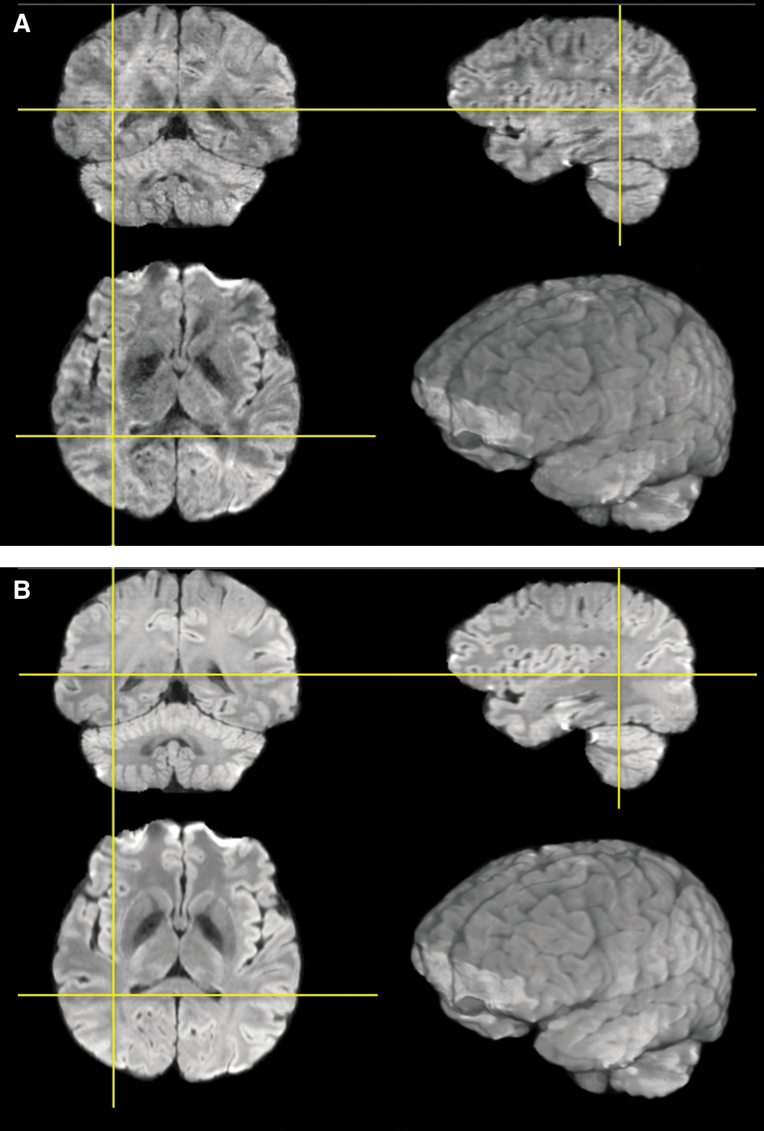
Three-plane view of diffusion weighted images at 0.85 mm isotropic resolution along one diffusion direction
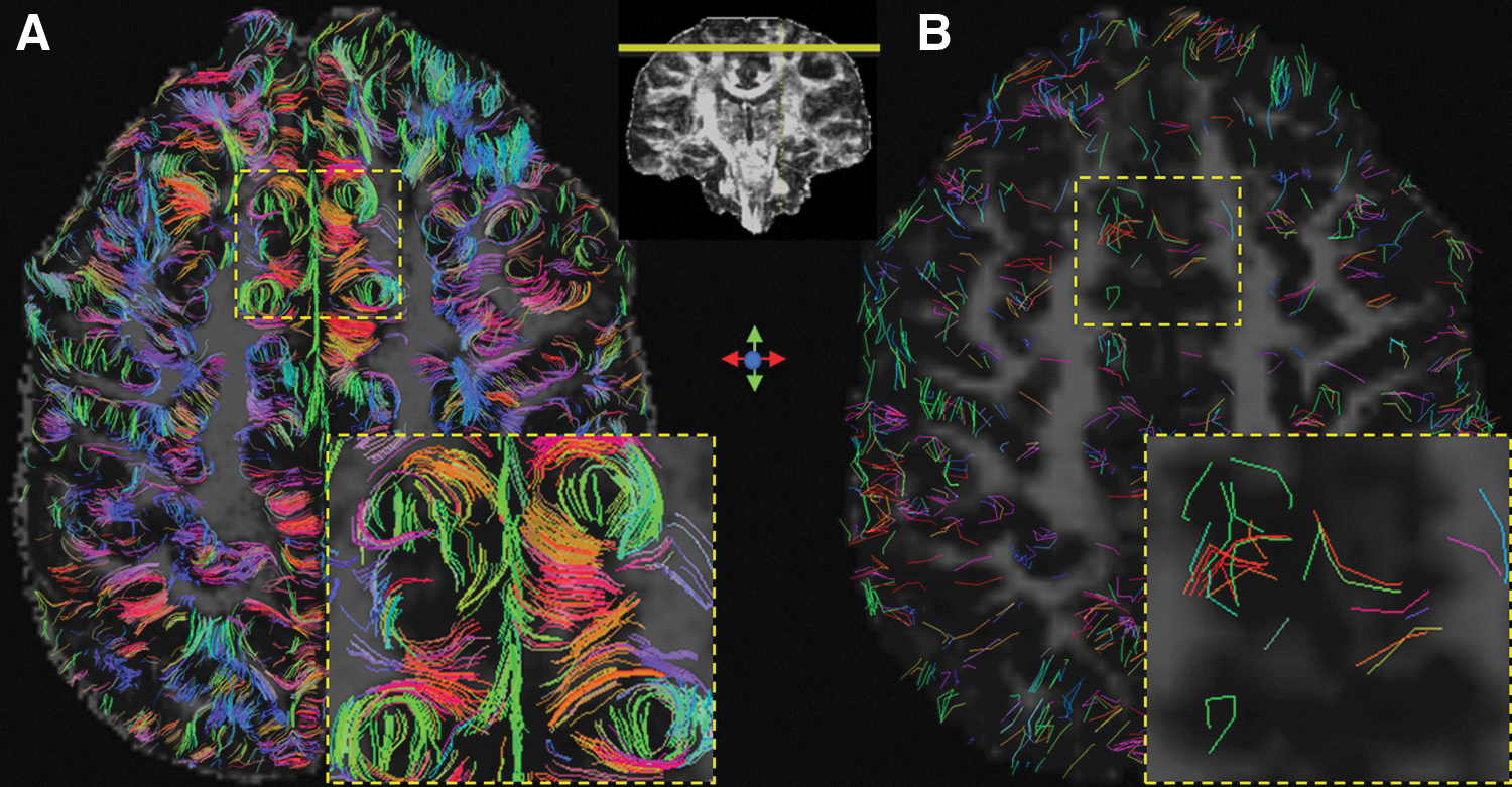
Whole-brain tractography was performed at both spatial resolutions. To best visualize the short and highly curved fibers, the authors restrict the fiber length to be between 3 and 10 mm (which covers the range of the shortest possible lengths for the curved portion of the U-fibers from one side of the sulcus to the other, assuming a cortical thickness of 3 mm). Figure 2 shows fiber illustrations at a superior brain location, where the short association fibers should be more readily visible. Indeed, at the native high resolution of 0.85 mm, these U-fibers closely hugging the valley of sulci were successfully identified and are clearly delineated in Figure 2A. In comparison, the majority of these fibers were not successfully tracked at the lower resolution at 2 mm and are illustrated in Figure 2B.
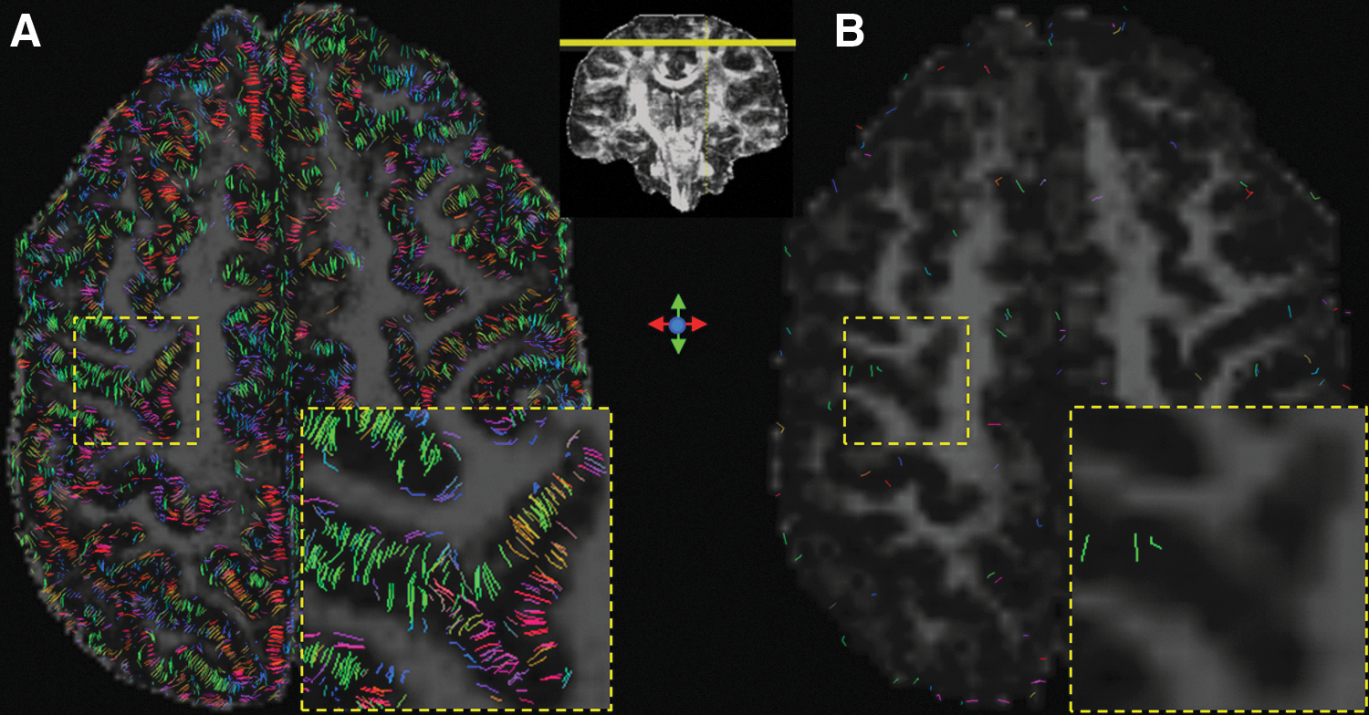
To further evaluate the benefit of a high spatial resolution on delineating the shortest intracortical fiber pathways within the gray matter and improving the gray/white matter boundary definition, the authors further display fiber tracts that are shorter, 3 mm in Figure 3. At the higher spatial resolution at 0.85 mm, an improved definition of gray matter fiber pathways with greatly increased density is shown (Fig. 3A). Many of these intracortical fiber pathways have a radial distribution and are perpendicular to the cortical surface, as seen in both in vitro and in vivo results (Heidemann et al., 2012; McNab et al., 2013; Miller et al., 2011; Truong et al., 2014). Importantly, these short fiber pathways delineated at the high spatial resolution also help better define the gray/white matter boundaries, which are beneficial in establishing connectivity with the white matter. In comparison, there is a clear lack of any fine-grained definition of the gray matter and the gray/white matter boundary at the lower spatial resolution of 2 mm (Fig. 3B).
A quantitative whole-brain analysis of the numbers and volumes of the fiber pathways at these different length ranges was carried out. At the 0.85 mm spatial resolution, the number of short association fiber pathways between 3 and 10 mm was found to be 580,857, whereas it was only 4657 at the 2 mm resolution. The increased number in fiber pathways greatly exceeds the increased number of voxels from low to high resolution (∼13 times). Indeed, the actual fiber volume was increased 33.9 times at the higher spatial resolution. Similarly, the number of intracortical fiber pathways (length <3 mm) at the 0.85 mm resolution was 553,777, whereas it was only 791 at the 2 mm resolution. Correspondingly, the fiber volume was increased 5.7 times at the higher spatial resolution. While neither the fiber pathway count nor the volume is an accurate representation of the underlying fibers, these numbers nevertheless help to illustrate the quantitative improvement of tract representational detail as the result of a higher spatial resolution.
Discussions and Conclusion
Using the improved 3D DTI technique that enabled a high spatial resolution with sufficient SNR, a significant direct finding in vivo is the greatly increased amount of reconstructed short association fibers, often with very high curvatures. This is consistent with a recent report illustrating a high-resolution tractography near the gray matter in humans at 7 T (Heidemann et al., 2012). It was shown that dMRI at 800 μm isotropic enables the disentanglement of adjacent fibers. However, these results were obtained with a partial FOV at ultra-high field (7 T) using a dedicated high-performance gradient system. While high-resolution dMRI at 7 T does have the advantage of higher SNRs, it is limited by the specific absorption ratio (SAR, a quantity indicating radiofrequency [RF] power deposition) issues, which necessitate the use of long TRs (and hence the long acquisition time). In comparison, high-resolution dMRI at 3 T using 3D acquisitions could reach comparably high SNRs, but without any significant SAR limitations. Thus, in this study, the authors aim to establish the feasibility of such high-resolution dMRI scans on widely available 3 T MRI scanners and to extend the coverage for the whole brain to evaluate the impact of spatial resolution on constructing the human connectome and other connectivity maps.
The impact of improved fiber delineation at the surface brain regions also proves to be more beneficial than just the improved characterization of the cortical association fibers or U-fibers. Indeed, a very significant increase in the connection density for mid- and long-range fiber pathways was also observed at a higher spatial resolution. This observation may be explained by an improvement in characterizing the connections at the gray/white matter boundaries, thereby allowing better association of functional gray matter regions over distance.
In the absence of a gold standard, it remains difficult to conclude what the optimal spatial resolution should be to construct the human connectome. It should also be pointed out that while other fiber tracking methods are available (e.g., probabilistic tracking), in this report, the authors only compared fiber tracts delineated using the FACT algorithm (Mori et al., 1999). While this algorithm has certain advantages such as its deterministic nature, it is prone to propagation biases and error accumulation. With this technical limitation in mind, and based on the findings in this report, the authors believe that a resolution sufficient to capture the shortest U-fibers, such as the 0.85 mm used in this report, would be required for human connectome imaging.
As it stands, the long acquisition time and physiological artifacts (e.g., brain pulsation) remain as the biggest obstacles for routinely acquiring DTI data at a very high spatial resolution as used in this report. But given the large potential benefit, more effort should be invested to remove these obstacles. Fortunately, imminent improvements in imaging hardware (e.g., strong gradient coils) and software (e.g., multiband imaging) will likely allow us to achieve such submillimeter spatial resolution within 10 min.
In conclusion, based on the concrete benefits in greatly improved delineation of human brain connectivities, the authors believe that high spatial resolutions can complement high angular resolutions to improve fiber tractography with greatly increased densities, especially in superficial cortical regions where highly curved fibers (e.g., U-fibers) are prevalent. In addition, the fine-grained delineation of the gray matter and gray/white matter boundaries can greatly improve the characterization of connectivities among functional brain regions, including those over a distance. Together these advantages can more effectively link functional regions in the gray matter (i.e., nodes) through white matter pathways (i.e., edges) for a more accurate construction of the human brain connectome.
Footnotes
Acknowledgments
The work presented in this report was, in part, supported by NIH grants (R01 NS 075017 to A.W.S., R01 NS074045 to N.K.C.). The authors thank Dr. Susumu Mori for helpful discussions.
Author Disclosure Statement
No competing financial interests exist.