
Abstract
The intra/extradimensional set-shifting task (IED) provides a reliable assessment of cognitive flexibility, the shifting of attention to select behaviorally relevant stimuli in a given context. Impairments in this domain were previously reported in patients with altered neurotransmitter systems such as schizophrenia and Parkinson's disease. Consequently, corticostriatal connections were implicated in the mediation of this function. In addition, parts of the default mode network (DMN), namely the medial prefrontal and posterior cingulate/precuneus cortices, are also being progressively described in association with set-shifting paradigms. Nevertheless, a definitive link between cognitive flexibility and DMN connectivity remains to be established. To this end, we related resting state functional magnetic resonance imaging (fMRI)-based functional connectivity of DMN with IED task performance in a healthy population, measured outside the scanner. The results demonstrated that greater posterior cingulate cortex/precuneus (DMN) connectivity with the ventromedial striatopallidum at rest correlated with fewer total adjusted errors on the IED task. This finding points to a relationship between DMN and basal ganglia connectivity for cognitive flexibility, further highlighting this network's potential role in adaptive human cognition.
Introduction
Cognitive flexibility, the ability to shift attention from a previously learned representation to select context-specific, behaviorally relevant stimuli, is an adaptive behavior, reportedly controlled by the corticostriatal circuitry (Cools et al., 2004). Impairments have been observed in patients with frontostriatal dysfunction, such as in Parkinson's disease (Cools et al., 2006) and schizophrenia (Jazbec et al., 2007), and a variety of neuropsychological tests and functional magnetic resonance imaging (fMRI) paradigms have been devised to define the neural correlates of this mental operation that includes simple stimulus–response associations (reinforcement/reversal learning), attentional set-shifting, and task switching (Kehagia et al., 2010).
In addition to frontostriatal associations (Cools et al., 2004), emerging evidence also implicates the default mode regions in cognitive flexibility (Dang et al., 2012a). Comprising the angular gyri, posterior cingulate, and medial prefrontal cortices, the default mode network (DMN) has been historically assumed to hinder task performance, mainly due to the observed deactivations in the network nodes during attention-demanding tasks (see Spreng (2012) for a detailed discussion on this topic). However, recent studies revealed extensive DMN functional and structural connectivity to the rest of the brain (van den Heuvel et al., 2009), a greater activity in a wide range of self-referential and memory-based paradigms (Andrews-Hanna et al., 2014; Buckner et al., 2008), task-induced alterations in its connectivity (Vatansever et al., 2015b), as well as altered network integrity in a variety of neuropsychiatric disorders (Whitfield-Gabrieli and Ford, 2012). Collectively, these findings indicate that the DMN may play a fundamental cognitive role that requires further definition.
In this regard, crucial evidence linking DMN and cognitive flexibility stems from a resting-state functional connectivity study that revealed the connection between superior ventral striatum and two major DMN hubs, the posterior cingulate and medial prefrontal cortices (Di Martino et al., 2008). Moreover, a combined fMRI and positron emission tomography task-based investigation have revealed that performance in an object-shifting paradigm is influenced by the effects of striatal dopamine on the medial prefrontal regions, widely thought to be part of the DMN (Dang et al., 2012a). In addition, in a variant of the classic Wisconsin Card Sorting Task, the DMN showed increasing activity in response to the prolonged application of a given rule (Provost and Monchi, 2015).
Although a consensus has not yet been reached as to whether DMN activity/connectivity is related to cognitive flexibility, DMN function has been previously linked to environmental vigilance and internal attentional control (Leech and Sharp, 2014), suggesting a potential role akin to a global integrator (Baars, 2002; Vatansever et al., 2015a), in the processing of behaviorally relevant stimuli. In this regard, the intra/extradimensional attentional set-shifting (IED) task provides a reliable assessment of cognitive flexibility with distinct stages that assess lower order stimulus–response learning and object shifts as well as higher order abstract rule shifts (Kehagia et al., 2010). Given the existing literature on the modulation of default mode regions through striatal input (Braskie et al., 2011; Dang et al., 2012a; Tomasi et al., 2009), we hypothesized that the ability to switch between internalized and novel environmental cues in the IED task (Dias et al., 1996) would require greater striatum and DMN functional interaction. To this end, we investigated resting-state functional connectivity of the DMN and its relationship with subject-specific performance on the IED task.
Materials and Methods
Participants and behavioral testing
Ethical approval was obtained from the local ethics committee and the participants provided informed consent before taking part in this study. Individuals were assessed for verbal intelligence as well as cognitive impairment using the National Adult Reading Test (NART) and Mini Mental State Examination (MMSE), respectively, and volunteers with any contraindication of MRI scanning were excluded. Consequently, the participant group consisted of 22 healthy individuals (19–57 years old, mean = 35.0, SD = 11.2, 9/13 females to male ratio) with an average NART score of 117.1 (SD = 5.76) and a mean MMSE score of 29.33 (SD = 0.85).
The IED task (Dias et al., 1996) is part of the Cambridge Neuropsychological Test Automated Battery (CANTAB) (
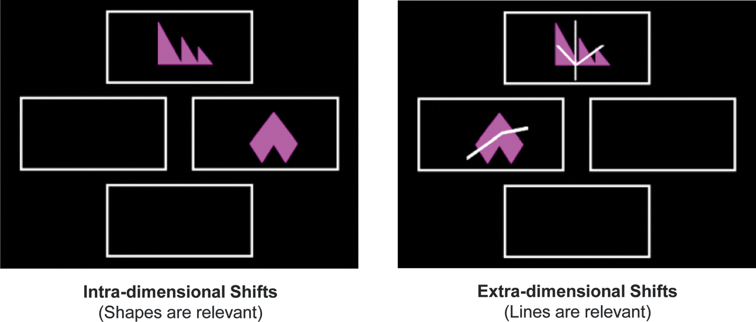
The simple and compound discrimination blocks of the intra/extradimensional set-shifting task (IED). Focusing specifically on cognitive/attentional flexibility, the IED task is part of the CANTAB battery of neuropsychological tests, designed to assess the integrity of frontostriatal connections. While the intradimensional (shapes are relevant) set-shifts represent lower order switching, the extradimensional (lines are relevant) set-shifts denote higher order abstract rule shifts. Color images available online at
Image acquisition
A Siemens MAGNETOM Tim Trio 3T scanner at the Wolfson Brain Imaging Centre, Cambridge, was used to scan participants with a high-resolution T1-weighted, magnetization-prepared 180° radio-frequency pulses and rapid gradient-echo (MPRAGE) structural scan (TR = 2300 msec, TE = 2.98 msec, slice thickness = 1.00 mm), followed by a 5-min eyes-closed resting-state fMRI sequence. Subsequently, four cognitive tasks and a DTI sequence were acquired, which will not be discussed in this study. The echo planar imaging parameters for the resting-state scans were as follows: 32 slices in each volume, 3.0 mm slice thickness, 3.0 × 3.0 × 3.0 voxel size, TR = 2000 msec, TE = 30 msec, flip angle = 78°.
Preprocessing
A standard preprocessing pipeline included slice timing and motion corrections, coregistration of the T1-weighted structural image to the mean motion-corrected functional image, segmentation of high-resolution structural image into gray/white matter and cerebrospinal fluid maps, normalization to the Montreal Neurological Institute (MNI) space using the segmented as well as an a priori gray matter template, and finally smoothing with an 8 mm FWHM Gaussian kernel. Statistical Parametric Mapping (SPM) Version 8.0 software (
Region of interest definition, functional connectivity, and behavioral correlation
Functional connectivity analyses have been previously shown to be influenced by the selected region of interest definitions (Smith et al., 2011). For this reason, we employed an objective approach using the NeuroSynth software (
A general linear model with six movement parameters, global white matter and cerebrospinal fluid signals as confounds, and the average BOLD signal from the PCC/PCUN mask as the variable of interest produced subject-specific connectivity maps. Second-level analysis included a t-test, to assess group-level PCC/PCUN connectivity, that is, DMN maps. Voxel p < 0.001 uncorrected and cluster familywise error (FWE) p < 0.05 corrected for multiple comparison thresholds were utilized. A linear regression analysis of PCC/PCUN connectivity was performed with total adjusted IED error scores (mean centered), denoting each subject's performance in this test. Age was included as a potential confound and the total IED errors were adjusted according to the number of chances that the subject had in making an error. In addition, the mean errors were calculated for the distinct blocks of the IED task. Specifically, the main stages of interest, namely the intra- and extradimensional set-shifting blocks, which denote lower order and higher order set-shifts, respectively, were used in the subsequent linear regressions. We report on the set of clusters that survived a random field threshold of p < 0.05 corrected for the striatal volume using FWE correction.
Results
Posterior cingulate cortex/precuneus connectivity at rest reveals an extensive DMN
With the aim of investigating whether DMN connectivity at rest is related to cognitive flexibility as measured by the attentional set-shifting IED task, we first conducted a seed-based functional connectivity analysis based on a PCC/PCUN mask acquired from a meta-analysis of three search terms included in the NeuroSynth database (Fig. 2A). Using the signal from this seed as the variable of interest, the group level t-test revealed an extensive DMN that included all the major hubs, namely medial prefrontal and posterior cingulate cortices, and bilateral angular gyri as well as the middle temporal lobe structures (e.g., hippocampus), superior frontal gyrus and cerebellar lobules, consistent with the current literature (Andrews-Hanna et al., 2014; Buckner et al., 2008) (Fig. 2B).
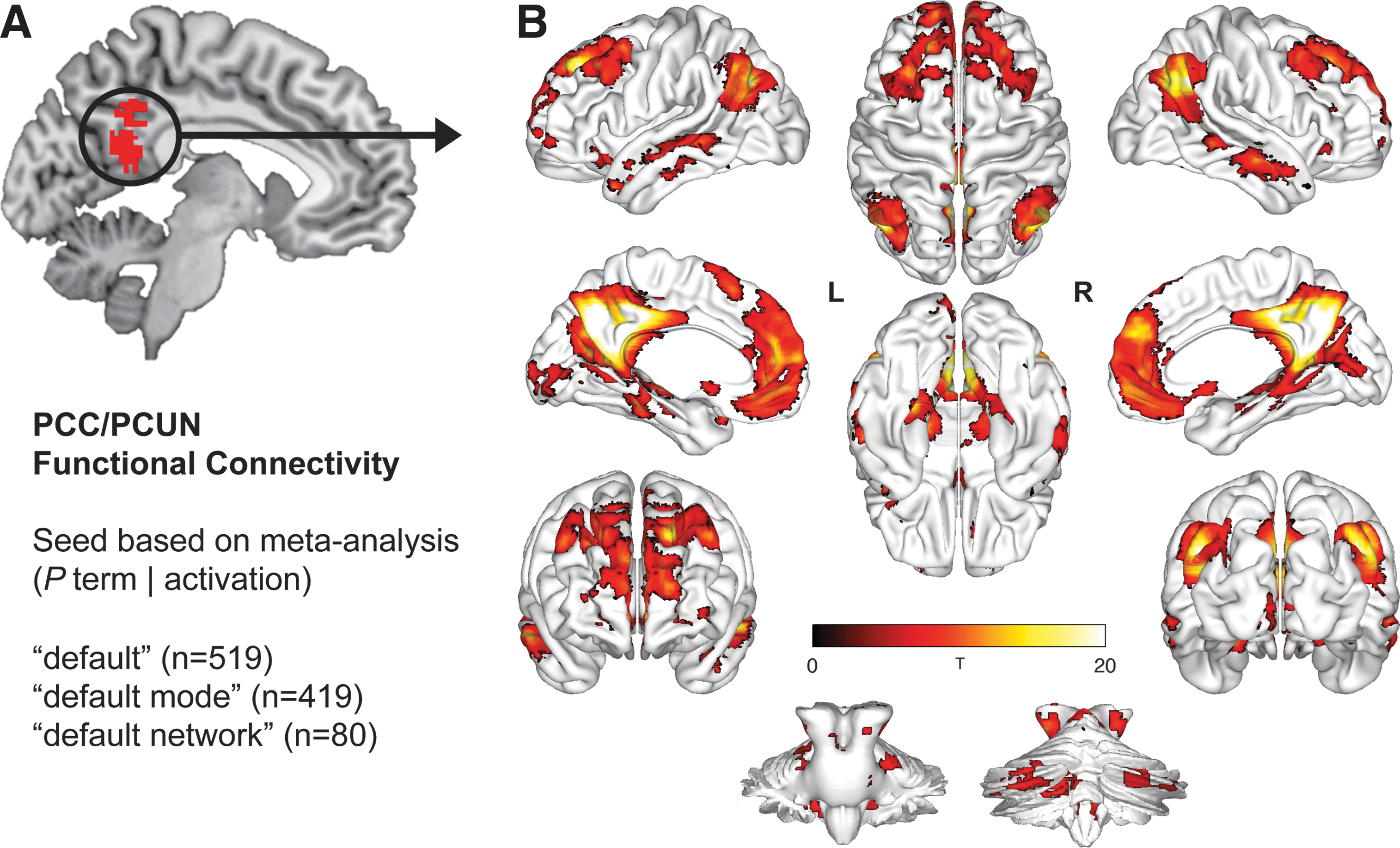
Posterior cingulate cortex/precuneus seed-based functional connectivity reveals an extensive default mode network (DMN).
Default mode-basal ganglia connectivity correlates with IED task performance
The participants' performance on the specific stages of the IED task is shown in Table 1. On average, the participants made 18.86 ± 4.02 total errors across the IED task (adjusted for the number of trials). In addition, we further delineated these behavioral results into the specific stages of the IED task spanning lower order simple stimulus–response learning (discrimination and reversal) and intradimensional set-shifting, as well as the higher order extradimensional set-shifting. A greater number of errors were made in the stimulus–response learning and extradimensional set-shifting blocks, suggesting that the variability in the total adjusted IED errors mainly arose from these distinct stages.
Descriptive Statistics Outlining the Performance of the Participants on the Different Stages of the IED Task
While the errors in the distinct stages of the IED task are raw errors, the total errors across the task have been adjusted for the number of trials in which the participants had an opportunity to make an error. The bold lines represent the participants' performance on the two main stages of interest in the IED task.
IED, intra/extradimensional attentional set-shifting.
Next, we investigated whether DMN connectivity with any other part of the brain during resting-state scanning would correlate with the IED task performance, measured outside the scanner using a touchscreen. We found a negative relationship between the total adjusted IED error scores and PCC/PCUN connectivity with ventromedial striatopallidum (age included as a potential confound) (Fig. 3A). In other words, participants with greater functional connectivity between the DMN and ventromedial striatopallidal regions displayed fewer total adjusted IED task errors, and thus achieved better performance (Fig. 3B). Specifically, the significant set of clusters included a global peak on the left ventromedial putamen [MNI: −28 6 0], with clusters extending to the right ventromedial putamen head and tail, bilateral ventromedial caudate (parts of the nucleus accumbens), and right ventral pallidum (p = 0.007). While the participants made a small number of errors in the intradimensional set-shifting stage (0.45 ± 0.10), on average 8.05 ± 1.72 errors were made in the higher order extradimensional set-shifting, for which we also found significant correlation with the PCC/PCUN and ventromedial striatopallidum functional connectivity. These ventromedial striatopallidal regions were previously related to IED task performance (Cools et al., 2004) and furthermore, the structural integrity of the basal ganglia has been linked to cognitive flexibility (van Schouwenburg et al., 2014).
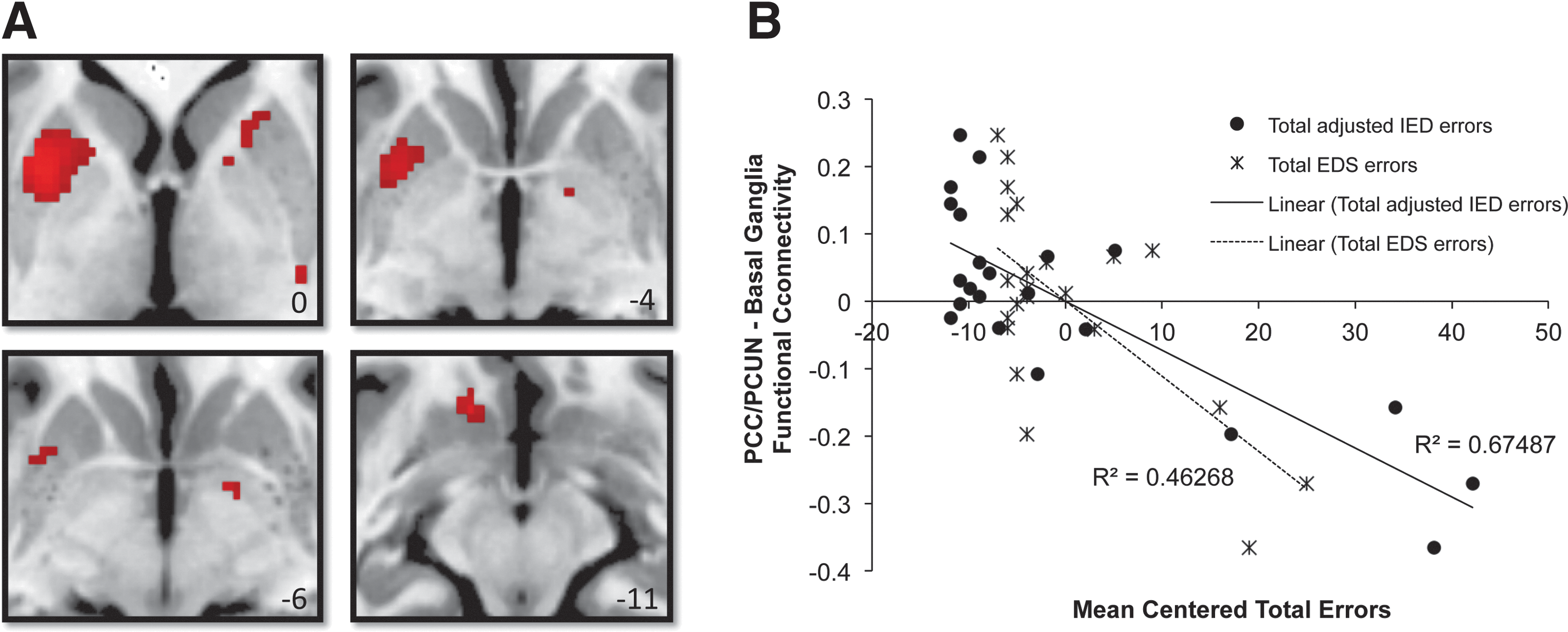
Posterior cingulate cortex/precuneus (PCC/PCUN) connectivity with the ventromedial striatopallidum correlates with IED task errors.
Discussion
Cognitive flexibility, the ability to shift attention from previously learned representations to select behaviorally relevant stimuli in a novel context, is a fundamental function that potentially provides an adaptive advantage for survival. Several neuropsychological tests and fMRI paradigms have been devised, including the Wisconsin Card Sorting, Stroop, stop-signal, and IED set-shifting tasks, to assess the neuroanatomical correlates of this cognitive process. Clinical populations, including Parkinson's disease (Cools et al., 2001) and schizophrenia (Jazbec et al., 2007) patients, have shown impairments in this domain, thus implicating corticostriatal connections and the dopamine system, as well as other neurotransmitters such as noradrenaline and serotonin (Kehagia et al., 2010). Therefore, deciphering the exact mechanism of this circuitry is not only important for understanding healthy brain function but also for discovering potential therapeutic avenues for neuropsychiatric and degenerative disorders (Insel et al., 2013).
The aim of our study was to investigate a potential relationship between cognitive flexibility as measured by an attentional set-shifting task outside the scanner and DMN connectivity during resting-state scanning. At first, our findings revealed the PCC/PCUN seed's extensive functional connection with the rest of the default mode regions, as has already been discussed in the relevant literature (Andrews-Hanna et al., 2014). The hubs displaying the greatest connectivity with PCC/PCUN were the medial prefrontal cortex and bilateral angular gyri. There was also involvement of medial temporal lobe structures such as the hippocampus, but also the superior frontal gyrus, cerebellar lobules, and vermis, all widely considered to be part of the DMN (Buckner et al., 2008).
The main DMN hubs have been previously referred to as rich-clubs with comprehensive structural and functional connections to the rest of the brain (van den Heuvel and Sporns, 2011), suggesting that DMN may facilitate the global integration of information from other large-scale brain networks for conscious processing (Braga et al., 2013; Smallwood et al., 2012; Vatansever et al., 2015a). This notion of DMN functionality, rooted in the global workspace framework (Baars, 2002; Dehaene, 2014), may provide a systematic explanation as to why a wide variety of tasks report DMN involvement (e.g., autobiographical memory retrieval, future planning, moral judgment, and narrative comprehension, see Andrews-Hanna and associates (2014) for a detailed review) and may allude to a fundamental cognitive role that still requires precise delineation.
In this context, the results of our study indicate that better IED task performance (fewer errors) is associated with greater resting-state functional connectivity between the DMN and ventromedial striatopallidum, and may signify a role for these brain regions in mediating cognitive flexibility in an attentional set-shifting task. It should be noted here that the correlated resting-state fMRI connectivity data and the behavioral IED task data were acquired separately, the former performed inside the scanner and the latter outside the scanner with a touchscreen computer. Thus, the ability to relate resting-state functional connectivity between the DMN and ventral basal ganglia with task performance is indicative of not only the sensitivity of the test but also the importance of analyzing spontaneous resting-state BOLD signal fluctuations that can be acquired from patient populations with relatively little effort.
Using this resting-state method, Di Martino and associates (2008) reported on extensive functional connectivity between the striatum and orbitofrontal and lateral prefrontal cortices, as well as posterior midline structures. During task execution, an earlier fMRI study on the contribution of frontostriatal dopaminergic circuitry to cognitive flexibility related low-order stimulus switching with ventral striatal input (centered on the putamen) (Cools et al., 2004). However, higher order abstract rule switching, which is increasingly associated with the noradrenergic system, was shown to engage a network of regions that include the dorsolateral prefrontal cortex (Kehagia et al., 2009). Moreover, a recent fMRI experiment using an alternative version of the Wisconsin Card Sorting Task suggested greater correlation of the frontoparietal network activity with greater numbers of rule shifts, whereas continuous object shifts with stable rule shifts were associated with greater DMN activity (Provost and Monchi, 2015). These findings implicate the DMN in stimulus–response associations and the intradimensional object shifts, reiterating the results of Dang and associates (2012a) who have shown that the striatal dopamine influences DMN to affect shifting between object features.
In light of these findings, we provide a twofold interpretation of our results: First, our finding for a significant relationship between behavior and DMN connectivity with ventromedial striatopallidal areas suggests a collective involvement of these systems in reward-based associative learning and memory. In fact, the main DMN hubs have been previously implicated in mnemonic processes (Shapira-Lichter et al., 2013) and working memory (Konishi et al., 2015). Moreover, increased striatal activation has been reported in retrospective reappraisal during associative learning (Corlett et al., 2004), and reward processing is suggested to be an integral part of the striatum's function (Humphries and Prescott, 2010). Given recent evidence on the breakdown of inhibitory striatal influence on the DMN in schizophrenia (Waltz et al., 2013; Wang et al., 2015), the DMN-ventromedial striatopallidal link we observed in our study might represent an important connection necessary for adaptive cognition.
Second, based on our observation that a large proportion of the variability in the total IED errors came from the extradimensional set-shifting stage, the observed correlation in our study may signify that a basal ganglia influence on the DMN is crucial for a successful extradimensional shift. In other words, basal ganglia input on the DMN may facilitate the cognitive flexibility for switching from attentional set maintenance to a new rule. More direct evidence for this basal ganglia influence would be a correlation between errors during the intradimensional set-shifting stage and DMN connectivity. However, we found no such correlation because of the small number of errors we observed at this stage of the task. This may be remedied by a greater sample size and the inclusion of further measures in the analysis such as reaction time. Nevertheless, previous studies have suggested that the striatal dopamine activity may drive coupling of the default mode, dorsal attention, and frontoparietal networks and such striatal gating of the dynamic interactions between these networks (Dang et al., 2012b; den Ouden et al., 2010; van Schouwenburg et al., 2010) is believed to be achieved through linear and nonlinear modulations (Cole et al., 2013). With its suggested influence on higher cognitive, attention-related networks (Dang et al., 2012b), the striatum may thus act as a switchboard directing the processing of external information either to the default mode or dorsal attention networks. Future research should explore the individual contributions of these networks to cognitive flexibility as well as their coupling with the striatum. Despite its utility in revealing important functional relationships in the resting brain, functional connectivity alone does not provide the means to infer causality. Thus, further studies with scanner-based paradigms and causal models are needed.
Although dopamine has long been the focus of research in cognitive flexibility, recent evidence has also highlighted the involvement of other neurotransmitter systems, specifically in higher order extradimensional set-shifting (Kehagia et al., 2009; Kehagia et al., 2010). For instance, a reduction in the main noradrenaline input to the medial prefrontal cortex through a lesion on the dorsal noradrenergic ascending bundle in rats was shown to impair extradimensional set-shifting (Tait et al., 2007). Furthermore, atomoxetine, a selective noradrenaline reuptake inhibitor, is currently under investigation as a potential treatment for the nondopaminergic cognitive deficits seen in Parkinson's disease (Kehagia et al., 2014; Vazey and Aston-Jones, 2012). Thus, future work should explore the potential link between DMN and different neurotransmitter systems in both patients and healthy controls through pharmacological interventions.
Although the manner in which resting-state networks dynamically change to process external stimuli is yet to be fully elucidated, it has been hypothesized that these functional connections might denote an internal representation of the external world, shaped through experience (Vincent, 2009), which can provide priors for future expectation and processing of incoming information (Friston, 2010). Thus, it is important to acknowledge the potential role that the DMN might play in learning and memory as a global integrator of information for conscious and predictive processing, especially with its core regions involved in reward associations and predictions (Pearson et al., 2011). Future research that investigates the role of DMN in learning, memory, motivated behavior, and the formation of habits will be necessary for deciphering the exact contribution of this network to adaptive human cognition.
Footnotes
Acknowledgments
The Evelyn Trust (RUAG/018) supported this research. In addition, D.V. received funding from the Yousef Jameel Academic Program; D.K.M. is funded by the NIHR Cambridge Biomedical Centre (RCZB/004) and an NIHR Senior Investigator Award (RCZB/014), and E.A.S. is supported by the Stephen Erskine Fellowship Queens' College, Cambridge. The authors also thank Dr. Guy Williams and Victoria Lupson and the rest of the staff in the Wolfson Brain Imaging Centre (WBIC) at Addenbrooke's Hospital for their assistance in scanning. Finally, they thank all the participants for their contribution to this study.
Author Disclosure Statement
No competing financial interests exist.