
Abstract
Diffusion tensor imaging (DTI) studies showed that microstructural alterations are correlated to reading skills. In this study, we aim to investigate white matter microstructure of a group of Portuguese speakers with poor reading level, using different parameters of DTI. To perform this analysis, we selected children ranging from 8 to 12 years of age, poor readers (n = 17) and good readers (n = 23), evaluated in the word-level ability based on a Latent Class Analysis (LCA) of Academic Performance Test (TDE). Poor readers exhibited significant fractional anisotropy (FA) reductions in many tracts of both hemispheres, but small and restricted clusters of increased radial diffusivity (RD) in the left hemisphere. Spatial coherence of fibers might be the main source of differences, as changes in FA were not similarly accompanied in terms of extension by changes in RD. Widespread structural alterations in the white matter could prevent good reading ability at word level, which is consistent with recent studies demonstrating the involvement of multiple cortical regions and white matter tracts in reading disabilities.
Introduction
P
Over the last 10 years, there has been increasing interest in understanding how specific neural fiber pathways connect the regions within these networks (Dick and Tremblay, 2012). DTI is a noninvasive technique that can be used for in vivo evaluation of structural pathways and connections in the human brain (Jones, 2008). DTI analysis relies on the fact that water diffusion is highly anisotropic in neural fibers (Beaulieu, 2002). The displacement distribution of water molecules can thus provide information regarding the structure and geometric organization of tissue (Le Bihan and van Zijl, 2002). The most commonly used measure that is derived from diffusion data is fractional anisotropy (FA). Differences in FA between groups reflect multiple sources of variability, including variations in axon diameter, packing density, myelination, and the coherence of fiber organization (Jones et al., 2013; Paus, 2010). To increase the specificity of FA results and to obtain a better characterization of tissue, complementary measures such as radial, axial, and mean diffusivities can be used (Alexander et al., 2007).
To the best of our knowledge, there is no study showing white matter differences between poor and good readers among Portuguese speakers. We identified subjects according to their performance on the Academic Performance Test (TDE) (Stein, 1994), which is frequently used to measure a subject's performance at the basic skills that are taught at school. Only the highly discriminative items of this test were used to perform a Latent Class Analysis (LCA) (Cogo-Moreira et al., 2013). After that, we investigated whole-brain differences using the tract-based spatial statistics (TBSS).
Materials and Methods
Subjects
A total of 40 children between 8 and 12 years of age (demographics in Table 1) were selected from a large school-based community study investigating psychiatric and neurocognitive aspects, through genetics and neuroimaging (Salum et al., 2014). Based on this sample, our study used several inclusion criteria to select children: no DSM-IV mental disorders, as confirmed through the Diagnostic and Well-Being Assessment (DAWBA) (Goodman et al., 2000); no abnormalities in auditory processing, as evaluated by Simplified Auditory Assessments (Engelmann and da Costa Ferreira, 2009) that were conducted by a hearing and speech pathologist; estimated IQ above 80, measured by the Wechsler Intelligence Scale for Children, 3rd edition (WISC-III) (Wechsler, 2002), using Brazilian norms (Figueiredo, 2001); at least 8 years of age, providing them a 2-year span to demonstrate failure in reading, since in Brazil, literacy education in public schools begins at 6 years of age; finally, to show a high-quality DTI scan.
Sum of the most discriminant reading and writing scores of TDE.
TDE, Academic Performance Test.
The division of the two groups (poor reader and good reader) was based on the Academic Performance Test (TDE) through an LCA (described in Section “Subject grouping”). The ethics committee of the University of Sao Paulo approved the study procedures (IORG0004884, 1138/08). The subjects’ parents provided written consent, and the subjects themselves provided verbal consent. Written consent was also obtained from the children who were able to read and write.
Subject grouping
Subjects’ scores on TDE were used as the basis for assigning groups. This psychoeducational test has been standardized and validated for use in the Brazilian population (Stein, 1994) and comprises reading, writing, and mathematical subtests. Reading performance is assessed by two subtests: the first requires reading aloud (70 isolated words; children can read each word correctly or not) and the second requires writing (34 dichotomous items) through dictation; performing both activities is important to obtain a complete analysis of the skills that pertain to the alphabetic principle. Comprehension is not evaluated because these subtests present only isolated words.
Although TDE is the most common assessment used to evaluate the basic skills taught at the Brazilian schools, recent studies have noted several concerns related to the item discrimination of the test. Therefore, to avoid biases that may be created by such item discrimination, our group performed an LCA that only included test items that could be reliably discriminated. These highly discriminative test items (61 items from the reading subtests and 10 items from the writing subtests) were identified by a discrimination parameter called “a,” which was required to be <1.7 for inclusion in our analysis [under the assumptions of Item Response Theory, see Baker (2001)]. The two-class solution that was developed by using our LCA had acceptable indices, and the methodological conception of this analysis has been previously described (Cogo-Moreira et al., 2013). Subjects were, therefore, classified as poor readers when they exhibited failures in the domains of word-level reading and orthographic dictation (writing skills), whereas subjects classified as good readers demonstrated the expected skills in these domains.
Image acquisition
An axial brain MR-DTI sequence (TR = 11,600 msec, TE = 99 msec, flip angle = 90°, matrix size = 128 × 128, NEX = 2, FOV = 24.0 cm, 15 directions and b0, b = 800 sec/mm2, thickness = 3 mm/0 mm gap, yield = 47 slices) was performed on each of the subjects using 1.5 T MR systems that were located at two different centers. Fourteen images were acquired in Signa HD scanner at the Porto Alegre Center, 26 images were acquired in Signa HDX scanner at the Sao Paulo Center (GE Healthcare, Milwaukee, WI).
Image processing and analysis
High-quality DTI images were rated based on an initial visual inspection, followed by inspection and detection of artifacts (spikes, noise, and geometric distortions induced through eddy currents and susceptibility effects such as motion). Images were than processed using FSL platform software, version 5.0.9 (Smith et al., 2006). After eddy current correction, non-brain voxels were removed. FA maps were subsequently analyzed using the TBSS (Smith, 2002). As our sample comprised children, each FA image was aligned to identify the most representative FA map to use as a reference image. The FA images for all subjects were nonlinearly registered to the most typical subject; subsequently, the entire aligned dataset was affine-transformed into a MNI152 standard space (1 mm3). The average of the aligned FA images was merged into a single four-dimensional (4D) mean FA image. A mean FA skeleton was derived from the generated 4D image, and the tracts from the spatially normalized FA map of each subject were projected onto this skeleton using a threshold of 0.2 (Smith et al., 2006). Nonlinear warps and skeleton projection were also applied to mean, axial, and radial diffusivities (mean diffusivity [MD], axial diffusivity [AD], and radial diffusivity [RD], respectively).
To extract the absolute head's displacement (in relation to b0) for each subject, we used avscale FSL tool (
Statistical analysis
We performed a voxel-based analysis by applying a generalized linear model in TBSS using as nuisance variables the site of acquisition and the head's subject movement. Permutation-based, nonparametric inferences were made on unsmoothed statistical maps using 10,000 permutations, and the cluster-like structures were enhanced using the threshold-free cluster enhancement (TFCE) algorithm (Smith and Nichols, 2009). This algorithm was also applied to the MD, AD, and RD maps. Data were corrected for multiple comparisons using the family-wise error (FWE) rate, setting the significance level at p < 0.05. After that, to extract the exact clusters, we have used the cluster tool (
One-tailed p-value.
FA, fractional anisotropy; F UF, left uncinated fasciculus; FMj, forceps major; FMn, forceps minor; L antTHR, left anterior thalamic radiation; L CG, left cingulum; L CGH, left cingulum (hippocampus); L CS, left corticospinal tract; L SLF, left superior longitudinal fasciculus; L/R IFOF, left/right inferior fronto-occipital fasciculus; R IFL, right inferior longitudinal fasciculus; R IFOF, right inferior fronto-occipital fasciculus.
Results
Comparing the PR group to the GR group, the voxel-based TFCE analysis showed a significant reduction of FA (p < 0.05, FWE corrected) in many tracts in both hemispheres. The most significant clusters (described in Table 2) were found in the left anterior thalamic radiation, left corticospinal tract, left and right inferior fronto-occipital fasciculus, left cingulum and left cingulum hippocampus, left inferior longitudinal fasciculus, left uncinated fasciculus, and forceps major and minor. Significant clusters showing increase of RD (p < 0.05, FWE corrected) were exclusively found in the left cingulum, left corticospinal tract, and left superior longitudinal fasciculus (Fig. 1). No significant differences were found related to the axial and mean diffusivities.
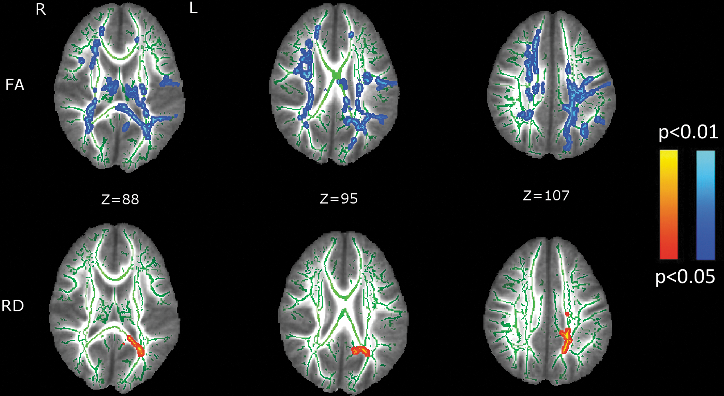
White matter differences between good and poor readers. Significant clusters (p < 0.05, FWE corrected). Blue depicts FA reductions in poor readers. Red depicts RD increase in poor readers. The mean FA skeleton is indicated in green. MNI coordinates. FA, fractional anisotropy; FWE, family-wise error; RD, radial diffusivity. Color images available online at
Discussion
In this study, poor readers at the word level showed significant FA reductions in both hemispheres in numerous white matter tracts when compared with the good readers. Increased RD was restricted to smaller clusters into three tracts of the left hemisphere. No differences were found regarding mean and axial diffusivities.
Recent functional studies have suggested that it is not actually possible to define a dedicated reading network when analyzing complex networks in the resting state (Vogel et al., 2013). Instead, it has been proposed that reading relies on the shared activation of various functional networks (He et al., 2013; Vogel et al., 2013). Recently, it has been demonstrated that scores on a variety of reading tasks (word reading, decoding, and reading fluency) are positively correlated with FA in bilateral brain regions (Feldman et al., 2012; Lebel et al., 2013). In our study, we identify multiple tracts with reduced FA in poor readers, many of them not only in the left hemisphere, but also in tracts in the right hemisphere. These findings show that many tracts can be involved in the word-reading task and also corroborate the involvement of multiple brain regions in this skill. Although reading relies on a wide network, it is well known that a mature reading network is functionally more confined, comprising the frontal, occipitotemporal, and temporoparietal cortices in the left hemisphere of the brain (Turkeltaub et al., 2003). Therefore, FA reduction in the right hemisphere of poor readers could be indicative of a less mature network. Horowitz-Kraus and associates (2014) demonstrated that right hemisphere activation during word recognition has been reported in individuals with reading impairment as a compensatory mechanism, as usually this hemisphere is more commonly associated with reading comprehension.
The observed changes in FA were not driven by changes in other DTI parameters, as the observed differences in RD were restricted to small clusters in the left hemisphere comprising the following tracts: cingulum, corticospinal, and superior longitudinal fasciculus. A reduction in FA without a similar increase in RD could be indicative of microstructural changes more closely related to the spatial coherence of fibers (Johansen-Berg and Behrens, 2014; Underhill et al., 2009), but not to the myelin content. Increased RD and MD has been suggested to have a potential association with decreased myelination, although the biological basis for this correlation remains unknown (Alexander et al., 2007; Engelbrecht et al., 2002; Johansen-Berg and Behrens, 2014; Song et al., 2002). As expected, no differences in AD were found, since decreased AD could be indicative of axonal degeneration in pathological conditions (Kim et al., 2006).
The main limitations of our study are associated with our modest sample size and the small number of gradient encoding directions, which likely led to some degree of inaccuracy when evaluating intergroup differences in measures such as mean and axial diffusivities. On the other hand, our findings demonstrated the existence of white matter alterations in various tracts in both hemispheres of the brain in subjects that performed poorly at word-level reading, which is consistent with recent studies that have shown numerous white matter pathways involved in reading disabilities.
In conclusion, our results contribute to the better understanding of the brain structures that underlies word-level reading in Portuguese speakers, showing the cross validation of results with other languages. Since FA reductions were found in both hemispheres children with poor reading skills at the word level and assuming that children with primary problems in word recognition represent a large proportion of poor readers (Catts et al., 2003), this study contributes to the understanding of neural basis of this disorder.
Footnotes
Acknowledgments
The authors would like to thank CAPES, INCT-MCT/CNPq (National Institute of Developmental Psychiatry for Children and Adolescents) for financial support. L.M.M. would like to thank CAPES for providing a fellowship (17930/12-0), and J.R.S. would like to thank FAPESP for financial support (2013/10498-6).
Author Disclosure Statement
No competing financial interests exist.