
Abstract
Meditation has a versatile nature to affect cognitive functioning of human brain. Recent researches demonstrated its effects on white matter (WM) properties of human brain. In this research, we aim to investigate WM microstructure of corpus callosum (CC) in long-term meditators (LTMs) of rajayoga meditation using diffusion tensor imaging. For this cross-sectional analysis, 22 LTMs and 17 control participants of age ranging from 30 to 50 years were recruited. Results show high fractional anisotropy values with low mean diffusivity in whole as well as different segments of CC in the LTM group. Also the experience of meditation was correlated with WM properties of CC tracts. Findings may suggest rajayoga meditation to bring potential changes in microstructure of CC segments. Further studies are suggested in clinical population to check its validity and efficacy against disorders involving agenesis of WM.
Introduction
M
Functional and anatomical changes have been observed to establish therapeutic effects of meditation practice. Subtle changes at macroscopic and microscopic level (Cao et al., 2010; Fox et al., 2014) were found in subjects undergone meditation training. Structural changes due to practice leading to neuroplasticity were inconsistently observed in meditation studies (Lazar et al. 2005; Luders et al., 2009). Inconsistent changes in both gray and WM were seen in brain wave vibration meditators compared with those in controls. Meditators with a mean 40 months training have significantly increased FA values in cuneus, precuneus, and majority of occipital regions in comparison with controls, although no correlation was found between meditation experience and FA values (Kang et al., 2013).
Pagnoni and Cekic (2007) have assumed that neuroplastic changes caused by meditation can be independent of training. In their experiments, gray matter volume was found to be reduced due to increasing age in control but no interaction was found in case of meditators.
Even short period of integrative body–mind training (IBMT) for 4 weeks (Tang et al., 2010) and 11 h (Tang et al., 2012) increases FA in anterior corona radiata associated with anterior cingulate cortex and genu of corpus callosum (CC). Overall, these changes in microstructure can predict the role of meditation in influencing cognitive processes and information flow between intrahemispherical and interhemispherical regions. WM related to CC lateralizes functional capacity of brain. In discrete meditators, enhanced FA was found along with thicker callosal regions (Pagnoni and Cekic, 2007), also the thickness of callosum was found to be correlated with intelligence (Luders et al., 2007). Role of rajayoga meditation in alteration of the temporal dynamics of brain activity was studied using simultaneous electroencephalogram/functional magnetic resonance imaging. Right middle frontal and left middle temporal gyri were more active in meditators while meditating. Progressively higher duration of the default mode network microstate was positively correlated with meditation experience (Panda et al., 2016). Along with the mentioned studies, very few were focused on long-term meditators (LTMs) and changes in their WM properties due to regular practice.
CC is an important role player in cognitive process and interhemispheric transfer of perceptual, cognitive, learned, and volitional information between the two brain hemispheres (Cao et al., 2010). Being the largest fiber network of human brain and having a crucial role in cognitive and affective processing, study of micro- and macrostructural WM changes in CC was proposed in the study. To ascertain the role of CC, meditation practice was correlated with measures of WM properties, that is, FA and diffusivities. To our knowledge, this is the first study to evaluate microstructural changes due to regular practice of rajayoga meditation by Brahmakumaris. We have hypothesized to find correlation between meditation experience particulars (daily frequency, total years, and total hours in lifetime) with WM properties. Laneri et al. (2015) have also correlated with reported meditation years by subjects, although daily practice hours can determine the real extent of meditation experience that is reported in our study. Daily frequency of meditation in minutes and accumulated lifetime experience were considered to correlate with FA values and other WM properties (Luders et al., 2011).
Materials and Methods
Subjects
The sample consists of 22 LTMs and 17 control subjects (demographics given in Table 1). Mean age of control group was 38.94 years (standard deviation [SD] = 6.35), whereas the mean age of the LTM group was 39.5 years (SD = 4.69). Approximate meditation experience was calculated by multiplying the subjective description of the daily practice duration of meditation and number of years since they were regularly practicing with the same duration. As all of them were devoted practitioners, they follow a certain routine/timetable for similar duration of meditation practice in a day. However, some meditators have exceeded their practice, except the morning and evening routine. Owing to their fixed time chart, it was precise to calculate approximate hours spent for meditation practice. Participants in the LTM group have already spent 10–30 years (mean 18.65 years, SD = 5.42) in practicing this technique after learning it. Approximate number of hours spent in meditation ranged from 5475 to 44,286 h throughout their life (mean = 17,824 h, SD = 9414.93) at the time of recording the MRI data. Controls subjects confirmed to have no prior experience in meditation. Subjects were comparable in education background and have similar number of years of education (mean of LTMs education years = 15.36, SD = 0.79; mean control education years = 15.94, SD = 1.02). All subjects in both groups were free from cardiac, pulmonary, and other nervous system dysfunctions. Also, they were found to be suitable for exposure under magnetic area. Experimental procedures were explained. A written information consent was obtained from each subject. The research protocol was approved by the Institutional Ethical Committee of the Institute of Nuclear Medicine and Allied Science before the experimental recording. The status of nonconsumption of alcohol, smoking, or any therapeutics by the subjects within past 6 months was confirmed.
p < 0.05 considered as significant, χ2 = value of chi square test.
LTMs, long-term meditators; SD, standard deviation.
Rajayoga meditation
Rajayoga refers to the Brahmakmaris school of thought. The practitioners make concerted daily spiritual efforts toward self-development while following a recommended set of lifestyle guidelines. This includes a daily timetable with early morning and evening time meditation, spiritual study, balance of work and service, spiritual outlook in relationships, maintaining celibacy, strict vegetarian diet, self-checking for progress, and early rest (Ramsay et al., 2010). The process of rajayoga is practiced in four stages, that is, initiation, meditation, concentration, and final steps in realization (Ramesh et al., 2013). In the process, while sitting quietly in a comfortable position with eyes open, subjects get involved in positive thinking about a universal force pervading light all over while remembering God and one's eternal form as point of light (Ramsay et al., 2010; Telles and Desiraju, 1993). Positive thought incorporation may reduce preservative behavior and help in facilitation of set switching. It provides cognitive flexibility and also has interaction with higher cognitive processes. Positive thought may modulate cognitive control and provide flexible updating of working memory (Dreisbach, 2006).
MR image acquisition
All MR images were acquired in the axial plane using a 3-Tesla MRI scanner (Megnetom, Skyra, Siemens) with a 20-channel head and neck coil and 25 mT/m actively shielded gradient system. The head was supported and immobilized within the head coil to minimize head movement and gradient noise. The conventional MRI was done before DTI to rule out any structural abnormality using routine T2-weighted turbo spin-echo sequence. DTI data were acquired using a single-shot echo-planar dual spin echo (SE) sequence in 30 directions with ramp sampling. Diffusion-weighted acquisition parameters were b-factor = 0 and 1000 sec/mm2, slice thickness = 3 mm with no interslice space, number of slices = 45, field of view (FOV) = 230 × 230 mm, matrix size = 128 × 128, flip angle = 90°, repetition time (TR) = 8800 msec, echo time (TE) = 95 msec, and echo train length (NEX) = 2.
DTI preprocessing
Eddy current and motion corrections were done in data using the automated image and registration package (Roger Woods, Los Angeles, CA). Removal of scalp to isolate the brain in the collected raw images was done by an automated stripping procedure. Subsequent DTI processing did not require any filtering, as justified by the absence of unprocessed voxels (Saksena et al., 2008).
DTI processing
The distortion-corrected data were then interpolated to attain isotropic voxels and decoded to obtain the tensor field for each voxel. The tensor field data were then diagonalized using the analytical diagonalization method to obtain the eigenvalues (l1, l2, and l3) and the three orthonormal eigenvectors (e1, e2, and e3). The orthogonality of the computed eigenvectors and the correctness of the eigenvalues were checked using random sampling at a number of voxels. The correctness observed was up to an order of 10–17, indicating that no iterative refinement of the computed eigenvalues/vectors was needed. The tensor field data were then used to compute the DTI metrics for each voxel (Saksena et al., 2008).
Segmentation of WM structures
Tensor field data obtained from the processing step were used for segmentation of WM structures. The key idea of this method is to segment the principal eigenvector field into stable voxels having a minimal e1 variation (curvature). Thus, a voxel P (i, j, k) is a member of the stable fiber mass (SFM), if there is a neighboring voxel Q (x, y, z) such that the principal eigenvectors e1's at P and Q point out to each other. Mathematically, it translates to the relation G (F (P)) = P, where F (P) = ROUND (P + e1(P) +0.5u), u = (1, 1, 1), and G (Q) = ROUND (Q−e1(Q) +0.5u), function ROUND representing component-wise “integral part” operation.
The method first generates SFM and then segments volume by coloring voxel P according to the values the components (l, m, n) of the vector joining P and Q take: (±1,0,0), red; (0, ±1,0), green; (0,0,±1), blue; (±1, ±1,0), yellow; (0, ±1, ±1), cyan; (±1,0, ±1), magenta; (±1, ±1, ±1), white. A nonstable voxel is colored gray and a voxel with FA <0.15 remains black. Typical segmented axial, sagittal, and coronal SFM color maps are generated, using that this method narrows down the region of interests (ROIs) selection for the standard tractography to pointing out to a color segment inside a broader ROI through a single mouse click (Rathore et al., 2011).
Diffusion tensor tractography
Different fiber bundles are easily reconstructed by the segmented components using fiber assignment by continuous tracking algorithm (Mori et al., 1999). This reconstruction allows the identification of the coordinates of specific WM fiber bundles and relates them to the anatomy. DTI measures were calculated for the entire fiber. FA threshold of 0.15 was used for fiber tracking.
The ROI for generating CC tracts was selected and subdivided into seven segments (rostrum, genu, rostral body, anterior mid body, posterior mid body, isthmus, and splenium) using the scheme proposed by Witelson (1989) in a manner that approximately represents the CC connections hypothesized across cortical brain regions (de Lacoste et al., 1985). From each subregion of CC, fibers were generated and DTI measures were quantified using in-house developed JAVA-based software (Fig. 1). Genu and rostrum were sampled as a single region after averaging, thus six segments of CC were studied for FA and mean diffusivity (MD) changes in both groups.
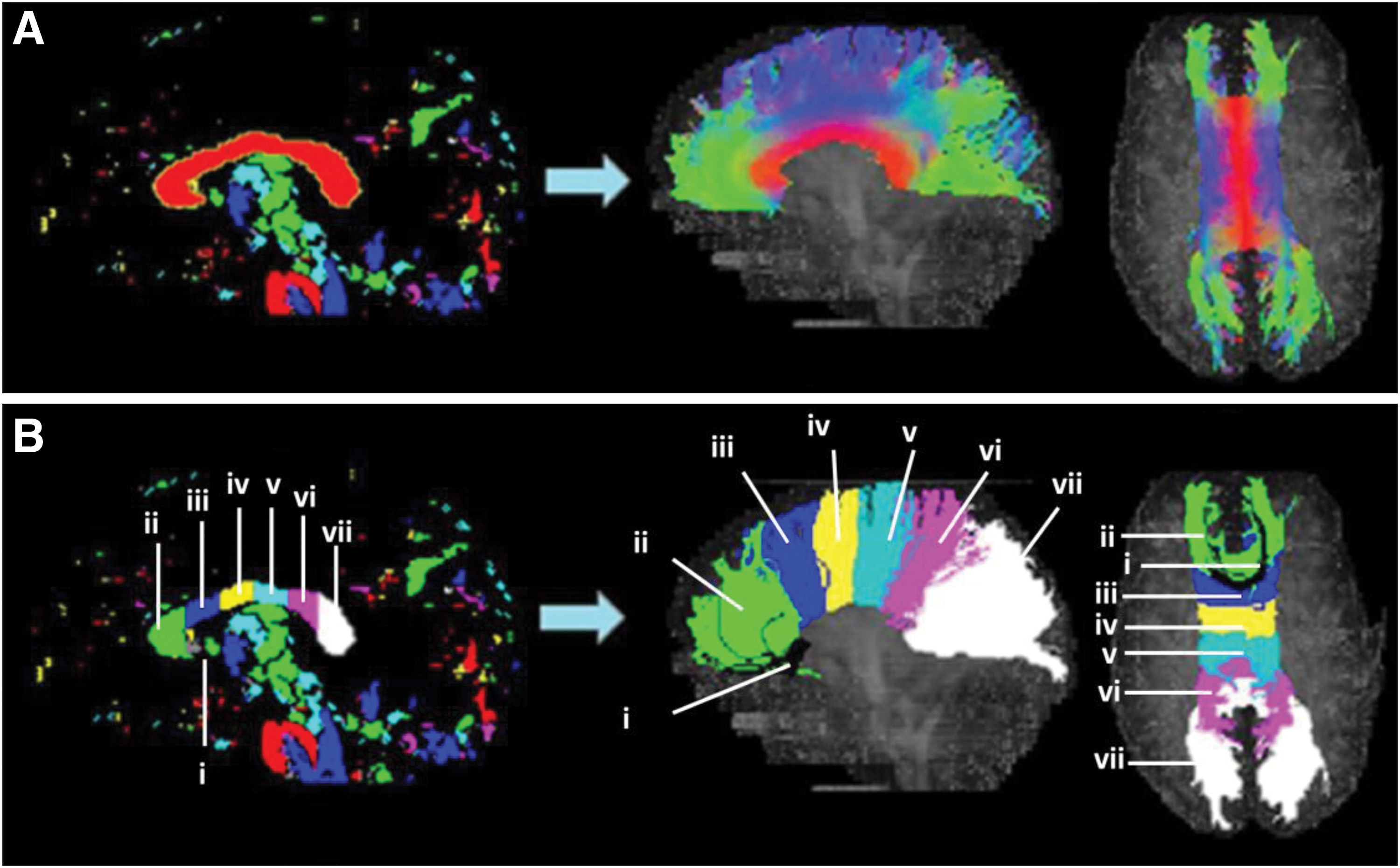
Figure shows a schematic presentation of the fiber tract reconstruction using FACT algorithm.
Statistical analysis
T-statistics with unequal number of observations, by the use of pooled mean square, was performed to study the difference in FA and MD values between controls and LTM group. Correlation analysis was performed between meditation experience variables, age and DTI correlates, and Spearman's rho (rs) was calculated. For multiple comparison, false discovery rate (FDR) corrections were applied using Benjamini Hochberg procedure. Provided p values survived FDR corrections. All statistical analyses were performed using SPSS (version 15.0; SPSS, Inc., Chicago, IL) statistical software.
Results
FA differences between LTM and control
As summarized (Table 2), significantly larger FA values were found in six callosal regions. Also the overall FA value of CC was found to be significantly larger in LTMs. Differences were found prominent in genu, rostral body, anterior mid body, posterior mid body, isthmus, and splenium. Decreased MD values were found in all regions of CC, where FA values were significantly high in LTMs.
p < 0.05 considered as significant.
CC, corpus callosum; RB, rostral body; AMB, anterior mid body; PMB, posterior mid body; FA, fractional anisotropy; MD, mean diffusivity.
Correlation between FA, MD, and meditation experience
Total hours of meditation in a lifetime were negatively correlated with MD of genu (r = −0.55, p = 0.033), rostral body (r = −0.52, p = 0.047), and anterior mid body (r = −0.543, p = 0.036), whereas whole CC was near to statistical significant level (r = −0.51, p = 0.062) Table 3.
Considered significant at p < 0.05.
Inverse relation was also found between daily frequency of meditation (minutes per day) and MD of whole CC (r = −0.53. p = 0.05), MD of genu (r = −0.552, p = 0.03), MD of RB (r = −0.497, p = 0.05), and MD of anterior mid body (AMB; r = −0.451, p = 0.091) although it did not reach statistical significant level. Except genu (r = 0.537, p = 0.039), no significant relation was established between FA values of other segments and meditation experience. FA value of genu was correlated positively with daily minutes of meditation frequency.
Discussion
In this study, highly significant increased FA values and reduced MD were observed in each fiber tract of CC between nonmeditating controls and LTMs.
In the LTM group, daily meditation frequency (minutes/day) was negatively correlated with MD of genu, rostral body, AMB, and whole CC. In addition to this, hours calculated in meditation throughout meditators' lifetime were also negatively correlated with MD of genu, RB, AMB, and whole CC.
DTI can measure cellular factors involved in formation of signals through WM. These factors include morphology of neurons (Beaulieu, 2002) and degree of myelination (Gulani et al., 2001). FA is a highly sensitive biomarker for WM integrity. This can effectively diagnose brain pathology due to demyelination, axonal damage, inflammation, and variations in cellular densities (Alexander et al., 2007). During development, myelination increases with high FA value in heavily myelinated fibers. Heavily myelinated fibers conduct neural signals with high velocity, which results in better cognitive functions (Aboitiz et al., 1992). FA and MD were correlated with cognitive performance (Madden et al., 2012), aging (Grieve et al., 2007), and experience in any functional task (Mackey et al., 2012). It was postulated that meditation experience can lead to increase in brain fibers and their myelination. Learning was found to influence axon diameter that ultimately regulates myelination and survival of axons (Zatorre et al., 2012).
Increased electrical activity at axons of CNS increases myelination. Increased myelination occurs due to increased firing in brain areas involved in training-based learning (Demerens et al., 1996). Consecutively, changes in axon diameter during learning can alter thickness of myelin sheath. Thus, regular meditation over a long term may induce myelinogenesis (Luders, 2014).
During the course of brain maturation, previous studies have reported gradual increase in FA along with decrease in MD in the WM, and this is believed to be associated with the process of myelination. Factors that influence MD include increased binding of water to macromolecules such as myelin, reducing free water content, and the formation of new structural barriers to water diffusion and progressive myelination within WM (Mukherjee and McKinstry, 2006). In our study, increased myelination may have contributed for high FA and lower MD values observed in CC of LTMs in comparison with controls.
Using seven-segment classification scheme of CC on LTMs (meditation practice ranged from 5 to 30 years) with different types of meditation, Luders et al. (2012) have shown high FA in middle region (RB and AMB) of CC in their segment-specific FA analysis but found no changes in anterior (genu and rostrum) and posterior segments (isthmus and splenium). In another longitudinal study, improved FA in anterior and middle segments of CC was also reported after 40 days of IBMT meditation practice compared with baseline study (Tang et al., 2010). Both of the studies orchestrate effects of meditation on CC. Our findings with high FA in entire CC can be explained in light of earlier studies as well as on the basis of type of meditation and duration of meditation practice.
Observation of increased FA in genu and callosal body by Tang et al. (2010) may consequently increase hemispheric transfer between ventral and dorsal anterior cingulate. Strong cingulate connections may indicate training of self-regulatory mechanisms (Posner et al., 2007) exercised by meditation practice.
Anterior mid body inclusively contains crossing fibers for motor, somatosensory, and auditory cortices (Ryberg et al., 2007). In detail, AMB associates dorsomedial prefrontal cortex (dmPFC) and lateral prefrontal cortex of both sides. Their role in conflict monitoring and emotion regulation, respectively, is revealed in previous studies of brain mapping (Catani et al., 2012; Viviani, 2014). However, dmPFC also regulates integration of emotional response during goal selection. Emotion regulation has been a critical trait of meditators that helps them to become resilient (Gray, 2004) and increase performance in any cognitive task.
Splenium carries posterior cortical connections (Aboitiz et al., 1992). Increased FA of splenium in LTMs indicates its role in strengthening the parietal networks involved in interhemispherical processing of attention networks (Niogi et al., 2010; Noudoost et al., 2006). Networks evident to be suitable for meditation accomplishment are strengthened due to training-influenced changes. This reaffirms the effects of concentrative meditation on brain areas mediating orientation of attention, that is, splenium. Distinctive processes of attention network (alertness, orientation, and conflict resolution) may become more efficient by tract-based microstructural changes (Niogi et al., 2010).
In previously attempted neuroimaging studies, microstructure changes in meditators brain at acute or experienced level of meditation were revealed. Meditation is often considered as a state training (Tang and Posner, 2014) and it may involve enhancement of cognitive functions. Training-induced effects were seen in healthy individuals after short period of training (Scholz et al., 2009) or in long-term practice effects (Bengtsson et al., 2005).
Meditation-induced changes were prominently observed in networks wherein degeneration occurred due to any disorder/lesion. In patients of migraine, reduced FA was found in genu, splenium, and body of CC. Similar findings were followed in mind cognitive impairment (Hanyu et al., 1999) or due to aging (Bennett et al., 2010, 2012). Also, these changes in WM can indicate possibility of larger gray matter in middle and superior frontal gyrus (Lazar et al., 2005) as development of more neurons accelerates more number of axonal processes for enhanced interhemispherical information flow.
Smaller splenial regions were found in attention deficit hyperactivity disorder (ADHD) (Semrud-Clikeman et al., 1994). Splenium is a possible/crucial ROI for orienting attention as it is involved in coordination of information across left and right posterior parietal regions (Niogi et al., 2010). Decreased area of entire CC, anterior mid body, and isthmus was found in ADHD children and reduced FA in isthmus was also seen (Cao et al., 2010). In substance-dependent subjects, tracts of rostral body and genu were found to be deteriorated (Moeller et al., 2005), which radiates to presupplementary motor regions, also found to be reduced in autistic population. Autistic children were observed for reduced FA in CC segments (Keary et al., 2009). With increased FA in meditators, regular practice of meditation might be used as potential therapy for autism to increase interhemispheric connections. Size reduction demonstrates poor connectivity between frontal and other brain regions' involvement of frontal CC connections, which results in poor executive functions, problem solving, and social deficits (Paul et al., 2007).
Age-related thinning of CC is more susceptible in anterior and mid part (Persson et al., 2006), which may lead to failed inhibition of hemispheres mediated by CC. Practicing meditation may reduce this thinning in CC fibers so that functional integrity remains exceptionally good in aged meditators. During high demands, attention resource allocation is interchanged between hemispheres. This interhemispheric transfer can provide independent regulation over information exchange by both hemispheres, which results in enhanced attention functioning (Banich, 1998). As the study is cross-sectional, it may not establish the exclusive effects of meditation practice. Rajayoga meditators also follow a routine life style that may have an effect on their metabolism, which can be separately studied.
Conclusion
Observed group differences were not confined to any specific region but to all parts of CC, which may establish the previously assumed theory (Luders et al., 2011) that meditation being a potential exercise has the capability to bring physical changes in brain structures. Meditation can prove to be effective against a patient population suffering with WM degeneration as well as against a training module for performance enhancement. Specifically, rajayoga being an easy-to-learn method can be examined on a larger population with different age groups.
Footnotes
Acknowledgments
This study was supported by project grant (INM 311/4.2) from the DRDO, New Delhi, India. Kanishka Sharma acknowledges the financial assistance as fellowship from the DRDO, New Delhi, India. The authors are grateful to BK Srikant and BK Jain for constant support in availability of LTMs from Brahmakumaris. Ms. Arpita supported in statistical validation of the data. The authors would also like to thank every participant involved in the study.
Disclaimer
Views expressed in the article are the authors' own and not an official position of the institution or funder.
Author Disclosure Statement
No competing financial interests exist.