
Abstract
Introduction:
The mental load caused by simultaneous multitasking can affect visual information processing and reduce its ability. This study investigated the effect of mental load caused by cognitive tasks simultaneously with visual task on the number of active voxels in the visual cortex.
Methods:
This study recruited 22 individuals with a mean age of 24.72 ± 5.47 years. 3-Tesla functional magnetic resonance imaging (fMRI) was used to examine the functions of the visual cortex and amygdala region during three different task conditions: visual task alone, visual task with an auditory n-back task, and visual task with an arithmetic task. The visual stimuli consisted of Gabor patches with a contrast of 55% at spatial frequencies of 0.25, 4, and 9 cycles per degree (cpd). These were presented in three trials of eight blocks with a stimulation time of 12 sec and a rest time of 14 sec.
Results:
Activated brain voxels in the primary, secondary, and associated visual cortex areas were reduced in response to the mental load imposed by the n-back and arithmetic tasks. This reduction was greater for a spatial frequency of 0.25 cpd in the n-back task condition and spatial frequency of 9 cpd in the arithmetic task condition. In addition, the amygdala was stimulated in 2-back task and arithmetic task conditions.
Conclusions:
This study revealed a decline in the number of activated voxels of the visual cortex due to the mental load caused by simultaneous cognitive tasks, confirming the findings of previous psychophysical studies.
Impact statement
Concurrent cognitive task-induced mental load can affect visual information processing and reduce its ability. The number of activated voxels of the visual cortex was investigated in three conditions of visual task alone and visual tasks simultaneously with 2-back task and arithmetic task. Visual stimuli consisted of Gabor patches with 55% contrast at spatial frequencies of 0.25, 4, and 9 cycles per degree (cpd). The results showed that the mental load imposed by n-back and arithmetic task decreased the number of activated voxels in the primary, secondary, and associated visual cortex areas.
Introduction
In numerous clinical and scientific studies, some visual responses, such as visual acuity, contrast sensitivity, and color vision, have been employed to examine visual functions (Male et al., 2022; Silva-Viguera et al., 2023). These properties have been investigated in early visual regions in previous studies (Duncan and Boynton, 2003; Zaretskaya, 2021). However, feedback from the higher-level region, such as attention, task-induced mental load, and distraction, can affect the processing of visual details in the early visual region (Huang and Dobkins, 2005; Lovell et al., 2022; Won et al., 2020).
In the real world, it is common to perform multiple tasks simultaneously, and the effects of these tasks on visual performance may be especially important during tasks that require good vision for safety. It has been discovered that mental load caused by concurrent tasks can impair visual function (Fadardi and Abel, 2018; Fadardi and Abel, 2015). Indeed, mental load refers to the demands that tasks place on a person's limited information-processing resources (Park et al., 2020). Due to this limitation in brain processing capacity (Marois and Ivanoff, 2005; Moray, 1967) or distractor interference (Lavie, 2005), concurrent tasks can reduce performance (Pongpipat et al., 2021).
Visual information is transmitted from the retina to higher processing centers through two main distinct visual systems, the magnocellular and parvocellular pathways, which have different structural and functional characteristics (Murav'eva et al., 2009; Sachs et al., 1971). The magnocellular pathway comprises large ganglion cells, with thick axons and large receptive fields, and is sensitive to low and intermediate spatial frequencies.
The parvocellular pathway has small ganglion cells, with fine axons and small receptive fields, and is sensitive to high spatial frequencies (Murav'eva et al., 2009; Sachs et al., 1971; Yoonessi and Yoonessi, 2011). Some visual functions, such as visual acuity and contrast sensitivity, are evaluated by sinusoidal grating stimuli at different spatial frequencies (Ferwerda, 1998).
Spatial frequency is expressed as the number of cycles per degree (cpd) of the visual angle, where each cycle consists of a pair of bright and dark stripes. Low spatial frequencies correspond to gross information of visual stimuli, such as the shape of an object, and high spatial frequencies correspond to details of visual stimuli, such as edges or small visual elements (Carretié et al., 2007).
Previous research has demonstrated spatial frequency tuning with arousal, emotion, and cognitive load in certain visual functions (Bocanegra and Zeelenberg, 2009; Lee et al., 2014). Involvement of the amygdala, which exhibits spatial frequency-tuned responses in internal states such as arousal (Bocanegra and Zeelenberg, 2009; Nicol et al., 2013) and working memory (Schaefer et al., 2006), may account for such interaction effects (Vuilleumier et al., 2003).
It has been discovered that mental load has a spatial frequency-tuned effect on sweep visual acuity as the cognitive load preferentially impairs the processing of higher spatial frequencies (Mahjoob et al., 2022). Moreover, previous research has indicated that task-induced mental load can impair contrast discrimination, while spatial frequency tuning has not been observed (Mahjoob and Anderson, 2019).
As such, reasonably small spatial frequency-dependent effects may be more readily perceived in the sweep visual evoked potential (VEP) signal than behavioral measures due to susceptibility to large-scale, higher-level distractor effects.
Functional magnetic resonance imaging (fMRI) is used as a noninvasive tool to measure anatomical neural activity through the blood oxygen level-dependent (BOLD) signal, which increases with activation of a brain region (Ghasemi et al., 2021a; Logothetis, 2002). The BOLD signal is generally accepted as an indicator of neural activity as it changes with blood flow to a given region of the brain and blood oxygen levels (Ghasemi et al., 2021b). For example, Blomberg et al. (2021) studied the effects of auditory distraction as a function of working memory load in adults with attention- deficit–hyperactivity disorder (ADHD) using fMRI.
Harrington et al. (2020) investigated abnormalities in context-dependent functional connectivity of parkinsonian working memory centers compared with cognitively normal controls when performing a visual–spatial working memory task under fMRI. According to the reviewed research, no study has studied the effect of mental load on visual cortex responses by task fMRI.
Considering the contradictory results of spatial frequency dependence with mental load in behavioral and VEP measures, this study aimed to investigate the effect of mental load induced by concurrent tasks with visual stimuli on visual cortex responses and spatial frequency dependence using task fMRI. This study hypothesized that the mental load induced by concurrent cognitive tasks reduces activities of the visual cortex, and the amount of this reduction depends on the spatial frequency.
Materials and Methods
In this part, the experiment design, participants, and the process of data processing and statistical analysis are described.
Experimental design
The Psychtoolbox (RRID: SCR 002881; version 3) (Brainard and Vision, 1997) in MATLAB R2012a (MathWorks, Natick, MA; RRID: SCR 001622) was used to generate visual stimuli. They were Gabor patches at a contrast of 55% and mean luminance of 45 cd/m2, randomly appearing in vertical 45° tilted directions to the right or left. A small red circle was used as the fixation point (0.1° visual angle).
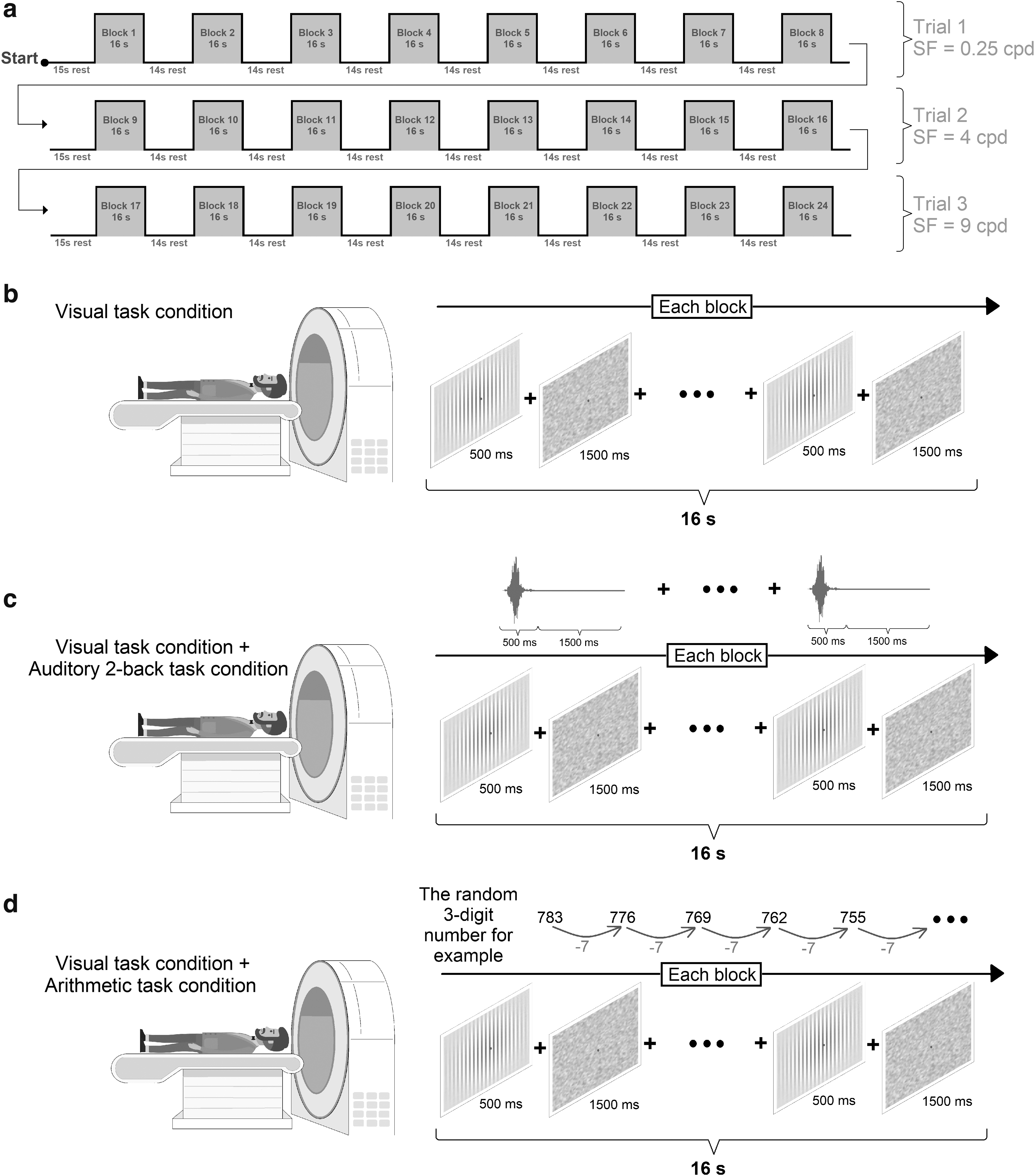
To evaluate spatial frequency dependence with mental load, visual stimuli were presented in three trials of spatial frequencies of 0.25 cpd (low spatial frequency [SF]), 4 cpd (intermediate SF), and 9 cpd (high SF). Each trial consisted of eight blocks, each block consisting of 16 sec of stimulation and 14 sec of rest. The visual task recording based on brain stimulation is depicted in Figure 1. In each excitation block, Gabor patches were presented for 500 msec, with an interval of stimulation using a gray screen for 1500 msec, eight times.
Experimental design and model evaluation.
In each block, the spatial frequency and contrast level were identical; the only variable was the direction. Using a Panasonic PT-EX620 LCD projector with a resolution of 1366 × 768 pixels, visual stimuli were displayed on a 60-cm-diameter screen at the back of the magnetic resonance imaging (MRI) gantry. Visual stimuli were reflected to the eyes of the participants through a 45 degree mirror placed on the coil. The viewing angle was 23.92°.
To attract sufficient attention, all participants had to press the A button on their left hand when they saw patches. Figure 1 depicts how the visual stimulus experiment was recorded when a person was in the gantry. fMRI times were synchronized with the visual task.
An auditory n-back task and an arithmetic task were performed concurrently with visual stimuli to impose a mental load. In the n-back task condition, an auditory 2-back task was synchronized within the visual task during the excitation phase of the visual stimulus. V, T, Q, P, K, G, D, and C are the letters used in the auditory n-back. The letters were presented in a 500-msec window with a 1500-msec response time. Accordingly, each stimulus block had eight letters.
In the 2-back test, participants were required to listen to a series of letters and press the C button on their right when the current letter matched the one from two steps earlier in the sequence.
The fMRI recording was repeated with visual stimuli and a concurrent arithmetic task to impose a task-induced mental load. While the participants looked at the cross-fixation in the center of visual stimuli, the arithmetic task required them to mentally subtract seven from a randomly selected three-digit number without a verbal response (talking would move the head and create artifacts in the images). fMRI was performed for three task conditions in a counterbalanced order.
Participants
The 22 participants (9 females and 13 males) selected were optometry students at the Iran University of Medical Sciences. Their mean age was 24.72 ± 5.47 years, average weight was 68.95 ± 14.06 kg, and mean height was 169.81 ± 9.18 cm. All of the participants were right-handed. Before the experiments were initiated, a general practitioner examined each participant's health.
All participants underwent a comprehensive ocular examination, which included refractive error evaluation using objective and subjective methods, binocular vision tests, direct ophthalmoscopy, and slit lamp examination. The inclusion criterion was uncorrected visual acuity of at least 20/20 in both eyes.
Exclusion criteria included any systemic or ocular disease, any metal in the body, a tattoo on the body, and a fear of empty spaces. Visual task, auditory 2-back task, arithmetic task, and fMRI acquisition instructions were provided, and practice runs for 2-back and arithmetic tasks were conducted before fMRI acquisition.
Participants were positioned within the MRI machine and their heads were secured within the coil using special sponges to prevent complete movement. For participants to hear the 2-back task, headphones were fully inserted into their ears.
MRI data acquisition
All participants were scanned at the National Brain Mapping Laboratory using a Siemens 3-Tesla (Magnetom Prisma Tim+Dot System) scanner with a 12-channel coil and a standard 20 head/neck. All fMRI image volumes were acquired using the T2-weighted echo-planar imaging (EPI) technique with time repetition (TR) = 3000 msec, time echo (TE) = 30 msec, flip angle = 90°, field of view = 200 × 200 mm2, matrix size = 64 × 64, and voxel size of 3 mm3.
To superimpose functional images on anatomical images and conduct statistical analysis based on volumetric high-resolution anatomical MRI, images were captured for each participant, including 3D-SPGR T1-weighted three-dimensional echo gradient sequence images with TR = 2300 msec, TE = 3000 msec, and flip angle = 9° with a 1 mm3 resolution.
MRI specialists visually examined Magnetization Prepared - RApid Gradient Echo (MRPAGE) anatomical images to analyze Magnetic Resonance (MR) images devoid of brain anomalies.
Preprocessing
All experimental images were preprocessed with the specialized FMRI Expert Analysis Tool (Woolrich et al., 2001) in the FMRIB Software Library (FSL 6.0.3, www.fmrib.ox.ac.uk/fsl; RRID: SCR 002823) (Jenkinson et al., 2012; Smith et al., 2004).
The preprocessing procedure included (1) removal of cranial and noncerebral tissue from structural images weighing T1 and functional images weighing T2 (using the FSL BET [brain extraction tool]); (2) removal of the first 12 sec of functional images (first four volumes); (3) motion correction with 6 degrees of freedom (DoF; motion correction linear image recording tool, version 5.0) (Jenkinson et al., 2002); (4) interleaved slice-timing correction; (5) spatial compression with a Gaussian core of Full Width at Half Maximum = 6 mm; (6) Independent Component Analysis melodic data exploration to identify artifacts and assist in activating data; (7) multiplicative mean intensity normalization of volume at every time point; (8) Gaussian-weighted temporal filter (Gaussian-weighted least squares straight line fitting with an inverse of 120.0 sec); (9) coregistration of functional images to self-high resolution utilizing FMRIB's linear image registration (FLIRT) (Esteban et al., 2019); and (10) nonlinear registration of structural T1 images to the MNI space with 12 DoF.
The results of the head correction of functional images in millimeters and radians are presented in Table 1.
A Summary of the Numerical Results of Head Motion Correction in Preprocessing Functional Images in Millimeters and Radians Between Tests
Statistical analyses
In this study, we utilized the FSL fMRI toolbox to conduct a two-level analysis of first-level data (between trials in each experiment) and higher-level (intergroup differences) data using the FEAT model and the general linear modeling (GLM) matrix (Woolrich et al., 2004). The onset of visual stimulation block times coincided with the GLM matrix. In the first-level analyses of blood flow per voxel for intragroup comparison, the difference in mean brain activation was compared with spatial frequencies of 0.25, 4, and 9 cpd using a Z-threshold >1.5 and p value <0.01.
A paired t-test between mean brain activation in the high-level analysis was conducted for the three task conditions (visual task alone, visual task with auditory 2-back task, and visual task with arithmetic task) utilizing the FMRIB (FMRIB's Local Analysis of Mixed Effects [FLAME]) local effects analysis tool (select images with the Z-threshold >1.5 and p value <0.01). For anatomical labeling and localization of brain activation using the Brodmann atlas and Automated Anatomical Labeling, MNI152 results were generated for four areas: the primary visual cortex (V1), secondary visual cortex (V2), associative visual cortex (V3, V4, and V5) (Garey, 1999), and amygdala (Long et al., 2018).
Figure 2 presents a schematic representation of our study's pipeline.

Overall procedures of the study. In the first step, the visual task alone, visual task with auditory 2-back task, and visual task with arithmetic task conditions were designed for fMRI recording. In the second step, fMRI was performed for 22 participants using a Siemens 3-Tesla machine. In the third step, necessary preprocessing was done using FSL software. In the fourth step, using statistical methods, differences in brain regions were noted for comparison between task conditions. fMRI, functional magnetic resonance imaging; FSL, FMRIB Software Library.
Results
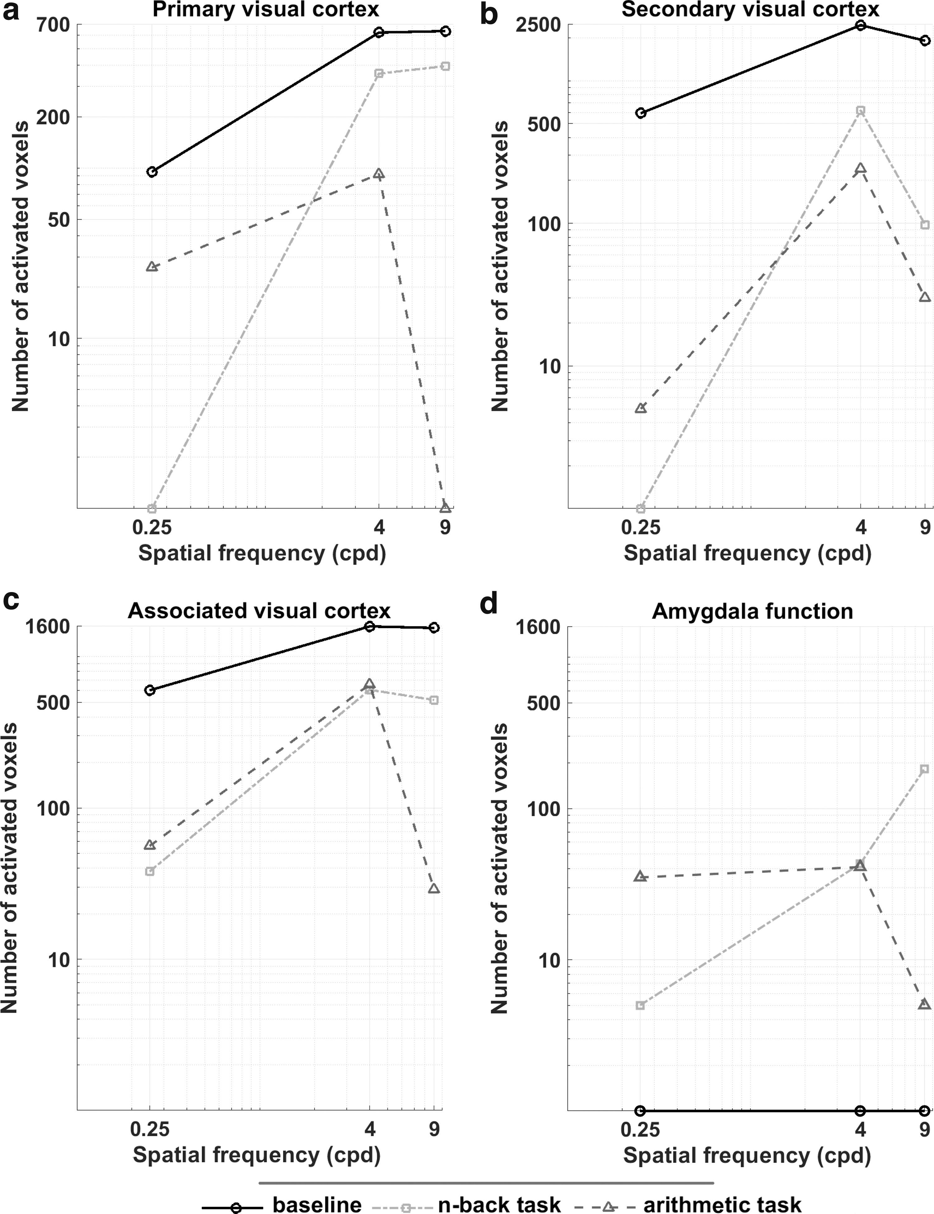
Significant differences in the visual cortex and amygdala regions were obtained by analyzing GLM in BOLD signals for the effects of spatial frequencies in each task condition based on the differences in brain activity in each task condition. Figure 3 depicts the number of activated voxels in the primary, secondary, and associated visual cortex regions and amygdala for each task condition. The number of activated voxels differed significantly across spatial frequencies in the three task conditions.
The number of voxels activated in the visual cortex regions and amygdala region for different tasks. The number of activated voxels in different regions of the
In the visual task alone condition, the number of activated voxels in the primary visual cortex increased significantly as spatial frequencies increased. The number of activated voxels increased when the spatial frequency was increased from 0.25 to 4 cpd, but reduced slightly when the spatial frequency was increased from 4 to 9 cpd, in the secondary and associated visual cortex regions in the three task conditions as well as in the primary visual cortex in the arithmetic task condition.
Figure 4 depicts the cluster activity in visual cortex regions for the visual task condition, n-back task condition, and arithmetic task condition. Two cognitive mental load tasks significantly decreased brain activity in the visual cortex compared with the visual task alone (p = 0.05 and Z-threshold >1.5). Changes in fMRI signal intensity with mental load were significantly different at spatial frequencies of 0.25, 4, and 9 cpd.

The degree of activation of brain areas in different tasks. The cluster activity in visual cortex areas for the visual task condition, n-back task condition, and arithmetic task condition at spatial frequencies of 0.25, 4, and 9 cpd (p < 0.05 and Z-threshold >1.5).
Significantly less brain activity was observed at spatial frequencies of 0.25 cpd compared with 4 and 9 cpd when performing the n-back task compared with when performing the visual task alone (p < 0.05 and Z-threshold >1.5). However, in the arithmetic task condition, the reduction was greater at the spatial frequency of 9 cpd than at spatial frequencies of 0.25 and 4 cpd. As can be seen in Figure 4, there was no stimulation in the amygdala in the baseline condition (visual task alone) and the number of activated voxels in this area was almost zero, but the amygdala was stimulated in the n-back task and arithmetic task conditions.
Discussion and Conclusions
This study investigated the effect of mental load through simultaneous arithmetic task and auditory n-back task with visual stimuli at different spatial frequencies on the visual cortex function in primary, secondary, and associated regions. Our results demonstrated a decrease in activated voxels with mental load. This decrease was more pronounced in the auditory n-back test at the spatial frequency of 0.25 cpd than at spatial frequencies of 4 and 9 cpd.
Moreover, the reduction was greatest at the spatial frequency of 9 cpd in the arithmetic task condition. The highest number of activated voxels was found for the spatial frequency of 4 cpd (Fig. 3), consistent with previous psychophysical research. Previous research has demonstrated that contrast sensitivity is greatest at intermediate spatial frequencies (Lovegrove et al., 1980).
Mental load represents the mental effort caused by concurrent tasks in the context of the brain's information-processing limitations (Park et al., 2020). This study revealed a decrease in the number of activated voxels in the visual cortex in response to the simultaneous task-induced mental load, which may be attributable to the brain's reduced capacity to process visual stimuli caused by the cognitive mental load. It is probably that the brain downregulates its visual processing, but upregulates other processing in other brain regions to integrate all concurrent tasks.
These findings align with previous psychophysical and electrophysiological investigations of vision (Mahjoob and Anderson, 2019; Mahjoob et al., 2022). A decrease in contrast discrimination and visual acuity with the auditory n-back test was reported due to limited brain information-processing capacity and distraction (Mahjoob and Anderson, 2019). Since mental load diminishes visual performance, regulating attention processes during simultaneous or dual tasks is necessary.
A previous study on the effect of mental load on attended visual stimuli revealed that the interaction between attention and mental load was not statistically significant and that the effect of mental load on attended and nonattended visual stimuli was statistically equivalent (Mahjoob and Anderson, 2023). Therefore, assigning attention by placing peripheral cues in visual stimuli to improve visual performance cannot compensate for the limitation of visual information processing caused by the mental load.
Attention, working memory, and executive functions are required for mental arithmetic calculations such as subtraction and addition, which were performed in this study. Besides, studies have demonstrated that mathematical calculations are associated with frontal and parietal involvement as well as increased amygdala activity (Hugdahl et al., 2004; Rosenkranz et al., 2022). The n-back task also involves the use of attention, working memory, and decision-making.
Previous research has revealed that the amygdala is stimulated in internal situations such as arousal and working memory (Schaefer et al., 2006). In line with previous studies, our results showed that the amygdala is stimulated in arithmetic and n-back task conditions, while no stimulation occurred in this area in the baseline condition (visual task alone).
The amygdala influences both parvocellular and magnocellular pathways (Bocanegra and Zeelenberg, 2009; Nicol et al., 2013). Due to the different effects of the amygdala on these two visual pathways, an increase in contrast sensitivity at high spatial frequencies and a decrease at low spatial frequencies have been observed during arousal (Lee et al., 2014).
Our previous psychophysical research has not reported interference between spatial frequency and mental load caused by an auditory n-back task (Mahjoob and Anderson, 2019). In visual acuity with sweep VEP, however, it was discovered that the reduction of VEP amplitudes after the concurrent cognitive task was greater at high spatial frequencies than at low spatial frequencies (Mahjoob et al., 2022). In addition, in this study, the number of activated voxels in the visual cortex decreased more at low spatial frequencies for the n-back task and high spatial frequencies for the arithmetic task.
Since involvement of the amygdala occurred in both of our mental load tasks, the reason for the different effects of mental load on visual cortex activity at various spatial frequencies could be due to the dual effects of the amygdala on the parvocellular and magnocellular pathways. However, more research is needed to investigate the functional connectivity between the amygdala and the visual cortex in the context of mental load.
The results of this study revealed a reduction in the number of activated voxels in the visual cortex in response to mental load caused by concurrent cognitive tasks, confirming previous psychophysical studies.
Ethical Approval
This clinical study was conducted according to the protocol and in compliance with Good Clinical Practice (GCP) and the Declaration of Helsinki (version 2008), with approval (ethics code: IR.MUMS.REC.1395.366) from Mashhad University of Medical Sciences (Ethics Committee) in Iran and other applicable regulatory requirements.
Informed Consent
Informed consent was obtained from all individual participants included in this study.
Data and Code Availability
The datasets analyzed during the current study are available from the corresponding author upon reasonable request. The statistical analysis and modeling code have been provided as Supplementary Software. fMRI data analyses were performed in FSL, v6.0.
Authors' Contributions
All authors listed have made a substantial, direct, and intellectual contribution to the work and approved it for publication.
Footnotes
Author Disclosure Statement
No competing financial interests exist.
Funding Information
No funding was received for this study.