
Abstract
Abrin is a heterodimeric toxin present in the seeds of the Abrus precatorius plant. The easily obtainable seeds can yield a highly toxic product that can be used in various types of biocrimes and terrorism-related activities, including “white-powder” letters. Although the vast majority of these threats are hoaxes, the lack of rapid and reliable detection assays for abrin, such as lateral flow assays (LFAs), can be an impediment to accurate and rapid hazard assessment. One of the complicating factors associated with LFAs is the use of antibodies of poor affinity and specificity that cross-react with near neighbors or that bind to plant lectins, which are capable of nonspecifically cross-linking the capture and detector antibodies. Because of the critical need to promote public safety and public health, we conducted a comprehensive laboratory evaluation of a commercial LFA for the rapid detection of abrin. This study was conducted using comprehensive inclusivity and exclusivity panels of abrin and near-neighbor plant materials, along with panels of lectins, related proteins, white powders, and environmental background material, to determine the sensitivity, specificity, limit of detection, dynamic range, and repeatability of the assay for the specific intended use of evaluating suspicious white powders and environmental samples for the presumptive presence of abrin.
Abrin is a heterodimeric toxin present in the seeds of the Abrus precatorius plant. The easily obtainable seeds can yield a highly toxic product that can be used in various types of terrorism-related activities, including “white-powder” letters. Although the vast majority of these threats are hoaxes, the lack of rapid and reliable detection assays for abrin, such as lateral flow assays (LFAs), can be an impediment to accurate and rapid hazard assessment. The authors conducted a comprehensive laboratory evaluation of a commercial LFA for the rapid detection of abrin.
A
Abrin is a heterodimeric type II ribosome-inactivating protein (RIP) consisting of an A-chain of 251 amino acids (Mr 30,000 Dalton) linked by a single disulfide bridge to a B-chain consisting of 268 amino acids. The B-chain (Mr 33,000 Dalton) is a lectin that binds to cell surface glycoproteins containing D(+)-galactopyranose moieties. The heterodimer is transported intact across the cell membrane by receptor-mediated endocytosis and is transported to the endoplasmic reticulum by the retrograde pathway.1,5-8 The reduction of the intersubunit disulfide bond is essential for the translocation of the A-chain to the cytosol, where it can interact with the ribosome. The A-chain is an N-glycosidase that cleaves the C-N bond of adenine at position 4324 in eukaryotic 28S ribosomal RNA. 9 This prevents the formation of a critical stem-loop configuration, thereby preventing ribosomes from binding to elongation factor (EF) 1 and 2, resulting in the inhibition of protein synthesis leading to apoptosis and cell death.3,4,10-12 The catalytic function of the abrin A chain may stem from highly conserved sequences clustered around Tyrosine-113, Glutamic Acid-164, Arginine-167, and Tryptophan-198. A single substitution of Glutamine for Glutamic Acid almost completely eliminates catalytic activity and results in a 1,600-fold increase in the infectious dose (ID50). 13 Hedge et al reported that abrin occurs in 3 isoforms, designated I, II, and III, 14 which can be separated by lactamyl-Sepharose affinity chromatography followed by gel filtration and DEAE-Sephacel chromatography; other researchers have reported 4 isoforms.12,15 The amino acid sequence of the isoforms varies slightly, and each has the same mechanism of action, although activity levels differ.
A. precatorius seeds also contain a related 120 kDa heterotetrameric protein, agglutinin (APA-1), comprised of 2 A chains and 2 B chains stabilized by hydrophilic and hydrophobic forces. 16 The LD50 of abrin in humans via injection is approximately 20 μg/kg body mass and via inhalation 3.3 μg/kg body mass. APA-1 has a much lower toxicity, with an LD50 of 5 mg/kg body mass (i.p.).14,17,18 APA-1 shares a high degree of sequence homology with abrin, with 44 (8%) similar amino acids and 382 (69.8%) invariant amino acids. In the abrin A-chain, an asparagine at position 200 is strongly associated with substrate binding. The reduced toxicity of APA-1 appears to be associated with a proline residue (proline-199) present at the corresponding position in APA-1.17,18 The presence of proline sterically hinders the formation of a critical hydrogen bond with glycine-4323 in 28S rRNA. Studies have demonstrated that using in vitro mutagenesis to change asparagine-200 in the A-chain of abrin to proline-200 in abrin resulted in a greater reduction in catalytic activity than with any other single mutation. 9
RIPs have been reported in up to 100 plant species belonging to 20 families, including Caprifoliaceae, Cucurbitaceae, Poacea, Euphoribiaceae (to which Ricinus communis belongs), and Leguminosae, the family to which the genus Abrus belongs. 13 It has been hypothesized that when plants are wounded, perhaps due to a virus infection, RIPs are released as a defense mechanism to prevent viral replication by inactivating protein synthesis and resulting in apoptosis. However, this defense mechanism may not be available to all plants, as only a limited number have been shown to produce RIPs. 19 RIPs have been investigated as potential immunotoxins. 6 They have also been tested as chimeras in which the A-chain has been conjugated to various ligands including growth factors or antibody fragments (or recombinantly expressed as fusion proteins) so they may be targeted to specific cells for selective elimination. Abrin in particular has been used in medicinal practices throughout the world, including as a treatment for malaria in Cambodia and Nigeria and as an emetic in the West Indies. 20 Both abrin and APA-1, while having disparate toxicities, have been found to have similar therapeutic indices. 18
Abrin can be introduced by several routes. Patients with abrin intoxication can present with gastrointestinal hemorrhaging following ingestion, muscle necrosis following injection, or acute pulmonary disease following inhalation. While most cases of abrin intoxication involve children who have ingested seeds of A. precatorius, the consequences of seed ingestion are generally not severe owing to the hard shell that protects the toxin. 16 If, however, the integrity of the shell has been compromised, abrin can affect the digestive tract even as digestive enzymes act to break down the toxin. Symptoms following ingestion can include nausea, vomiting, and diarrhea, which in more severe cases can cause dehydration and death. In some cases, symptoms do not occur until 1 to 3 days following ingestion, while the clinical course of toxicity can last up to 10 days. There is no specific treatment for ingested abrin. Current treatment involves inducing emesis, gastric lavage, activated charcoal, and whole bowel irrigation. 4
First responders are often faced with unknown white powders and are called on to test for the presence of potential biological agents of concern in the field. The purpose of this study was to conduct a multicenter evaluation of the sensitivity, specificity, reproducibility, and limitations of a lateral flow assay (LFA) for abrin (Abrin BioThreat Alert® Test Strip, Tetracore®, Inc., Rockville, MD) that could be used in the field to screen for abrin. To ensure an unbiased evaluation, blinded samples were prepared by a third party and provided to the testing sites to support data generation.
The goal of this study was to determine whether the Abrin LFA can provide reliable results, so that appropriate and effective decisions can be made by first responders and public health officials to protect the public, avoid unnecessary disruption of civil society, and reduce economic impacts. The primary outcomes of this evaluation were an understanding of assay performance, including false-positives (assay is positive but the analyte is not present), false-negatives (assay is negative but the analyte is present at amounts at or above the limit of detection [LOD]), and assay repeatability (assay works consistently and reproducibly) results.
Materials and Methods
Abrin BioThreat Alert® Test Strips and Rapid BioAlert® Readers were obtained from Tetracore, Inc. (Rockville, MD). The performance of the Abrin LFA and reader was evaluated using a single lot of strips at 5 test sites: the Centers for Disease Control and Prevention (CDC); the Food and Drug Administration, Center for Food Safety and Applied Nutrition (FDA-CFSAN); the Massachusetts Department of Public Health (MDPH); the Texas Department of State Health Services (TDSHS); and the Virginia Division of Consolidated Laboratory Services (DCLS). Samples for analysis were prepared at Lawrence Livermore National Laboratory (LLNL, Livermore, CA), and OmniArray Biotechnology (Rockville, MD), coded, and shipped (with cold packs) to the participating laboratories, where they were stored at 4°C until use, then diluted and analyzed according to a standard protocol provided by Tetracore. Abrin LFA results were read both visually and with the BioAlert® Reader according to directions provided by the manufacturer—that is, between 15 and 30 min after adding the sample (150 μL) to the lateral flow strip. The BioThreat Alert® Reader measures the ratio of incident light and absorbing light intensity on the surface of the lateral flow strip. The resulting ratio, converted into a BioThreat Alert® Reader value by the instrument, is expressed without units. The manufacturer's cutoff for the Abrin LFA was determined as follows: 50 Abrin LFA strips were tested with negative controls (BTA buffer). These were then compared against the readings for abrin and near neighbors at various concentrations. The cutoff is determined to be the value that indicates a positive for abrin at least to the lowest concentration that is positive by visual inspection. Samples with readings of ≤125 were considered negative, and test strips on which the control line failed to develop were noted and discarded. The study consisted of 7 phases, which are enumerated below. At least 5 negative controls (BTA buffer) and 2 positive control (purified abrin, 1 μg/mL) LFAs were run at each test site during each phase of the study.
Crude Extracts
Seeds of A. precatorius were weighed and placed in a biological safety cabinet. Using proper precautions, the seed coats were cracked with pliers and then the seeds were soaked in PBS for 6 to 12 hours at 4°C in the dark (1.7 mL/g of seeds). During this time, the seeds expanded and absorbed all of the liquid. The expanded seeds were transferred to a mortar and pestle. After grinding, the material was transferred to a 50 mL conical centrifuge tube and PBST added in the ratio of 5 mL/g of seed. The tube was vortexed and then incubated on a rocker platform for 16 to 19 hours at 4°C in the dark. After this incubation period, the tube was centrifuged for 4 minutes at 3,000 rpm (room temperature) in a Sorvall GSA rotor. The middle aqueous layer was removed, aliquoted into cryovials, and stored at −80°C until used.
Purified Abrin
A modification of the method developed by Hedge et al 14 was used to prepare abrin for this study. Briefly, crude extract prepared from the Banana Tree Red cultivar of A. precatorius was centrifuged at 13,000 rpm for 5 minutes and the supernatant removed for purification. The sample was exchanged into 10 mM potassium phosphate buffer (pH 7.0) using a Millipore Amicon Ultra spin column (10,000 MWCO). The sample was then loaded onto a Tricorn 10/50 glass column packed with lactamyl-Sepharose 4-B and purified using a GE Healthcare AKTA Purifier. After washing the column, abrin was eluted with 50 mM lactose and concentrated with a Millpore Amicon Ultra spin column (10,000 MWCO). Samples of abrin were separated on a NuPAGE Bis-Tris 4-12% precast gel (Invitrogen, Carlsbad, CA) following manufacturer's procedure, and then stained with Pierce Imperial stain (Thermo Scientific, Rockford, IL). Visible bands were excised, washed 3 times with 50% acetonitrile/10 mM ammonium bicarbonate, and then subjected to in-gel tryptic digestion overnight with 0.5 μg trypsin in 10 mM ammonium bicarbonate. The digested peptides were extracted with 60% acetonitrile/10 mM ammonium bicarbonate, dried by speed vad rotary evaporation, and reconstituted in 4 μL 60% formic acid and 16 μL 2% acetonitrile/0.1% formic acid/0.05% TFA. Peptides were analyzed by nano-LC-MS/MS using Dionex UltiMate 3000 and Bruker MaXis mass spectrometer as described elsewhere. 21 The resulting data were entered into NCBInr datAPA-1se using Bruker ProteinScape software and Mascot search engine (data not shown).
Plant Sources
Seeds from 11 A. precatorius cultivars were obtained from the following sources. Banana Tree Brown and Banana Tree Red were obtained from the Banana Tree Seed Company (Easton, PA). EG 13625 24840 and EG 13625 24530 were obtained from B & T World Seeds in Aigues-Vives, France, and provided by E. A. E. Garber, FDA. Seeds with the following designations were obtained from the Desert Legume Project (Tuscon, AZ): DES, AUS, CBFL, MelFL, and ZMB. Seeds designated ONA were purchased from Onalee's Seeds (Madeira Beach, FL). Seeds designated TRO were purchased from Tropilab, Inc. (St. Petersburg, FL). Seeds from near neighbors of A. precatorius and other plants were obtained from various sources: Abrus laevigatus, Adriana quadripartita, Bryonia dioica, Canavalia gladiata, Canavalia rosea, Canavalia virosa, Cinnamomum camphora, Cucurbita moschata, Dianthus caryophyllus, Jubernardia globifera, Luffa acutangula, Luffa cylindrical (aegyptica), Lychnis chalcedonica, Momordica charantia, Phytolacca americana, Phytolacca dioica, Plukenetia volubilis, Sambucus ebulus, Sambucus nigra, Saponaria officinalis, Senna occidentalis, Trewia nudiflora, and Trichosanthes kirilowii were obtained from B & T World Seeds. Fatsia japonica and Saponaria officinalis were obtained from Plant World Seeds in Newton Abbott, Devon, United Kingdom. Macaranga grandifolia was obtained from Top Tropicals in Ft. Myers, Florida. Seeds of Mallotus philippensis and Mercurialis annua were obtained from the US Department of Agriculture (USDA), Agriculture Research Services (ARS), in Pullman, Washington. Leaves from Acalypha rhomboidea and Manihot esculenta were obtained from the Botanical Gardens in Washington, DC. Abrus schimperi subs. Africanus, Galactia striata, and Galactia wrightii were obtained from the Desert Legume Project in Tucson, Arizona. Iris hollandica bulbs were purchased from American Meadows in Williston, Vermont.
Lectins
The following lectins were purchased from E Y Laboratories, Inc. (San Mateo, CA): A. precatorius, Agaricus bisporus, Aleuria aurantia, Allium sativum, Amaranthus caudatus, Arachis hypogaea, Autocarpus integrifolia, Arum maculatum, Bauhinia purpurea, Bryonia dioica, Canavalia ensiformis, Caragana arborescens, Cicer arietinum, Colchicum autumnale, Cytisus scoparius, Datura stramonium, Dolichos biflorus, Euonymuse europaeus, Galanthus nivalis, Glycine max, Griffonia (Bandeiraea) simplicifolia Lectin I, G. B. simplicifolia Lectin II, G. simplicifolia, Hippeastrum hybrid, Iberis armara, Iris hybrid, Jacalin, Laburnum alpinum, Lens culinaris, Lotus tetragonolobus, Lycopersicon esculenntum, Maackiaa amurensis Lectin I, Maclura pomifera, Mangifera indica, Narcissus pseudonarcissus, Peanut agglutinin, Phaseolus lunatus, Phaseolus vulgaris, Phaseolus vulgaris Agglutinin, Phaseolus vulgaris Erythroagglutinin, Phytolacca Americana, Pisum sativum, Psophocarpus tetragonolobus, P. tetragonolobus Lectin I, P. tetragonolobus Lectin II, Robinia pseudoacacia, Salvia sclarea, Sambucus nigra Agglutinin I, Sambucus nigra Agglutinin II, Solanum tuberosum, Sophora japonica, Soybean Agglutinin, Trichosanthes kirilowii, Trifolium repens, Tulipa sp., Ulex europaeus Agglutinin I, Ulex europaeus Agglutinin II, Urtica dioica, Vicia fava, V. graminea, V. villosa, V. radiata, Wheat Germ Agglutinin, Wisteria floribunda Agglutinin, and W. floribunda Lectin.
Toxins/Proteins
Purified ricin (RCA60), ricin A chain, ricin B chain, and ricin agglutinin (RCA120) were purchased from Vector Laboratories, Burlingame, CA. Formalin-inactivated ricin toxoid, formalin-inactivated abrin toxoid, and purified Shiga toxin were obtained from Toxin Technologies, Inc., Sarasota, FL. Deglycosylated ricin A chain and the vaccine candidate rRTA1-33/44-198 were obtained from Martha Hale (US Army Medical Institute for Infectious Diseases [USAMRIID], Ft. Detrick, Frederick, MD). RiVax, a candidate ricin vaccine consisting of a recombinant ricin A chain containing residues 1-267 with 2 substitutions, V76M and Y80A, to reduce toxicity, 22 was obtained from P. Legler, US Navy.
White Powders
Powdered milk, powdered coffee creamer, powdered sugar, talcum powder, baking powder, cornstarch, and popcorn salt were purchased from Raley's Grocery Store, Pleasanton, CA. Rice flour was purchased from Ranch 99, Pleasanton, CA. Wheat flour and soy flour were purchased from Van's, Livermore, CA. Baking soda, baby powder, chalk dust, and powdered infant formula (iron fortified and low iron formulation) were purchased from Target Stores, Livermore, CA. Powdered toothpaste was purchased from Walmart Pharmacy, Livermore, CA. Brewer's yeast was obtained from GNC Stores, Livermore, CA. Drywall dust was obtained from Home Depot, Livermore, CA. Gamma aminobutyric acid, L-glutamic acid, kaolin, chitin, chitosan, magnesium sulphate, and boric acid were purchased from Sigma-Aldrich Corp, St. Louis, MO. Bacillus thuringiensis (Dipel) powder was purchased from Summerwinds Nursery, Palo Alto, CA.
BioWatch Filters
Thirty BioWatch filters, after being subjected to environmental aerosol collection for 24 hours, were extracted by shaking with PBST and the extracts pooled. The protein concentration of the pooled extract was adjusted to 200 ng/μL with PBSTB prior to testing.
Protein Determination
Protein concentrations were determined using Bradford Assay Reagent (Pierce Chemical Company, Rockford, IL) using a standard curve prepared with bovine serum albumin (EM Sciences, Cole-Parmer, Vernon Hills, IL).
Phase 1: Repeatability Study
The repeatability of the Abrin LFA was determined using purified abrin provided by Dr. Jan Pohl at CDC. Stock solutions of abrin were prepared at concentrations of 250 ng/mL and 500 ng/mL in PBSTB—phosphate-buffered saline (Life Technologies #20012-050) containing 0.1% (v/v) Tween-20 (Promega #H5152) and 0.1% (w/v) bovine serum albumin (EM Sciences #EM2930)—and shipped to the testing sites, where they were centrifuged for 3 minutes at 3,000 rpm in a microfuge. The stock solutions were diluted 10-fold using BioThreat Alert® (BTA) buffer provided by Tetracore®, Inc., and after mixing, 150 μL of each diluted toxin solution was added to lateral flow strips (final concentration 25 ng/mL and 50 ng/mL [3.75 ng/test and 7.5 ng/test], respectively). Twelve replicates of each concentration were tested by 2 operators per site (24 total replicates/site) at each of the 5 sites, for a total of 120 LFAs tested at each concentration.
Phase 2: Inclusivity Panel
In order to determine whether this assay could detect abrin in cultivars from diverse geographic locations, crude extracts of 11 A. precatorius cultivars (Table 1) were prepared as described below. The extracts were diluted in PBSTB to a final protein concentration of 20 μg/mL and then shipped to the test sites. Before testing, the sample tubes were vortexed and centrifuged for 3 minutes at 3,000 rpm in a microfuge. The extracts were subsequently diluted 2-fold with BTA buffer and, after mixing, a 150 μL volume was added to each test strip. The final protein concentration of the diluted extract was 10 μg extract protein/mL (1.5 μg/test). Each cultivar was tested once per site.
Abrus precatorius Cultivars (n=11) Used for Testing
Reference cultivar.
Phase 3: Near Neighbor Panel
Crude extracts were prepared from the seeds or leaves of 35 near neighbors of A. precatorius (see Plant Sources above). The extracts were diluted in PBSTB to a protein concentration of 20 μg/mL and shipped to the test sites, where they were subsequently vortexed and centrifuged for 3 minutes at 3,000 rpm. The supernatant was then diluted 2-fold in BTA buffer to a final concentration of 10 μg protein/mL. A 150 μL volume containing 1.5 μg extract protein was added to each test strip. Each near neighbor was tested once per site.
Phase 4: Lectin Panel
Stock solutions of 65 lectins (see below) were prepared in PBSTB to yield a concentration of 10 μg lectin/mL and shipped to test sites, where they were subsequently vortexed and centrifuged for 3 minutes at 3,000 rpm, diluted 2-fold in BTA buffer, and a 150 μL volume containing 750 ng of lectin was added to each LFA. Each lectin was tested once per site.
Phase 5: Toxin/Protein Panel
In order to determine the specificity of the abrin LFA, solutions of abrin toxoid, abrin agglutinin (APA-1), Shiga toxin, ricin (RCA60), ricin agglutinin (RCA120), ricin A chain, ricin B chain, ricin toxoid, deglycosylated ricin A chain, and 2 ricin vaccine candidates were prepared at 2 μg/mL in PBSTB. The protein solutions were shipped to the test sites, where they were vortexed and centrifuged for 3 minutes at 3,000 rpm and then diluted 2-fold in BTA buffer. After mixing, a 150 μL volume containing 150 ng of protein was added to each LFA. Each sample was tested once per site.
Phase 6a: White Powder Panel
A stakeholder panel consisting of representatives from state public health laboratories, CDC, the Department of Defense (DoD), the Environmental Protection Agency (EPA), the Federal Bureau of Investigation (FBI), and other federal entities created a list of the 26 white powders (see White Powders above) most commonly encountered by first responders and the CDC Laboratory Response Network (LRN) reference laboratories. These materials were evaluated for their ability to affect the performance of the assay. Five mg of each of the 26 white powders (see above) were sent to the test sites. After the addition of 500 μL of BTA buffer (final concentration=10 mg/mL), each tube was vortexed for 10 seconds. The suspension was allowed to settle for at least 5 minutes, and then 150 μL of the supernatant was removed and added to the Abrin LFA. Each powder was tested once per site.
Phase 6b: White Powders Spiked with A. precatorius Extract
The 26 most commonly encountered white powders were also evaluated for their ability to interfere with or inhibit the detection of abrin by the assay. Five milligrams of each of the 26 white powders along with a tube of crude extract of the Banana Tree Red A. precatorius cultivar (prepared in PBSTB at a concentration of 100 μg extract protein/mL) were shipped to each test site. Approximately 2.4% of protein extracted from whole abrus seeds is lectin (ie, abrin+APA-1). 23 Operators added 450 μL BTA buffer and 50 μL of the Banana Tree Red extract to each tube containing powder (final white powder concentration=10 mg/mL). After vortexing for 10 seconds, the suspension was allowed to settle for 5 minutes, after which 150 μL of the supernatant was tested on the Abrin LFA. Each spiked powder was tested once per site.
Phase 7a: BioWatch Filter Extract
BioWatch filter extracts collected from filters from BioWatch collectors operating in several US cities and containing 200 ng protein/μL were shipped to each test site, where they were vortexed and centrifuged for 3 minutes at 3,000 rpm. Operators added 250 μL BTA buffer to 250 μL extract (2-fold dilution to 100 ng protein/μL). After mixing for 10 seconds, the suspension was allowed to settle for at least 5 minutes, followed by removal of 150 μL of supernatant (15 μg total protein) for testing using the Abrin LFA. The filter extract was tested once at each test site.
Phase 7b: BioWatch Filter Extract Spiked with A. precatorius Extract
A 250 μL volume of BTA buffer and 55 μL of the diluted Banana Tree Red extract (see above) were added to a second tube containing 250 μL BioWatch filter extract (final concentration 100 ng filter extract protein/μL). After mixing for 10 seconds, the suspension was allowed to settle for at least 5 minutes, followed by removal of 150 μL supernatant containing 1.5 μg of Banana Tree Red protein for testing on the Abrin LFA. The spiked filter extract was tested once per site.
Dynamic Range
In addition to the above 7 phases, purified abrin was diluted in BTA buffer to concentrations ranging from 320 to 0.25 ng/mL. After dilution, 150 μL of sample was tested using the Abrin LFA. Each concentration was tested either 5 or 10 times at 1 site.
Standard Deviations
All standard deviations (SD) were calculated with the STDEVPA function in Microsoft Excel 2010.
Results
Controls
Of the 156 negative control LFAs run during the course of this study, all were negative when read visually. When the results were read with the Rapid BioAlert® Reader, 87.2% (n=136) gave readings of 0 and 12.8% (n=20) gave readings between 30 and 74. The mean and standard deviation for the readings of all negative controls was 6 and 15, respectively (Figure 1). The manufacturer's cutoff for a positive reading is 125. Thus, all of the readings for the negative controls were below the cutoff value, yielding a negative result as expected. Forty positive control LFAs were run with purified abrin at 1 μg/mL. When the results were read visually and with the Rapid BioAlert® Reader, 100% were positive, with an average reading of 1,473 and a standard deviation of 449.

BTA Reader Value as a Function of Abrin
Repeatability
The results from 240 abrin LFAs (120 at 25 ng/mL and 120 at 50 ng/mL) performed by 10 operators were used to assess repeatability. When read visually, both the control and sample lines on all 240 LFAs were positive. When the test strips were read with the Rapid BioAlert® Reader between 15 and 30 minutes after the addition of 150 μL sample to the sample well, the means of the readings were 436 (SD 95) and 698 (SD 168) for 25 ng and 50 ng abrin/mL, respectively (Figure 1). The mean readings for an abrin concentration of 25 ng/mL varied across sites from a low of 410 to a high of 491, while those for a concentration of 50 ng/mL varied from a low of 582 to a high of 806. There was also variation in readings within each site; coefficients of variation (%CV) ranged from 9% to 26% and 14% to 24% at 25 ng/mL and 50 ng/mL, respectively (Table 2).
Results of Repeatability Study Using BioThreat Alert® Reader by Performance Site
CDC, Centers for Disease Control and Prevention; CFSAN, Center for Food Safety and Applied Nutrition, FDA; MDPH, Massachusetts Department of Public Health; TDSHS, Texas Department of State Health Services; DCLS, Division of Consolidated Laboratory Services.
SD=standard deviation.
CV=coefficient of variation.
Inclusivity Panel
Extracts prepared from the seeds of 11 cultivars of A. precatorius (Table 1) were tested at a final total protein concentration of 1 μg/mL. All LFAs were positive when read visually or with the BioThreat Alert® Reader, suggesting that the assay would give a positive result with Abrin-containing extracts from geographically diverse sources.
Near Neighbor Panel
Both types I and II RIPs are widely distributed throughout the plant kingdom. In order to investigate whether any of these could give false-positive results with the Abrin BioThreat® LFA, we tested extracts (1.5 μg protein/150 μL) prepared from the seeds or leaves of 35 near neighbors of A. precatorius. Thirty-four of 35 near neighbors yielded negative results at all 5 test sites, while 1 near neighbor, Abrus laevigatus, yielded a positive result at all 5 test sites. All of the LFAs (n=175) exhibited a positive reading for the control line.
Lectin Panel
High concentrations of lectins have been shown to cause false-positive results 24 by simultaneously binding to carbohydrate residues on the capture and detector antibodies. In order to investigate this possibility, 65 lectins, including wheat germ agglutinin, which has been shown to cause false-positive results, 24 were tested at a concentration of 5 μg/mL. All of the LFAs (n=325) had positive readings for control lines and negative readings for the presence of abrin (both visually and by the reader). Thus, the lectins, at the concentration tested and in the buffer used, did not interfere with the development of the lateral flow assay positive control line nor give false-positive results.
Toxin/Protein Panel
Abrin toxoid, Shiga toxin, ricin, and the ricin-associated proteins (Table 3) were tested at a concentration of 1 μg/mL, once each per test site. Eight of the 11 proteins yielded negative results visually and by BioThreat Alert® Reader. Abrin agglutinin (APA-1) yielded a positive result as expected; the Abrin LFA was designed to detect the presence of either abrin toxin or abrin agglutinin. Of interest was that both ricin A chain and ricin B chain yielded positive test results, while the intact ricin molecule (RCA60) and deglycosylated ricin A chain yielded negative results at all 5 test sites.
Reactivity of Abrin BioThreat Alert® LFA with Abrin- and Ricin-Related Proteins and Shiga Toxin
Proteins were tested at 1 μg/mL.
White Powder Panel
Unknown white powders are often encountered in the field and tested for the presence of biothreat agents by law enforcement officers or first responders. Thus, it is important to determine whether any of these powders could interfere with the detection of abrin by this assay. Twenty-six white powders commonly encountered in the field were tested as described above. Some of the powders (eg, powdered sugar) were soluble to some degree in the BTA diluent, while others (eg, chitin) were insoluble. However, after settling for at least 5 minutes, 18 of the powder suspensions produced a clear supernatant with a white precipitate, while the remaining 8 were opaque. After testing, none of the powders interfered with the development of the positive control line, and all gave a negative result for the presence of abrin when read visually and with the BioThreat Alert® Reader. The powder suspensions were subsequently spiked with an extract of A. precatorius Banana Tree Red (10 μg protein/mL) and retested. Twenty-four of 25 (n=120) yielded a positive reading at all 5 test sites for the presence of abrin when read either visually or with the BioThreat Alert® Reader. However, the presence of the powder reduced the mean reading by 1 to 82% percent (Table 4) when compared with the mean value for the Banana Tree Red extract alone. The powder having the greatest effect on the reading was powdered toothpaste (82% reduction); however, the degree of reduction was influenced by the fact that negative results were observed at 2 of the 5 test sites. The mechanism by which these powders reduced the readings is unclear.
Effect of White Powders and BioWatch Filter Extracts on the Performance of the Abrin BioThreat Alert® LFA
Mean readings were calculated for 5 sites.
SD=standard deviation calculated using STDEVPA function in Excel 2010.
Mean reading was calculated from 5 repetitions of Banana Tree Red crude extract.
BioWatch Filter Extracts
Thirty BioWatch filters were extracted and pooled as described above. The pooled filter extracts were tested before and after spiking with Banana Tree Red crude extract. The filter extracts alone did not affect the performance of the assay, and positive results were obtained visually and with the BioThreat Alert® Reader after the addition of Banana Tree Red crude extract (1.5 μg total protein/150 μL).
Limit of Detection
Five or 10 replicates of Abrin at concentrations ranging from 320 ng/mL to 0.25 ng/mL were tested to obtain an estimate of the limit of detection (LOD). The results are presented in Figure 2. Using the manufacturer's recommended reader cutoff value of 125, this assay can detect ≥10 ng abrin/mL.
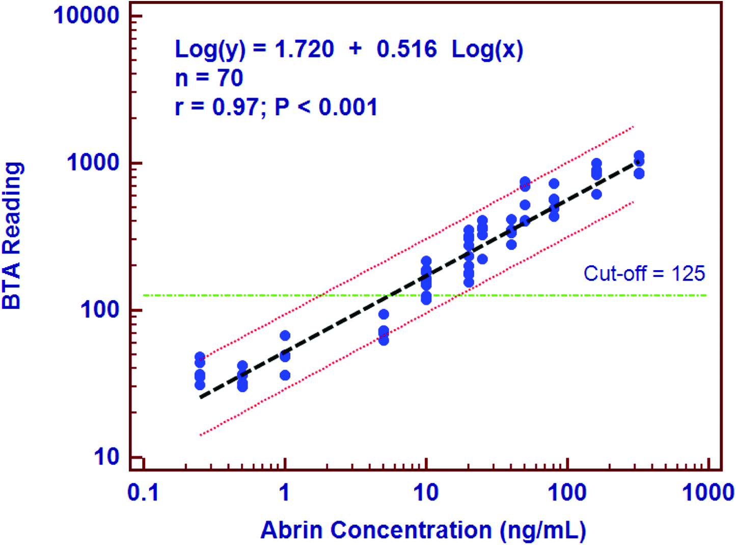
Rapid BioAlert reader values at different abrin concentrations in the abrin lateral flow assay. Reader cut-off value of 125 is shown as the horizontal line on the Y-axis. The limit of detection of the abrin lateral flow assay is ≥10 ng/mL. Color images available online at www.liebertonline.com/bsp
Sensitivity and Specificity Determinations
A receiver operator characteristic (ROC) curve was plotted with the study data (571 positive samples and 872 negative controls). In a ROC curve, the true-positive rate (ie, sensitivity) is plotted as a function of the false-positive rate (ie, 100% specificity) for different cut-off points. Each point on the ROC curve represents a sensitivity/specificity pair corresponding to a particular decision threshold. A test with perfect discrimination (no overlap in the 2 distributions) has a ROC curve that passes through the upper left corner (100% sensitivity, 100% specificity). The ROC curve for the Abrin LFA (Figure 3) shows that the area under the curve is 0.988 at a BTA reader threshold greater than 107 (maximal sensitivity and specificity). The data from the study are also depicted as an interactive dot diagram (Figure 4), which shows the BioThreat Reader reading for all positive and the negative samples tested. The samples were tested at 5 different sites by 10 operators on at least 2 different days at each site. From this interactive dot plot, it is clear using a cut-off value of 107, the assay has 98.2% sensitivity and 98.7% specificity. The default threshold cut-off value for the Rapid BioAlert reader is 125, with sensitivity of 98.1% and specificity of 98.2%.
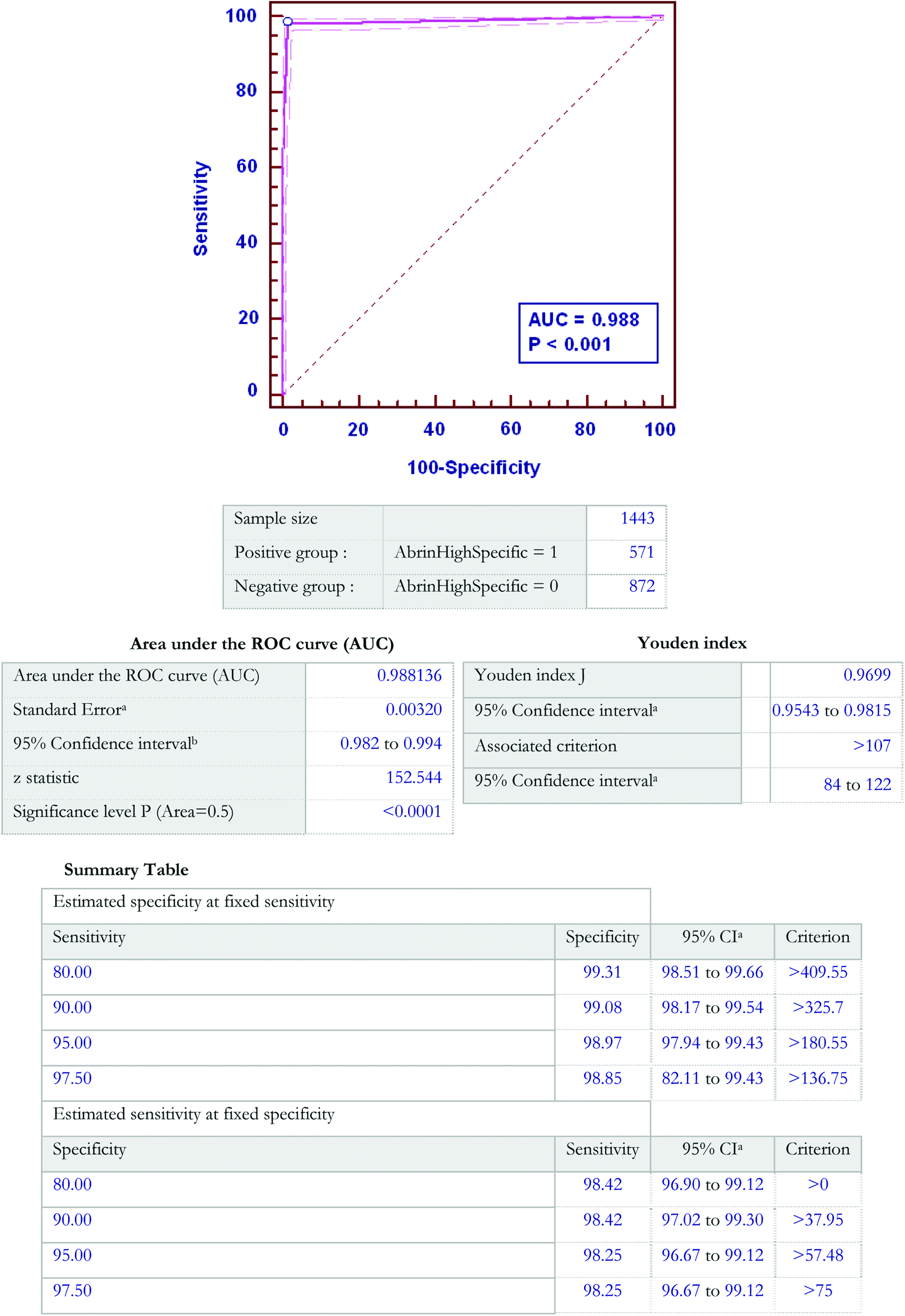
Receiver Operator Characteristic (ROC) Curve for Abrin Lateral Flow Assay. Color images available online at www.liebertonline.com/bsp
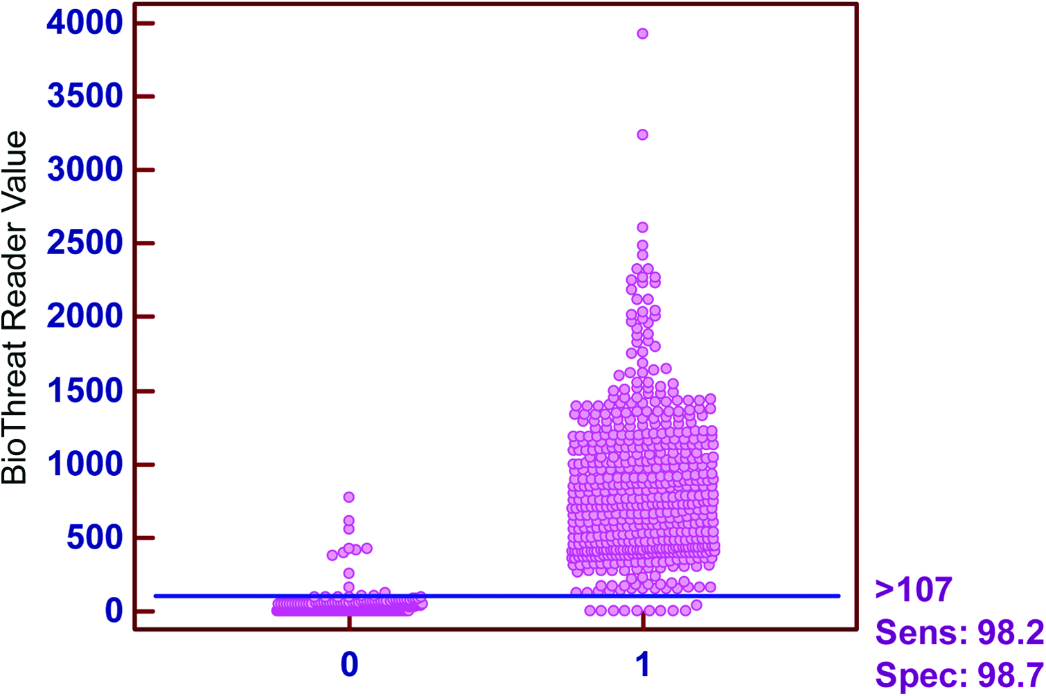
Interactive Dot Plot for Lateral Flow Assay of Abrin (cut-off at 107); X axis: 0 are negative samples and 1 are positive samples; each dot is a data point for a single result. Color images available online at www.liebertonline.com/bsp
Discussion
Methods to detect abrin include real-time PCR with an LOD of approximately 1.2 copies of DNA in crude preparations of abrin. 2 However, this method can only detect the presence of the targeted DNA sequences and not the presence of abrin. Additional methodologies such as the identification of tryptic peptides by liquid chromatography electrospray ionization with tandem mass spectrometry (LC-ESI MS-MS) can detect approximately 64 ng (1 pMol) per injection. 16 An aptamer-based assay was able to detect abrin at 64 ng abrin/mL (1 nM); an increased sensitivity of 35 ng/mL was reported using surface plasmon resonance (SPR). 16 ELISAs, however, remain the gold standard for the identification of abrin in both environmental and food samples, with limits of detection ranging from 0.1 to 0.5 ng/mL in buffer samples and 0.5 to 2 ng/mL in most food matrices, although in certain liquids, such as cola, the LOD may increase to 10 ng/mL. While the sensitivity of ELISA for abrin is lower in food matrices than in buffer, the LOD is still well below clinically significant amounts.16,25,26
Lateral flow immunochromatographic assays (LFA) were commercially introduced for pregnancy testing in 1988. 27 These “hand-held” assays (HHA) are simple to use and require minimal training, 28 making them ideal for use by first responders and law enforcement to test suspicious powders in a field setting. BioThreat Alert® Assays have previously been evaluated for other biothreat agents including orthopoxviruses, 29 ricin (manuscript submitted, 2013), and Yersinia pestis. 30 Limited evaluations have also been conducted with assays for Bacillus anthracis, Francisella tularensis, botulinum neurotoxins, and staphylococcal enterotoxin B (unpublished data). The Abrin BioThreat Alert® Test Strip is a rapid qualitative test to detect the presence of abrin in environmental samples. The test uses a combination of a polyclonal capture antibody to selectively capture and a monoclonal antibody to specifically detect the presence of abrin (and abrin agglutinin) in aqueous samples. The purpose of the current study was to evaluate the performance of Abrin LFA in order to understand its sensitivity, specificity, reproducibility, and limitations for field use and to determine whether this assay could also be used as a screening assay in a laboratory setting.
Using the BioThreat Alert® Reader and the manufacturer's recommended cutoff of 125, we estimated the LOD of the Abrin BioThreat Alert® Test Strip to be approximately 10 ng/mL or 1.5 ng/reaction (Figure 4), which is well below clinically relevant levels (LD50 3.3 μg/kg inhaled and 20 μg/kg ingested17,18). While the sensitivity is lower than that seen with ELISA, it is appropriate for a field-based test to give a rapid qualitative result that can be confirmed with further analysis in a laboratory.
Because of the widespread geographical diversity of A. precatorius, we wanted to determine whether the Abrin LFA would detect the presence of abrin in extracts from seeds of 11 geographically diverse cultivars. The Abrin LFA was positive for all cultivars when tested with 1 μg extract protein/mL. If one assumes that 2.4% of the extract protein is abrin+APA-1, 23 then 1 μg of extract protein contains approximately 24 ng abrin+APA-1.
The Abrin LFA was also tested against extracts from 35 near neighbors and other plants and 65 lectins, including wheat germ agglutinin, which had been shown previously by Dayan-Kenigsburg et al 24 to cause false-positive results in a ricin LFA. Crude extracts of the near neighbors were tested at a protein concentration of 10 μg/mL, while the lectins were tested at 5 μg/mL. All 65 lectins, including wheat germ agglutinin, yielded negative results at the concentration tested. Of the near neighbors and other plants, 34 of 35 yielded negative test results, while 1, A. laevigatus, yielded positive test results at all 5 test sites. It is well known that other plants, including Abrus pulchellus, produce RIPs.31,32 However, an extract of seeds from A. pulchellus did not produce a positive test result. Ramos et al 31 found that the type II RIP from A. pulchellus, pulchellin, exhibited only partial sequence identity with abrin. Because of the highly specific nature of the antibodies used in this assay, we considered the possibility that A. laevigatus produces abrin or a close homologue. A crude extract of A. laevigatus seeds underwent purification by the procedures described in Materials and Methods, including SDS-PAGE and GC-MS. The purified toxin (100 ng/mL) was then tested with the Abrin LFA and a positive result obtained (data not shown).
The Abrin LFA was also tested against 11 related proteins and toxins including APA-1, abrin toxoid, ricin and ricin subunits, and ricin vaccine candidates at a concentration of 1 μg/mL. APA-1, which shares a high degree of homology with abrin, produced a positive result, suggesting the presence of epitopes that are shared by both the toxin and agglutinin, which are recognized by the antibodies used in this assay. However, purified ricin A chain and ricin B chain both yielded positive test results as well. These false-positive results could be eliminated by the addition of powdered milk to the sample buffer, although this also caused a reduction in sensitivity to abrin toxin (data not shown). The LFA did not, however, yield positive test results with RCA60, deglycosylated ricin A chain, or with a crude extract of Ricinus communis seeds. One possible explanation is that the anti-abrin antibodies used in this LFA bind weakly or nonspecifically to carbohydrate-containing epitopes that are present on ricin A-chain and ricin B-chain but that are not accessible on RCA60. We previously showed that Ricin BioThreat Alert® Test Strip did not exhibit cross-reactivity with an extract of A. precatorius seeds (manuscript in press). Because neither the A-chain nor B-chain of ricin is a public health threat, this should not impede the use of this assay in the field. Additionally, all samples yielding positive results are forwarded to an LRN laboratory for additional testing and confirmation. 33
The Abrin BioThreat Alert® Test Strip has been used in the field to identify the potential presence of abrin in powders and other environmental samples. As part of this study, we evaluated the ability of this assay to detect abrin in commonly encountered powders spiked with an extract of the Banana Tree Red cultivar of A. precatorius. The LFA gave positive results with 25 of 26 powders tested, while inconsistent results were observed with powdered toothpaste. However, spiked suspensions or solutions of these powders gave reduced readings when compared to Banana Tree Red extract in BTA buffer. It is not clear whether this reduction is due to (1) an inhibition of abrin (or APA-1) binding to one of the antibodies; (2) a reduction in the flow rate of the strip due to increased viscosity of the sample; or (3) binding of abrin (or APA-1) by the powder and its subsequent removal during centrifugation prior to loading on the test strip. White powders and BioWatch filter extract alone yielded negative results at all test sites.
Because this assay does not discriminate between abrin and APA-1, it can only be used as a qualitative screening assay when testing unknown samples. Positive LFA results must be confirmed at a laboratory with the capacity to specifically identify the presence of abrin. The performance, cost, shelf-life, ease of use, and rapidity of results suggest this test is suitable for field use as a screening assay for abrin.
Footnotes
Acknowledgments
The authors wish to acknowlege the important contributions to this manuscript from Dr. Douglas L. Anders, Hazardous Materials Response Unit, Federal Bureau of Investigation Laboratory, Quantico, Virginia, for subject matter expert advice and consultation. We express our thanks and appreciation for his assistance in the preparation and execution of this project.