
Abstract
Background:
Palatal petechiae are 95% specific for streptococcal pharyngitis. Despite this, and despite prior research demonstrating that Group A Streptococcus (GAS) is a common antecedent to pediatric acute-onset neuropsychiatric syndrome (PANS) episodes, we anecdotally observed a low rate of documented GAS in patients with PANS and palatal petechiae. This retrospective chart review was conducted to formally report the rate of palatal petechiae and concurrent GAS in a cohort of patients with PANS and investigate other etiologic factors.
Methods:
The clinical notes of 112 patients seen at the Stanford PANS Clinic who met PANS research criteria were reviewed for mention of palatal petechiae. The medical records of patients who demonstrated palatal petechiae on physical examination were reviewed for signs of infection, a clinical history of trauma, and laboratory results that could indicate other causes of petechiae.
Results:
Twenty-three patients had documented palatal petechiae on physical examination (ages 5–16, 13/23 [57%] male). Fifteen patients had a rapid GAS test and GAS culture in the Stanford PANS clinic, all with negative results. Evidence of recent GAS infection was found in 8/23 (32%) patients (elevated GAS titers [n = 6] or documentation of a positive rapid GAS test at another facility [n = 2]), one of whom also had potential herpes simplex virus (HSV) infection. One patient had potential HSV infection and recent palatal trauma. No patients had thrombocytopenia. 14/23 (61%) of patients with palatal petechiae had no discernable cause of petechiae. 10/19 (53%) of patients had antihistone antibodies.
Conclusions:
Despite the established relationship between palatal petechiae and GAS, no patient with palatal petechiae in our clinic tested positive for GAS and only 32% had evidence of recent GAS. Most did not have an identifiable cause for the palatal lesions. This finding suggests the potential for alternative causes of palatal petechiae or undetectable GAS in our patient population. The high prevalence of palatal petechiae without GAS infection suggests that the pathogenesis of PANS is multifactorial and may involve disruption or inflammation of the microvasculature. Additional research is needed to further elucidate these findings.
Introduction
P
At our institution, we have recognized a high rate of palatal petechiae in patients presenting with acute neuropsychiatric symptoms who meet criteria for pediatric acute-onset neuropsychiatric syndrome (PANS). Children with PANS are defined by established research criteria as having an abrupt, dramatic onset of obsessive compulsive disorder (OCD) or OCD-related eating restriction accompanied by an equally abrupt onset of comorbid neuropsychiatric symptoms, which include anxiety, emotional lability, depression, irritability, aggression, oppositionality, deterioration in school performance, behavioral (developmental) regression, sensory amplification, movement abnormalities, sleep disturbance, and urinary issues (Swedo et al. 2012; Chang et al. 2015). The causes of PANS are unknown and are currently under investigation.
The criteria for Pediatric Autoimmune Neuropsychiatric Disorder associated with Streptococcal infection (PANDAS) differ from PANS in that a diagnosis necessitates a preceding or concurrent infection with Group A Streptococcus (GAS) (Swedo et al. 1998). Additionally, tics are often a cardinal symptom. Nevertheless, previous studies have demonstrated that over half of all patients who meet PANS criteria have a concurrent and/or recent GAS infection (Frankovich et al. 2015; Murphy et al. 2015; Toufexis et al. 2015).
Despite GAS being a common antecedent to PANS episodes and the finding of palatal petechiae in our PANS patients, we anecdotally observed a low rate of documented GAS in patients with palatal petechiae at the Stanford PANS clinic. We have also noted a high rate of histone antibodies in this patient population (Frankovich et al. 2015). Although the etiology of PANS (or possibly etiologies) is unknown, PANS is hypothesized to be an inflammatory disorder, potentially involving blood vessels in the blood brain barrier (Cutforth et al. 2016; Dileepan et al. 2016). We therefore feel that palatal petechiae, suggestive of vascular injury, are an important clinical finding in this unique patient population.
The unexpectedly low rate of GAS in our patients with petechiae served as an impetus for further investigation, with the hope of shedding light on the underlying pathogenesis of PANS.
The purpose of this review is to formally report the prevalence of palatal petechiae in the Stanford PANS cohort, and to further elucidate additional etiologic factors associated with these lesions. Additionally, since antihistone antibodies have been reported in systemic lupus erythematosus (SLE) (Fritzler and Tan 1978; Sun et al. 2008; Vedove et al. 2009), a disorder associated with microvascular disease and petechiae (Norton et al. 1968; Riccieri et al. 2005), we evaluated the association between palatal petechiae and antihistone antibodies in our PANS cohort. Furthermore, among patients with SLE, antihistone antibodies have been shown to be associated with neuropsychiatric disease, which is another reason we decided to evaluate histone antibodies in our PANS population (Sun et al. 2008).
Methods
We reviewed the clinical records of 112 patients who presented to the Stanford PANS Clinic between September 2012 and February 2016, consented to be in our research database, and met full PANS or PANDAS research criteria, including acute-onset neuropsychiatric deterioration at any point during their clinical course. Of note, some patients had a subacute initial onset but after recovery subsequently had acute deteriorations and thus met PANS criteria.
At every medical visit, patients underwent a full medical examination by nurse practitioners or physicians, including oropharyngeal examination, and palatal petechiae were noted if found. All patients had a medical visit during initial work-up and subsequent relapses, and most were also seen during periods of remission.
In this study, petechiae are defined as flat, red, discrete lesions less than four millimeters in diameter (Reyes and Eichenfield 2012; Fett 2016; George and Arnold 2016). Petechiae-like lesions, called doughnut lesions, have been occasionally seen with GAS pharyngitis. These lesions are described as 3–4 mm red, raised, small follicular lesions with a 1 mm yellow center (Breese and Hall 1978). These lesions were not seen in any of the patients in this cohort.
The clinical notes and laboratory results of patients who presented with petechiae were reviewed for signs of bacterial or viral infection, particularly GAS, a clinical history of trauma, and laboratory results that could indicate other causes of petechiae.
A complete GAS work-up entailed a rapid GAS throat screen, a throat culture, and a perianal swab culture, when indicated. The throat culture examined both GAS polymerase chain reaction (PCR) and Group C/G (S. dysgalactiae ssp. equisimilis) using selective broth enrichment procedure and PCR/nucleic acid amplification. This PCR test was developed by the Stanford Microbiology Laboratory, and shown to be more sensitive than conventional methods and highly specific.
Specimens were obtained by vigorous swabbing of the posterior pharynx and both tonsils (or tonsillar fossae in patients who had undergone tonsillectomy) by nurse practitioners in the Stanford PANS clinic. The swabs were moved in and out of the mouth without touching the tongue or the buccal mucosa. The importance of obtaining an adequate specimen is emphasized in clinic, as the sensitivity of both rapid antigen detection test and culture correlate with density of colonization (Kurtz et al. 2000).
On physical examination, physicians or nurse practitioners systematically evaluated each patient for signs of infection or inflammation, including elevated temperature (>100.4°F or 38°C), swollen and tender cervical lymph nodes, tonsillar exudates or swelling, rashes, and arthritis. All parents were asked to complete a symptom questionnaire at each visit, which specifically asked about signs of recent illness or infection, sore throat, fevers, cough, vomiting, and/or rashes. Physicians and nurse practitioners asked about possible recent infections at every visit.
Centor scoring is a clinical decision-making tool designed to predict the likelihood of GAS in the setting of pharyngitis (McIsaac et al. 2004; Choby 2009). The tool is age-adjusted, validated in 600 adults and children, and validated retrospectively (McIsaac et al. 2000; Fine et al. 2012). A score ≤0 predicts a 1%–2.5% risk of GAS, 1 predicts 5%–10% risk, 2 predicts 11%–17% risk, and 3 predicts 28%–35% risk. Scores ≥4 predict a 51%–53% risk of GAS and suggest that patients should be empirically treated with antibiotics (McIsaac et al. 2000, 2004). Centor scores were retrospectively calculated for all patients who presented with palatal petechiae and reported a recent sore throat.
Antistreptolysin O (ASO) and anti-DNAse B (ADB) levels were obtained in all patients willing and able to obtain a blood draw in our institution. Titers were processed by Mayo Clinic Laboratories (Kaplan et al. 1998). A single elevated antibody titer was considered diagnostic if elevated for age (Kaplan et al. 1998; Gerber et al. 2009).
Additional bloodwork obtained when clinically indicated included antihistone antibodies, complete blood count with differential (CBC-D), Epstein Barr viral capsid antibody test, antinuclear antibody, and antiphospholipid antibodies.
All laboratory personnel processing all tests were unaware of the presence or absence of palatal petechiae.
To evaluate association between palatal petechiae and antihistone antibodies, we calculated the odds ratio using chi-squared analysis.
This chart review study was approved by the Stanford University Institutional Review Board, under protocol number 26922. All participants gave informed consent. Written informed consent for the publication of clinical images was obtained from the parent of the patient in Figure 1. A copy of the consent form is available for review by the editor of this journal.
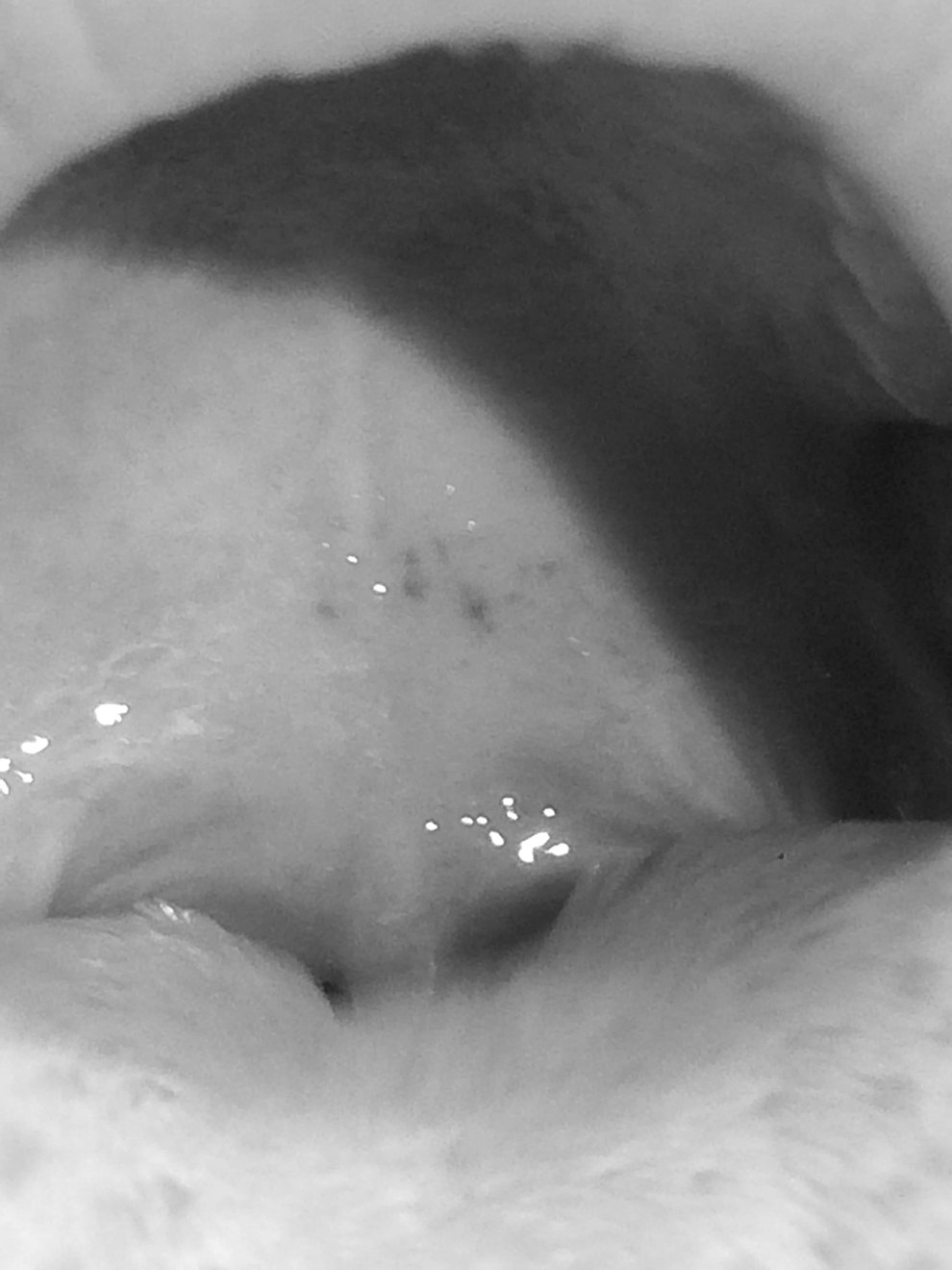
Photograph of palatal petechiae in one patient that is typical of the rest of the cases in this cohort.
Results
During the time period reviewed, 23/112 (21%) patients had palatal petechiae one or more times on physical examination in the Stanford PANS clinic (ages 5–16 years old, 13/23 (57%) male). All but one patient (22/23, 96%) met strict PANS criteria, and 15/23 (65%) of patients met PANDAS criteria. The one patient who did not meet PANS research criteria met PANDAS research criteria (Swedo et al. 1998).
One patient had petechiae on the palate and abdomen that resolved and then relapsed on the abdomen. The remaining 22 cases of petechiae were confined to the palate: 10 confined to the soft palate, one confined to the hard palate, 8 on both the soft and hard palate, and 3 unspecified (Fig. 1).
Of the 23 patients with palatal petechiae, 21 returned for follow-up. Of these, two patients had palatal petechiae on follow-up physical examination, 41 and 42 days later, respectively. The remaining 19 patients' follow-up times ranged from 5 to 151 days, with an average of 47 days. None of these 19 patients had palatal petechiae at their next follow-up physical examination.
Three of these 19 patients experienced resolution of petechiae within 1 week, 2 weeks, and 3 months, respectively. This subset of patients then experienced recrudescence of petechiae, at ∼2 weeks, 4 months, and 6 months, respectively, which resolved within 3–4 weeks. One of these patients had three episodes of palatal petechiae within a 6-month period. This patient also had strawberry tongue, but despite numerous throat swabs and frequent GAS serology testing, we were unable to detect GAS. This patient reported occasional sore throat, but there was no physical examination finding of pharyngitis. No other patient in this cohort had strawberry tongue.
Patient history, physical examination findings, and laboratory results are presented in Table 2.
present anytime.
ADB, anti-DNAse B; ASO, Antistreptolysin O; GAS, Group A Streptococcus; OCD, obsessive compulsive disorder; PANS, pediatric acute-onset neuropsychiatric syndrome.
GAS evaluation results
Fifteen out of 23 (65%) patients had a pharyngeal rapid GAS test and GAS culture obtained in the Stanford PANS clinic, nine of whom also had a perianal GAS culture obtained in the Stanford PANS clinic. One patient had a perianal swab but not a throat swab. None of these 16 patients had evidence of GAS based on a rapid GAS screen and/or culture at the time of the palatal petechiae. The remaining seven patients declined a GAS swab. Of the 16 patients who had a rapid GAS test or culture in the Stanford PANS clinic, 12 were taking antibiotics (a beta-lactam or azithromycin) at the time of the swab.
Three patients had GAS tests at either their pediatrician's office or an urgent care center 1 week or less before their presentation with petechiae (one negative, two positive on rapid, culture not done). Clinical notes indicated that providers were concerned for GAS due to: GAS exposure at school (n = 1), fever (n = 1), and fever and sore throat (n = 1). One patient who was positive on a rapid test at an outside facility tested negative on a rapid GAS test and GAS culture in the Stanford PANS clinic.
Fourteen patients had ASO and ADB titers measured at the time of palatal petechiae or within 6 weeks after documented petechiae. 6/14 (43%) patients had one or more elevated titer suggesting possible recent GAS infection or colonization.
Other potential causes of palatal petechiae
One patient with elevated ASO and ADB titers at the time of palatal petechiae also reported herpes labialis 3 weeks earlier, and at 2 months follow-up. Unspecified HSV work-up at the patient's pediatrician's office was negative, but the patient had another episode of herpes labialis 4 months later and a positive HSV-1 IgG at 5 months follow-up.
Another patient presented with mouth sores at the time of his presentation with petechiae, then developed genital sores and high HSV titers at 3 months follow-up. This patient also reported tooth extraction 4 days before presentation with petechiae.
Unknown causes of palatal petechiae (n = 11)
In most patients (14/23, 61%), history, physical examination, and laboratory work-up failed to reveal possible causes of palatal petechiae.
No evidence of thrombocytopenia was found in this cohort. Ten patients had CBC-D within 2 weeks of petechiae, and six had CBC-D within 6 months after having petechiae.
Antihistone antibodies
Compared to patients in our PANS cohort without palatal petechiae, patients with palatal petechiae were significantly more likely to have antihistone antibodies. The prevalence of elevated histone antibody in patients with palatal petechiae was 10/19 (53%), compared to 12/73 (16%) in patients without palatal petechiae (OR: 5.6, 95% CI 1.89–16.84, p = .0019).
Arthritis
Seven of 23 (30%) patients had arthritis, with six having had arthritis at the time of palatal petechiae. All seven patients with arthritis were male, and six patients were older than age six at the time of diagnosis of arthritis. Two had psoriatic arthritis, three had post-streptococcal arthritis primarily affecting knees, ankles, and hips, and two met criteria for spondylarthropathy.
SLE criteria
Of those with palatal petechiae, three patients met three out of the needed four criteria for the classification of SLE (Riccieri et al. 2005). Two met criteria based on the presence of chronic arthritis, persistent leukopenia, and psychosis. One met criteria based on chronic arthritis, elevated antiphospholipid antibodies, and psychosis. All three patients who met 3/4 lupus criteria had elevated histone antibodies but negative antinuclear antibodies. We continue to monitor all three patients for SLE.
Discussion
In many instances (11/23, 48%), history, physical examination, and laboratory work-up failed to reveal possible causes of palatal petechiae. Palatal petechiae have been found to be 95% specific for streptococcal pharyngitis. It is noteworthy that in our sample, no patients with PANS tested positive for GAS on a rapid GAS screen and/or culture in our clinic when they had palatal petechiae on physical examination. It is possible that the patients' GAS infections were missed by laboratory examination, or that antibiotic treatment eliminated GAS, but a GAS-related process led to palatal petechiae. It is also possible that another infection or inflammatory process contributed to the alteration of the microvasculature on the palate.
Previous studies comparing the results of GAS swabs and tonsillar core analysis have documented that throat swabs can frequently fail to reveal GAS (Gul et al. 2007; Hembrom et al. 2014). Similarly, some of our patients may have harbored GAS or other bacteria deep in the tonsillar tissue that was not found on throat swab. It is also well documented that some children do not have a rise in ASO and ADB following GAS infections. This has been reported to be as high as 38% in a cohort of children with PANS, PANDAS, Tourette's disorder, and other tic disorders (Johnson et al. 2010). Furthermore, single titers are notably unreliable indicators of recent infection. Our patients may include some of the many children with recent infection who do not demonstrate positive ASO or ADB titers even in the context of GAS pharyngitis or perianal GAS infection (Johnson et al. 2010). Only 1/14 (7%) patient had elevated ASO and ADB titers for age at or within 6 weeks after the time of palatal petechiae, which is similar to what is expected for the general population (McIsaac et al. 2000). Therefore, it is possible that our patients harbored GAS infection that was not found on careful physical and laboratory examinations. Arguing against this, no patient had a Centor score above a three, indicating that all patients with pharyngeal pain had a 35% or lower risk of GAS. The average Centor score of patients in this group (2.17) corresponds to only a 11%–17% average risk of GAS pharyngitis.
Arcanobaterium Haemolyticum is another bacterial cause of palatal petechiae. A. haemolyticum is rare, particularly in the United States. It is often associated with erythema and exudates, and/or a scarlet-fever like rash, and is most common in teenagers (Bisno et al. 1997). No patients had a scarlet-fever type rash at the time of palatal petechiae, and only four were above age 13. However, throat culture assays performed at our institution do not evaluate for A. haemolyticum, which may have gone undetected.
Viral infections, specifically infectious mononucleosis, rubella, and HSV have also been reported to be associated with palatal petechiae. Infectious mononucleosis was an unlikely trigger, based on the absence of atypical lymphocytes and normal white blood cell counts (n = 10), and negative Epstein-Barr virus antibody test (n = 1) (Sprunt and Evans 1920). One additional patient who did not have a CBC did have a negative Epstein-Barr IgG and IgM virus antibody test. No patients had clinical history or physical examination findings suggestive of rubella. Only two patients tested seropositive for HSV (with one also having elevated ASO and ADB titers). Although patients did not present with vesicles or report previous cold sores, it is possible that more patients harbored HSV, as HSV is often asymptomatic (McMillan et al. 1993; Klein 2014); however, we feel this to be unlikely, as palatal petechiae without development of vesicles in HSV has not been previously described.
It is possible that thrombocytopenia, defined as a platelet count of less than 150,000/microL (Long 2012), played a role in the etiology of petechiae in some patients. While none of the 10 patients who had a CBC had low platelet counts, the 13 patients who did not have a CBC may have had undiagnosed thrombocytopenia. Furthermore, thrombocytopenia has been associated with naproxen and ibuprofen use (Reese et al. 2010). Of the eight patients on daily naproxen or ibuprofen, six had a CBC within 2 weeks of having petechiae, none of whom had low platelet counts. Although unlikely, the two patients on daily naproxen or ibuprofen who did not have a CBC-D may have had a higher risk of undiagnosed thrombocytopenia; this remains unknown.
The palatal petechiae in patients with GAS is most likely not due to bacterial invasion, but is more likely a reactive process in the microvasculature. It is unknown whether this process could also affect the microvasculature of the blood-brain barrier. Interestingly, these patients had a high rate of arthritis [30% vs. worldwide pediatric prevalence of 0.007%–0.401% (Manners and Bower 2002)] and autoantibodies (Table 2). We were especially interested to note the high prevalence of elevated antihistone antibodies among the patients with palatal petechiae (10/19, 53%), and to note that histone antibody was associated with palatal petechiae in our cohort (OR: 5.6, 95% CI 1.89–16.84). Antihistone antibody elevation is also found in SLE (Fritzler and Tan 1978; Sun et al. 2008; Vedove et al. 2009), a disorder associated with microvascular disease and petechiae (Norton et al. 1968; Riccieri et al. 2005). Although antihistone antibodies are associated with SLE, only one patient in our cohort of 112 patients with PANS met SLE criteria, and this patient did not have palatal petechiae, although she did have a painless palatal ulcer and nasal sore.
The high prevalence of palatal petechiae without GAS infection in this cohort of patients with PANS may be an important clue regarding the pathogenesis of PANS if it reflects disruption or inflammation of the microvasculature. Recent evidence from an animal model suggests that PANDAS is an inflammatory disorder that effects the microvasculature of the blood-brain barrier (Cutforth et al. 2016; Dileepan et al. 2016). Further research is needed to understand the factors driving the high prevalence of palatal petechiae in the absence of GAS infection seen in this cohort of patients with PANS.
Limitations
Some infectious sources may have gone undetected. Three patients had GAS tests completed at an outside facility, and we cannot attest to the method used to collect the samples. Additionally, not all tests were performed on all patients, ASO and ADB titers were not measured serially, and each test has limitations in sensitivity and specificity.
In the context of disabling psychiatric and behavioral symptoms, parents may not have noticed milder physical complaint, such as pharyngeal pain. Naproxen and ibuprofen usage may have also contributed to less pain reporting and less febrile episodes. This under-reporting of symptoms may have artificially lowered our Center scores. Furthermore, some of these patients were oppositional and/or combative during physical examination, making the oropharyngeal examination and pharyngeal swabbing challenging. Three patients fiercely opposed being swabbed and thus, we were unable to attain a culture. It is conceivable that these children may have had GAS as it is our observation that when children are positive for GAS they are much more resistant to being swabbed. Therefore, it is possible that there were some overlooked cases of palatal petechiae and/or GAS.
Conclusions
Although palatal petechiae usually signal GAS infection (specificity = 95%), our study found a high prevalence of palatal petechiae in the absence of GAS in a cohort of patients with PANS. Additionally, over half of our patients with palatal petechiae also had elevated antihistone antibodies. These clinical findings may be important clues to the pathogenesis of PANS, if they reflect disruption or inflammation of the microvasculature. Further research is needed to better understand the biological mechanisms underlying the high prevalence of palatal petechiae and elevated antihistone antibodies in patients with PANS.
Clinical Significance
If a psychiatrist suspects PANS or PANDAS, we recommend referral back to the primary medical doctor for full medical examination based on the 2013 PANS evaluation guidelines (Chang et al. 2015). In addition to these guidelines, we recommend an oropharyngeal examination noting the presence of petechiae, Centor scoring, and evaluation for GAS and other relevant organisms.
Footnotes
Acknowledgments
The authors would like to acknowledge Dr. Susan Swedo, National Institute of Mental Health, the PANDAS Physician Network, the PANDAS Network, Dr. Janell Sherr, and all the faculty and staff at Stanford Children's Health and the Stanford PANS clinic who make caring for children with PANS possible.
Disclosure
No competing financial interests exist.