
Abstract
Aim:
The aim of this study was to observe the biological distribution and anticancer effect of 32P-chromic phosphate colloid (Cr32PO4, 32P-CP) after intratumoral injection to Pc-3 human pancreatic carcinoma-bearing nude mice.
Methods:
Eighty-four (84) BALB/c nude mice with transplanted tumor were allocated to 11 groups. Groups 1–5 (n = 6) were intratumorally injected with 14.8 MBq of 32P-CP and sacrificed at 2, 24, 48, 72, and 168 hours, respectively. Groups 6–11 (n = 9) received injections of 3.7, 7.4, 14.8, 18.5, 29.6, and 0 MBq of 32P-CP, respectively, and the tumor volume on body surface was measured daily. The animals (n = 6) were sacrificed at 14 days after administration. The dynamic distribution of radioactivity in body (percentage of injected dose per g), morphological changes, the tumor-inhibiting rate (TIR), proliferating index (PI), proliferating cell nuclear antigen (PCNA) tumor microvascular density (MVD), continuous counting of white blood cells (WBCs) and platelets (PLTs) in venous blood, body weight, and toxic reactions were observed.
Results:
The injected 32P-CP mainly accumulated in the tumor mass and was retained for a long time. The TIR of each dosage group in order was 21.68%, 39.73%, 50.43%, 71.18%, and 74.09% (F = 159.74; p < 0.001), PI was 70.85, 67.90, 46.70, 20.66, 10.75, and 90.11 (F = 509.54; p < 0.001), and MVD count was 39.19, 28.33, 17.45, 8.89, 8.10, and 64.80 (F = 643.26; p < 0.001), respectively. The data for WBC, PLT, and body weight observed for 28 days in the treatment groups did not indicate significant differences compared with those of the control group.
Conclusions:
Interstitial injection of 32P-CP seems to be a safe and effective interventional nuclide therapy for pancreatic carcinoma-bearing nude mice.
Introduction
Pancreatic carcinoma is listed fourth in cancer-related fatalities in the United States, where almost 37,000 new cases are registered annually. 1 The annual incidence of pancreatic carcinoma in China is 5.1 per hundred thousand, a number that has tripled over the past 20 years. 2 About 80% patients presenting with inoperable tumors arising from local or remote metastasis at the time of diagnosis are candidates for receiving palliative treatment to improve their quality of life. About 10 years ago, gemcitabine became the medicine of choice in treating progressive pancreatic carcinoma, as this agent was found to be superior to fluorouracil. 3 However, despite these advances in treatment, the median survival time for progressive pancreatic carcinoma remains 3–4 months. Investigators in this field have applied considerable effort toward improving the therapeutic outcome for patients with pancreatic carcinoma by introducing measures, such as combining radiotherapy and chemotherapy, and applying of antiangiogenesis drugs and/or epidermal growth-factor receptor (EGF) inhibitors. 4
In recent years, the therapeutic application of internal irradiation, which utilizes radioactive isotopes for treating solid tumors, has received considerable attention. Malmud 5 has emphasized the importance of ß -ray emitters for treating tumors, of which 32P-chromic phosphate colloid (Cr32PO4, 32P-CP) is a promising one; preliminary clinical data have revealed that interstitial injections of 32P-CP were effective for treating refractory solid tumors, such as pancreatic carcinoma and brain tumor, as well as some metastatic tumors. 6,7 According to some recent reports, prevention and inhibition of lymphatic metastasis could be achieved by interstitial administration during resection of lung cancer and carcinoma of gastric cardia. 8,9 Although previous studies were conducted on human subjects, some information may be easier to obtain from animal models. The aims of this study were to observe the biodistribution of 32P-CP and its anticarcinogenic effects and to determine the range of effective dosage and safety profiles. In addition, the mechanisms involved in the treatment of solid tumors with 32P-CP were explored.
Materials and Methods
Animal models
All animal experiments were approved by the governing Animal Welfare Committee and conducted in accordance with the regulations of the institution.
The pancreatic carcinoma cell line Pc-3 was used to establish tumor xenografts in nude mice, which were purchased from the Department of Pathology, Peking Union Hospital. Cells were routinely cultured in a medium containing 5% fetal bovine serum (FBS; Gibco) and RPMI1640 (Gibco) with gentamicin 50 U/mL, at 37°C with sufficient humidity. The culture medium was changed every 2–3 days. The cells were detached with 0.25% trypsin when they reached 80% confluence. The cells in a logarithmic growth phase were resuspended in phosphate-buffered solution (PBS) and collected after centrifugation at 1500 rpm/minute at room temperature for 5 minutes. Cell viability was determined using trypan blue exclusion. A single-cell suspension was made when cell viability exceeded 99% and was adjusted to 1 × 10⁷ cell concentration; 0.2 mL of this suspension was then injected subcutaneously (s.c.) into the dorsum of BALB/c (nu/nu) nude mice, which were obtained from the Chinese Academy of Science, Shanghai Experimental Animal Center (with certificates, age 6 weeks, male; body weight 18–22 g). The animals were bred in clean airflow racks in a barrier system (specific pathogen free) at room temperature (25°C ± 1°C) at a relative humidity of 40%–60%. When tumor mass in these mice reached 1.0 cm in diameter, the animals were sacrificed via cervical dislocation. Under aseptic conditions, the tumor mass was dissected and sliced into pieces of 2.0 mm diameter and rinsed in RPMI1640 solution (without FBS). The tumor tissue was then implanted s.c. with an 18G trocar into the right axilla of 84 BALB/c-nu/nu nude mice. Thereafter, the animals were randomly allocated to 11 groups, and the tumors grew to 0.8–1.0 cm in size after 10 days.
Administration of 32P-CP
32P-CP was obtained from the Chinese Institute of Atomic Energy, Beijing, China. The particle size of 32P-CP used was ∼20–50 nm; its other attributes included a radioactive concentration of 222–370 MBq/mL, a radiochemical purity of ≥98%, pH of ∼ 6.0–8.0, a physical half life of 32P 14.28 days, and an average energy of 0.695 MeV (maximum energy of 1.711 MeV); in this form, 32P-CP has an average penetration distance of 3–4 mm in soft tissue. After allowing the substance to stand for 140 days, it served as a cold colloid, bereft of radioactivity. The tip of the needle was carefully directed to the distal edge of the tumor along its long axis, and then 32P-CP was injected into the tumor slowly, while the needle was withdrawn carefully. Once the pressure decreased in the tumor, the needle was withdrawn from injection point to avoid solution overflow.
Grouping and experiments
Eighty-four (84) nude mice bearing Pc-3 tumors were randomly allocated to 11 groups. Groups 1–5 comprised 6 mice each and received 14.8 MBq 32P-CP by injection into the tumor mass, following which the animals from groups 1 to 5 were sacrificed at 2, 24, 48, 72, and 168 hours postinjection, respectively. From the animals in these groups, the tumor, liver, spleen, lung, kidney, stomach, intestine, brain, and heart were harvested, wet-weighed with an electronic balance (DF-200A; Changzhou Weigher Factory) and homogenized; and ß emissions were measured with an automatic liquid-scintillation counter (FJ-2115; Xi'an Nuclear Instrument Factory). The percentage of injected dose per g (%ID/g) and ratio between tumor and normal tissue (T/N) were calculated. There were 9 mice each in groups 6–11; doses delivered to animals in groups 6–11 were 3.7, 7.4, 14.8, 18.5, 29.6, and 0 MBq, respectively; cold colloid-injected tumors served as the control group. Six (6) mice from each group were sacrificed at 14 days postadministration, and tumors were resected and weighed. Tumor inhibiting rate (TIR) was calculated as follows:
in which W
1 and W
2 denoted the average mass of tumors in control and treatment groups, respectively. The maximal diameters (a, b) in two perpendicular directions were measured before and after intratumoral injection. The tumor volume was calculated using the following equation:
in which the tumor growth inhibition curve plots the changes in average tumor volume against time.
Bremsstrahlung imaging with a gamma camera
Three (3) mice from each group were selected randomly for imaging at 2 hours, 24 hours, and 1 and 2 weeks after administration. Single-photon emission computed tomography (SPECT, FORTE; ADACA) fitted with a low energy, general-purpose collimator was used for Bremsstrahlung imaging. A single 20% energy window was set at 70 keV and 300 K.
Toxic reaction
In 3 mice each from groups 6 to 11, blood was collected from the tail vein, or alternately, from the left and right orbital veins, at 0, 7, 14, 21, and 28 days after treatment; blood counts for white blood cells (WBCs) and platelets (PLTs) were recorded, and the body weight was determined simultaneously.
Calculation of absorbed dose
Internal radiation absorbed dose was calculated according to the following formula
10
:
in which D is absorbed dose (cGy), A is radioactivity (MBq), W is tumor mass (kg), and 3.6 is the unit absorbed dose per kg of tumor (cGy/37 MBq).
Histological observation
The tumor mass was dissected; two pieces of tumor tissues without necrosis, 1 mm3 in size, were taken, fixed in 0.5% glutaraldehyde, and dehydrated; and sections of 100 nm thickness were double stained with lead–uranium, followed by observation under TEM H-600 (Hitachi). The remaining tumor tissues were immediately fixed in 10% formaldehyde and embedded in paraffin, and 5-μm-thick sections were prepared for light microscopy (Olympus).
Immunohistochemical studies
All of the paraffin slides were subjected to microwave retrieval with tissue antigen and dyed using the SABC method. Immunohistochemical reagent kits were obtained from Zymed Company. Contrasts for positive expression of mouse against human proliferating cell nuclear antigen (PCNA) were carried out. A positive cell was defined by clear-cut yellowish-brown staining of the tumor cell nucleus, against a clear and unstained cytoplasmic background. Areas dominated by abundant tumor cells and fewer necrosed cells were selected in a low-power field, and tumor-cell numbers were counted in a high-power field. Five (5)–10 areas of different tissue structure were carefully observed in high-power fields for each slide. The proliferating index (PI) was calculated as follows:
CD34 was measured as the specific surface marker for endothelial cells within tumor blood vessels; positive CD34 expression denoted the presence of a microvessel, allowing the estimation of number of microvessels in the tumor tissue. Each yellow-stained cell or cell cluster was assigned 1 microvascular density (1 MVD) value. Each slide was examined by two pathological experts per a double-blinded protocol.
Statistical methods
TIR, PI, and MVD values, body weight, WBC, and PLT were expressed as mean value plus standard deviation (mean ± SD). The results of TIR, PI, and MVD values were subjected to multiple comparisons using one-way analysis of variance (SAS, 9.1). When the difference revealed statistical significance, the Student–Newman–Keuls' method was used. Data for body weight, WBC, and PLT were tested with Student's t-test; p < 0.01 was considered statistically significant.
Results
Biodistribution of 32P-CP after treatment
For groups 1–5, the uptake rates (%ID/g) for tumor and organs at 2, 24, 48, 72, and 168 hours after treatment are summarized in Figure 1. The T/N ratio is shown in Table 1. Evident 32P-CP uptake by tumor was observed in SPECT images taken at a prolonged residual time after injection (Fig. 2).
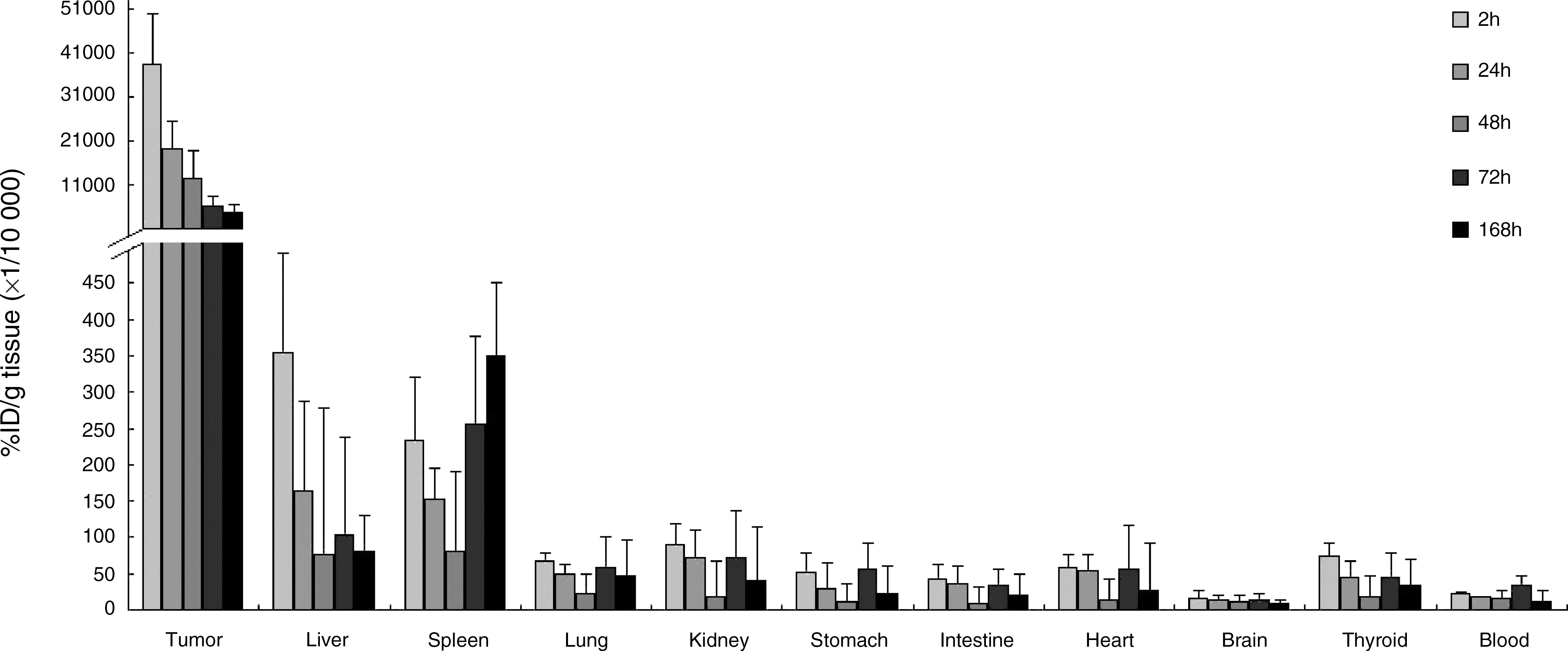
Biodistribution of 32P-chromic phosphate colloid in nude mice (14.8 MBq).
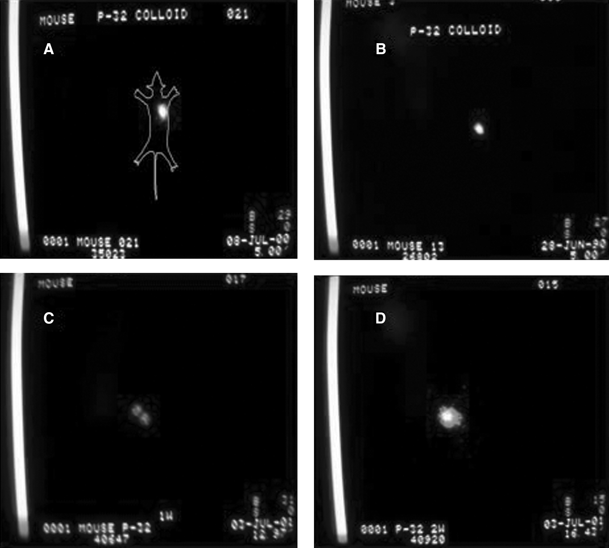
Dynamic scintigrams of tumor at various timepoints after intratumoral injection of 14.8 MBq 32P-chromic phosphate colloid: (
Tumor absorbed dose and suppressing effects
For groups 6–11, average absorbed dose in tumor, TIR, PI, and MVD at 14 days are shown in Table 2.
Different capital letters mean significant difference of mean values between groups (p < 0.05).
TIR, tumor inhibiting rate; PI, proliferating index; MVD, microvascular density; SD, standard deviation; SNK, Student–Newman–Keuls' method.
Gross presentation
After tumor transplantation, the tumor gradually presented as a prominent solid nodular mass protruding from the body surface, covered with smooth, light red skin; tumors were oval and attained a maximal length of 0.8 cm by the 10th day, when measured along the long axis. After interstitial injection of 32P-CP, localized redness and swelling appeared at the injection sites, these symptoms vanished within a day. Two (2)–3 days post-treatment, the nude mice had a slight decrease in appetite; however, they recovered appetite and movement later. In the control group, tumors were markedly enlarged and were solid and nodular in shape. In contrast, however, tumor growth was significantly reduced in the treatment groups and scattered ulceration with bleeding appeared on the tumor surfaces within 5 days, while the tumors shrunk in size and flattened out. In mice from groups 9 and 10, which received doses equivalent to 18.5 and 29.6 MBq, respectively, scattered small petechiae appeared on the dorsum and hip of 3 nude mice at 1 week after medication, while 2 mice became thinner; however, all were alive at 14 days. Interestingly, those signs were absent in groups 6–8 (3.7–14.8 MBq). A graph plotting the time-dependent changes in tumor volume is shown in Figure 3.
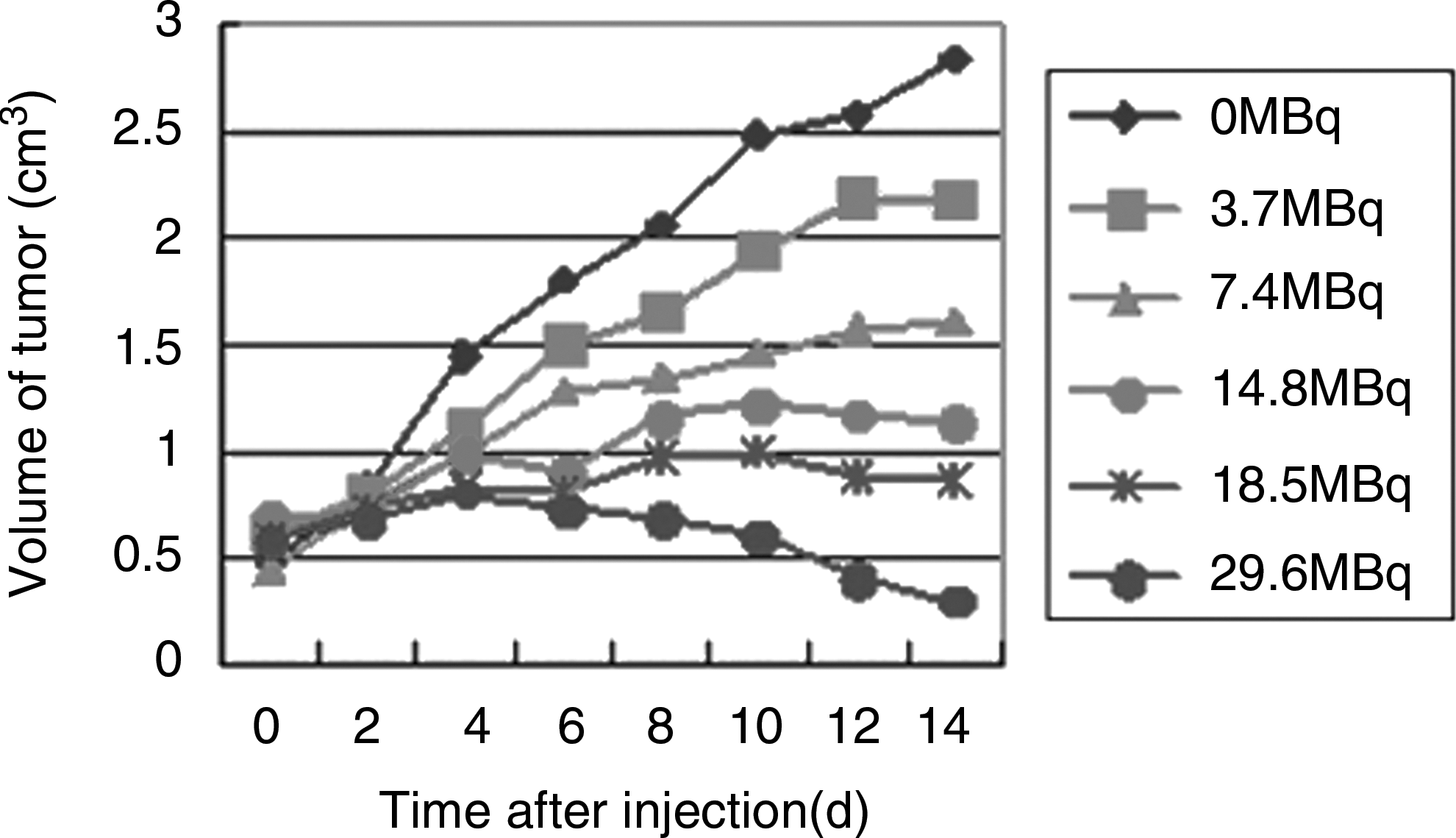
Curves of tumor volume after injection of different doses of 32P-chromic phosphate colloid.
Toxic reactions
For groups 6–11, the dynamic changes in WBC and PLT counts and body weight are listed in Table 3.
p < 0.01 (p = 0.009), compared with control group.
WBCs, white blood cells; PLTs, platelets.
Light microscopy and immunohistochemistry
In samples collected from the control group, the Pc-3 cells were densely arranged and had high rates of cell division and proliferation. Several malformed nuclei were observed, while the tumor core presented with abundant blood sinuses and focal necrosis. Varying degrees of necrosis were observed in a dose-dependent manner. Some tumor cells were divided by fibrous connective tissue cords (Fig. 4). After treatment, proliferation of Kupffer's cells in liver and lung-tissue fibrosis of varying severity were observed. In group 10 (29.6 MBq), dedifferentiation was observed in epidermal tissue adjacent to tumors, whereas normal muscular and nervous tissues were observed around the shrunken and scabby tumors. In the control group, a strong positive expression of PCNA and CD34 was observed, whereas their expression was obviously weaker in the treated groups (Figs. 5 and 6).
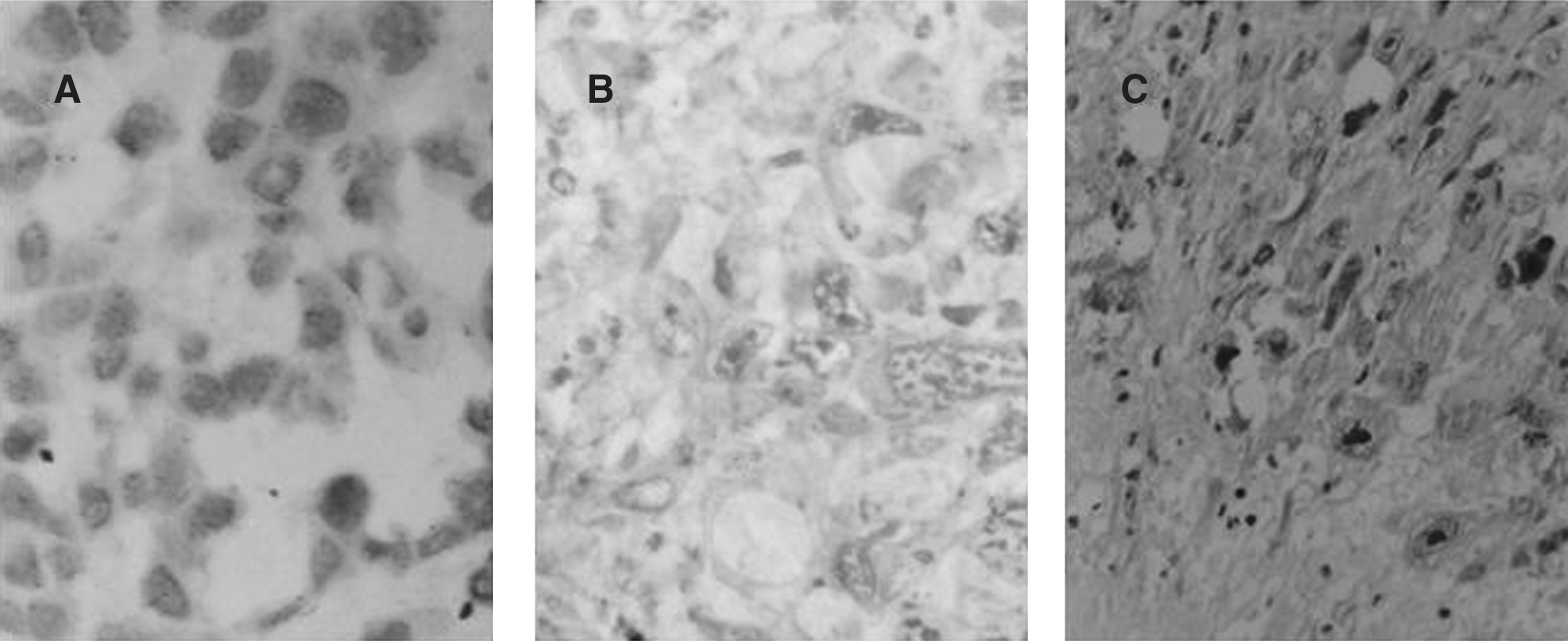
Light microscopic image shows changes in Pc-3 tumor cell: tumor cells were cloudy and swollen, and volume increased with an intact cell membrane. Nuclei pyknosis, rupture, and karyolysis were common. Those changes become notable with an increase of dose of 32P-CP and time. (
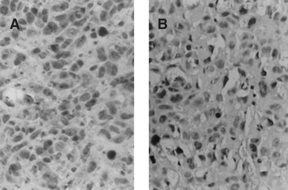
Results of immnohistochemistry assay of proliferating cell nuclear antigen expression in Pc-3. (

Results of immnohistochemistry assay of CD34. (
Transmission electron microscopy
In the control group, Pc-3 cells were round and had large nuclei containing pseudoinclusion bodies, several nucleoli, scanty cytoplasm, and an abundance of free ribosomes. A few microvilli appeared on the cell surface with desmosomes, while some pseudoglandular lumen were visible between cells. In groups that received larger doses of 32P-CP, tumor cells were damaged, where only the contour of nuclei remained and several vacuoles appeared in the cytoplasm, whereas neutrocyte infiltration, collagen fibrils, and proliferation of fibroblasts were observed in the connective tissue surrounding the cells. In the medium-dose group, bleeding was detected in mesenchymal tissue surrounding the Pc-3 cells containing small nucleoli, swollen mitochondria, dilated endocytoplasmic reticulum, and more lipid droplets than normal in the cytoplasm. The groups receiving lower doses of 32P-CP presented with viable tumor cells, with many free ribosomes in the cytoplasm. In addition, well-differentiated Pc-3 cells with small nucleoli, long microvilli on cell surfaces, and glandular lumen between the cells were observed (Fig. 7).

Transmission electronic microscopy of tumor. (
Discussion
This study demonstrates the concentration of radioactivity within the tumors in a majority of mice after injection; the T/N ratios for some vital organs, such as liver and spleen, exceeded 100 within 2 days after administration. In addition, dose-dependent increase in toxicity was observed, and delivered doses approximating 444–558 Gy were found to be most effective.
Radioactive nuclide colloids have played an important role in interventional nuclear medicine. 32P-CP, with ∼20–50 nm particle size, is a radioactive agent used in treating malignant thoracic effusion or ascites 11 –13 ; this agent has an average penetration distance of ∼3–4 mm in soft tissue, and the agent's linear-energy transfer and relative biological efficiency are higher than those of X- and γ-rays.
As early as the 1960s, 32P-CP had been used for treating malignant tumors. In 1964, McFee et al. 14 reported the use of 32P-CP to prevent liver metastasis in mice. In addition, 32P-CP has been used for treating multiple osseous metastasis 15 and disseminated ovarian cancer. 16 The safety profile for i.v. injection of 32P-CP for treating bone metastasis of prostate cancer has been studied by Potsaid and his colleagues in 1978. 17
Several clinical studies reveal that interstitial injections of 32P-CP have definite efficacy for treating refractory malignant solid tumors, or lymphatic metastases of cancers of pancreas, lung, and other sites. 8,9,18,19 However, defining a standardized 32P-CP regimen has remained an unsolved problem and has severely limited its acceptance in clinical practice. Thus, a study of the biological distribution of 32P-CP after interstitial administration is of great significance. In this study, SPECT imaging clearly revealed the concentration of radioactivity mainly within the injection site of the tumor, without visible traces in other organs. The majority of 32P-CP was concentrated in the tumor, whereas the radioactivity in other tissues and organs was low, at times undetectable over the background. Although the uptake of radioactivity was higher in spleen, liver, and lungs than in other organs, the T/N ratios for these vital organs exceeded 100 within 48 hours of treatment.
To date, the dosing for 32P-CP interstitial injection treatment of solid tumors remains arbitrary, determined by the tolerance of each patient, and different opinions exist among specialists. 7,20 –22 Therefore, to standardize optimal dosage for achieving maximal tumoricidal efficacy while minimizing the radiation damage to adjacent normal tissues and organs as far as possible, it is crucial to investigate the biodistribution, toxicity profile, and therapeutic efficacy of 32P-CP. In this study, it was found that increased 32P-CP dose resulted in greater concentration of radioactivity in the liver, spleen, and other organs, leading to unexpected toxicity to the liver and bone marrow. In groups 6 and 7, tumor-inhibition rates were 21.68% and 39.73%, respectively, and histological observation revealed that focal necrosis was localized within the tumor cores, with many proliferating tumor cells in the tumor peripheries. The antitumor efficacy for the doses of 32P-CP delivered to these groups was found to be inadequate. However, tumor-inhibition rates for groups 8, 9, and 10 were 50.43%, 71.18%, and 74.09%, respectively. Morphological examination revealed that coagulative necrosis existed in most tumor tissues, in which nuclei broke and disintegrated within the cells of the residual adenocarcinoma. Although apparent antitumorigenic effects were observed in these groups, group 10 had signs of dedifferentiation in tissues adjacent to the tumor, where keratotic pearls appeared in the epidermis. In addition, petechiae appeared on the hip and dorsum for some mice in this group after medication. It is possible that these radiation injuries to normal tissues resulted from excessive 32P-CP exposure. This study demonstrates that absorbed doses between 444 and 558 Gy achieve the desired therapeutic effects without serious systemic toxicity or significant side-effects in experimental mice.
Blood cell counts and body weight are important parameters that reflect general toxicity. This study revealed that 32P-CP exerts transient inhibition of bone marrow vitality. However, these parameters reverted to pretherapeutic levels by the 28th day, signifying that these radioactive injuries were reversible and recovery may be possible in a short time span.
PCNA is an accessory protein of DNA polymerase δ, which is necessary for DNA synthesis. Therefore, PCNA can reflect proliferation of tumor cells and is viewed as an index of cell replication. 23,24 This study demonstrated positive PCNA expression in tumor cells, whereas tumor PI was found to be inversely correlated to the absorbed dose. Thus, an increase in delivered 32P-CP dose led to proportionally elevated TIR in various groups, whereas PI values decreased inversely with statistical significance (p < 0.05). It is possible that ß-rays emitted by 32P-CP may inhibit the priming 25 of the PCNA gene, which is essential for DNA synthesis, thus impeding the proliferation of Pc-3 tumor cells. It might be one of the mechanisms involved in 32P-CP–dependent growth inhibition of the transplanted Pc-3 human tumor cells.
It is well-known that angiogenesis is one of the major prerequisites for tumor growth, invasion, and metastasis, 26,27 because the newly developed blood vascular system provides not only sufficient nutrients for tumor growth, but also access to blood circulation. Previous studies have shown that angiogenesis is closely related to pathological progression and poor prognosis. 28 –30 Inhibition of angiogenesis has become a new modality for cancer treatment. 31,32 CD34, a marker of vascular endotheliocytes, 33,34 can reveal minute new vessel growth within tumors, with high specificity and a sensitivity superior to other labels of endothelial cells. 35,36 Ren et al. reported that proliferation of vascular smooth muscle cells of rat was effectively inhibited by low dose of β-irradiation, 37 whereas higher doses of radiation (absorbed dose >20 Gy) induced apoptosis in many of those cells. With the help of CD34 analysis, the quantity of microvessels in tumor tissue can be determined accurately and objectively, allowing insight into the mechanisms involved in hematogenous dissemination of cancer cells. Quantitative analysis of CD34 expression in tumor tissues was adopted in this research, and the results showed that MVD values for treated groups were significantly lower than those in the control group, and the changes in MVD value reflected a dose–effect relationship with absorbed doses in tumors. The data suggest that intratumoral injection of 32P-CP might have led to localized occlusion of vessels, inhibition of angiogenesis, scarcity of nutrients in tumors, suppression of metastasis via blood vessels, and finally, accelerated tumor-cell death. This may be another important mechanism by which 32P-CP acts to inhibit tumor growth and prevent metastasis.
Conclusion
In conclusion, intratumoral injection of appropriate doses of 32P-CP appears to be a safe and reliable strategy for treating pancreatic cancer-bearing nude mice in a minimally invasive procedure. This method holds immense promise in treating pancreatic carcinoma, once the procedure is validated by further studies involving multicenter clinical trials.
Footnotes
Acknowledgments
This study was supported by the National Advanced Technique Investigation and Development Project of China (863 Project, 2007AA02Z471) and Jiangsu Province Society Development Foundation of China (BS2004020).
Disclosure Statement
No competing financial interests exist.