
Abstract
99mTc-DTPA-bis(His) conjugate has been synthesized and evaluated as a potential radiopharmaceutical for tumor imaging. The compound was synthesized by the covalent coupling of DTPA bis(anhydride) with L-histidine and was characterized on the basis of infrared, nuclear magnetic resonance, and mass spectroscopy. 99mTc-labeled compound was found stable for about 24 hour under physiologic conditions with a more than 96% radiolabeling yield. A blood kinetic study of this complex showed a biexponential pattern as well as quick washout from the blood circulation. The biologic t1/2(F) and t1/2(S) was found to be 45 ± 0.041 minutes and 6.5 hours ± 0.039 minutes, respectively. Imaging and biodistribution studies were performed in mice bearing Ehrlich ascites tumor (EAT) tumors in the right thigh. The EAT tumors in the mice were readily visible in the γ-images and showed major accumulation of the radiotracer in the kidney. Biodistribution studies revealed a high accumulation at the tumor site. Tumor-to-muscle ratios were 5.07 ± 0.08 and 4.2 ± 0.01 at 1 and 4 hours, respectively. The receptor binding of the 99mTc-DTPA-bis(His) by an established human tumor cell line (U87-MG) showed K D = 1.08 nM. The preliminary studies of the 99mTc-DTPA-bis(His) are encouraging to carrying out further in vivo experiments for targeted tumor imaging.
Introduction
In recent years, several biomolecules have been labeled with either gamma-radiation-emitting radio nuclides or positron-emitting radio nuclides and widely used for the diagnosis of tumor. 1 –3 Among them, amino acids are most frequently used molecules. 4 –6 They are retained in tumor cells because of their higher metabolic activities, including incorporation into proteins, than most normal cells. 7 –10 It has been observed that malignant cells have an increased amino-acid transport, compared to normal cells, and this property has been used in the development of single-photon emission computed tomography and position emission tomography (SPECT/PET) tracers for tumor detection and assessment of anticancer therapy. 11 Amino acids are transported within cells by various membrane-transport systems, which are either sodium-dependent or-independent systems. Once inside the cells, amino acids can be either incorporated into proteins after enzymatic conversion into amino-acyl-tRNA by the enzyme amino-acyl RNA synthetase or can be transformed into nonprotein metabolites. 12 As the fate of the amino acid intimately depends on the amino acid being used, some labeled amino acids are markers for protein synthesis and others only reflect intracellular amino-acid transport. However, the tumor concentration is limited by efflux of the tracer or its labeled metabolites. To minimize this phenomenon, compounds with limited efflux and minimal metabolism to nonproteins may be chosen. Compared to Fluorodeoxyglucose (FDG), which shows increased uptake during inflammatory process, radiolabeled amino acids have an advantage due to their low level of protein synthesis. 13 –15 Amino-acid analogs have been labeled with both PET (carbon 11 and fluorine 18) and SPECT tracer for the imaging of tumors. Methylmethionine (MET) is the most extensively studied amino-acid analog, and studies of 11C-MET have been shown that this radiotracer may be useful to monitor chemotherapy 16 or evaluate recurrence or progression after therapy in gliomas. Another amino acid that have been widely studied are [18F]fluorotyrosine, l-[11C]leucine, and [18F]fluoro-α-methyl tyrosin. 17 –19
DTPA (diethylenetriamine pentaacetic acid) is a well-known chelating agent and can be easily and efficiently labeled with radionuclides, such as 99mTc and 111In, with high radiochemical purity and stability. However, it has been found to exhibit passive, nonspecific distribution in vivo and has been conjugated with various biovectors to make it target specific without inhibiting or reducing the biologic activity of the biovectors. 20 –22 Among them, 99mTc-DTPA-bis (amide) derivatives have been successfully used as target-specific radiopharmaceuticals. 23 –27 Amino acids have been found to show different uptake in various parts of the body as per their chemical structure and properties. Histidine is a basic amino acid, which contains an imidazole moiety. It is the precursors for various neurotransmitters (e.g., histamine and carnocine) and show significant accumulation in the tumor. These various biologic properties of histidine prompted us to conjugate it with DTPA for the complexation with metal ions, such as 99mTc, to be used as a potential marker for tumor in SPECT. The corresponding conjugate of DTPA-bis(His) exhibited the properties of native amino acid as well as showing strong binding affinity toward various M+3 ions. In this present work, we have reported the synthesis, characterization, and biologic evaluation of a novel DTPA-Bis(His) derivative as a potential marker for tumor.
Materials and Methods
Experimental
All chemicals used in the present study were of analytic grade and purchased from Sigma (Darmstadt, Germany). All the solvents were used after distillation. Thin-layer chromatography (TLC) was run on the silica-gel-coated aluminum sheets (silica gel 60 F254; E Merck, Germany) and visualized under ultraviolet (UV) light. Fourier transform infrared (FT-IR) spectra were recorded on the FT-IR Perking Elmer Spectrum BX spectrophotometer (Beaconsfield, UK) with KBr discs. Nuclear magnetic resonance (NMR) spectra were recorded in D2O on a Bruker (St. Louis, MO) 400 MHz system with Me4Si as an internal standard Electrospray ionization (ESI) spectra were recorded on a 6310 Agilent (Germany) system with ion-trap detection with positive and negative mode. Radio complexation and radiochemical purity was checked by instant thin-layer chromatography (silica-gel-impregnated paper chromatography) with ITLC-SG (Gellman Sciences, Ann Arbor, MI). The gamma-scintillation counting was done at ECA (Electronic Corporation of India Ltd., Hyderabad, India) on a gamma ray spectrometer (K 2700 B). All the reaction steps were monitored by TLC. Millipore double-distilled water was used during the whole of the procedure.
Preparation of ligand
The synthetic route of ligands is shown in Scheme 1. Diethylenetriamine-N,N′,N″-triacetic-N,N′-dianhydride was prepared, as previously described. 21 The product was obtained as a white solid, and its final spectroscopic characterization is given in Table 1.
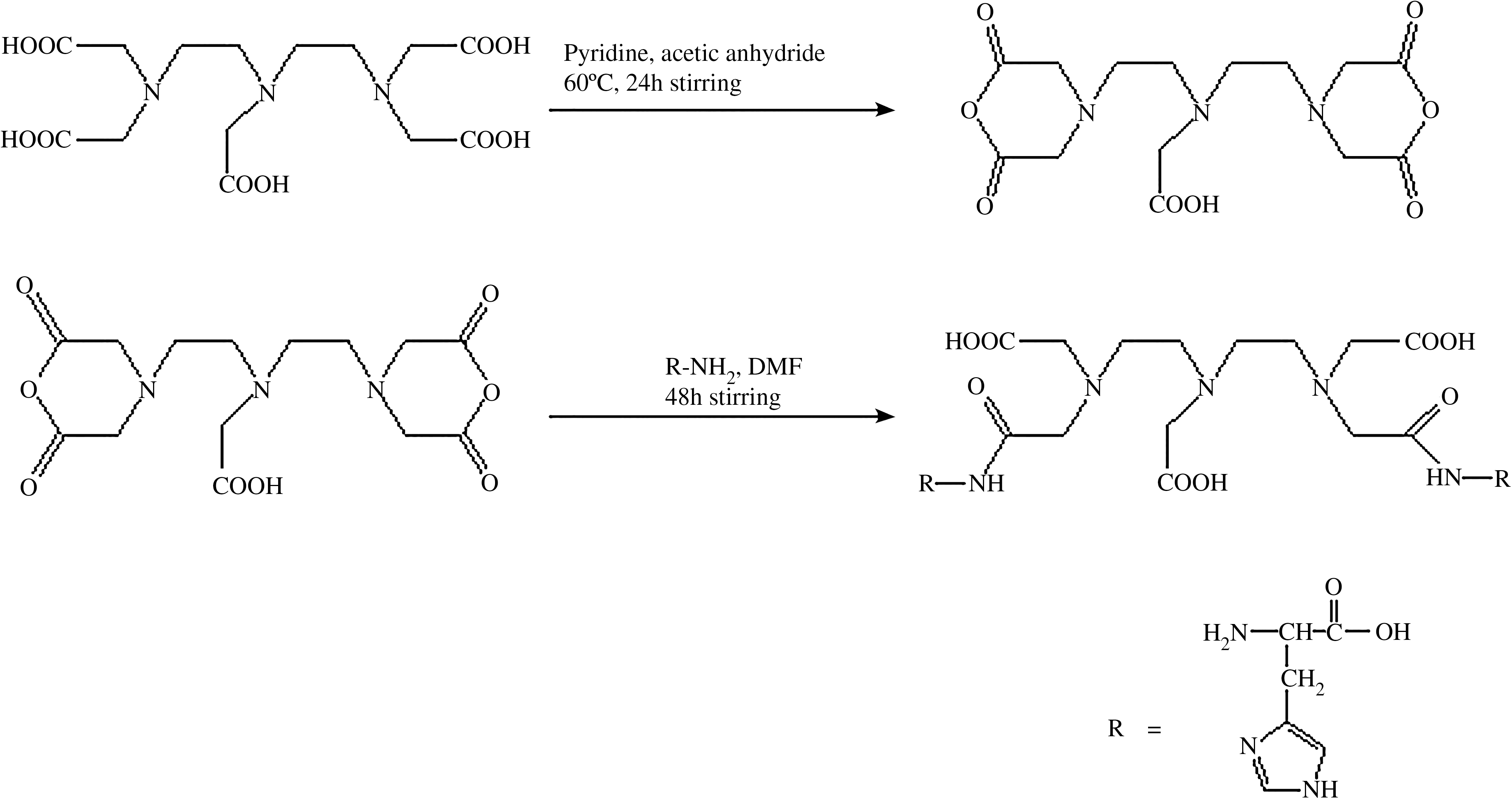
Synthesis scheme of DTPA-(His)2.
IR, infrared; NMR, nuclear magnetic resonance.
DTPA-bis(His)
First, 35.7 g (0.1 mol) of DTPA dianhydride was dissolved in 50 mL of dry dimethylformamide (DMF). A 10% excess of L-histidine (34.1 g, 0.22 mol) dissolved in DMF was added dropwise. Triethylamine was used to adjust the pH of the reaction mixture to around 8. The reaction mixture was stirred for an additional 12 hours at 60°C under a nitrogen atmosphere. To the reaction mixture, 100 mL of diethyl ether was added and left to stand for 6 hours. After filtration and drying in vacuo, DTPA-(His)2 was obtained as a light-yellow amorphous solid (yield: 53.3 g; 80.0%).
Animal models
Animal protocols were approved by the Institutional Animal Ethics Committee. New Zealand rabbits (2–3 kg) and BALB/c (22–28 g) were used for blood clearance and imaging and biodistribution studies. Rabbits were housed under conditions of controlled temperature of 22 ± 2°C and a normal diet.
Radiolabeling of the compounds with technetium (99mTc)
Radiolabeling of the compound was done following the procedure described in the literature. 28 –30 It was performed by taking 100 μL of 1.5–2 mg of solution of the compounds dissolved in water and taken in a shielded vial. Further, 60 μl of 1 × 10⁻² M of SnCl2 · 2H2O (dissolved in N2-purged 1-mL 10% acetic acid) was added, followed by freshly eluted saline solution of sodium pertechnetate (NaTcO4) (74 MBq, 100 mL). The pH of the reaction mixture was adjusted to 6.5 with 0.1 M of NaHCO3 solution. The vial was allowed to incubate for 20–30 minutes at room temperature. Labeling of the compounds, radiochemical purity, as well as R f of the 99mTc-based complex was determined by ITLC-SG strips, using 0.9% NaCl aqueous solution (saline) as developing solvent and simultaneously in acetone and PAW (pyridine, acetic acid, and water in a 3:5:1.5 ratio). Each ITLC was cut in 0.1-cm segments, and counts of each segment were taken.
In vitro serum stability assay
The fresh human serum was obtained by allowing blood collected from healthy volunteers to clot for 1 hour at 37°C in a humidified incubator maintained at 5% carbon dioxide, with 95% air. Then, the sample was centrifuged at 400 rpm, and the serum was filtered through 0.22-micron syringe filter into sterile plastic culture tubes. The above freshly prepared technetium radio complexes were incubated in fresh human serum at physiologic conditions (i.e., at 37°C) at a concentration of 100 nM/mL and then analyzed by ITLC-SG at different time intervals to detect any dissociation of complex. Percentage of free pertechnetate at a particular time point, that was estimated by using saline and acetone as the mobile phase, represented a percentage dissociation of the complex at that particular time point in serum.
Blood kinetic studies
The blood-clearance study was performed in normal rabbits (weighing 2–2.5 kg). First, 18.5 MBq of the 99mTc-labeled compounds (0.3 mL) was administered intravenously (i.v.) through the dorsal ear vein. At different time intervals, about 0.5-mL blood samples were withdrawn from the dorsal vein of the other ear and radioactivity was measured in the gamma-counter. The data from the experiment were expressed as the percentage of administered dose at each time interval (Fig. 1).
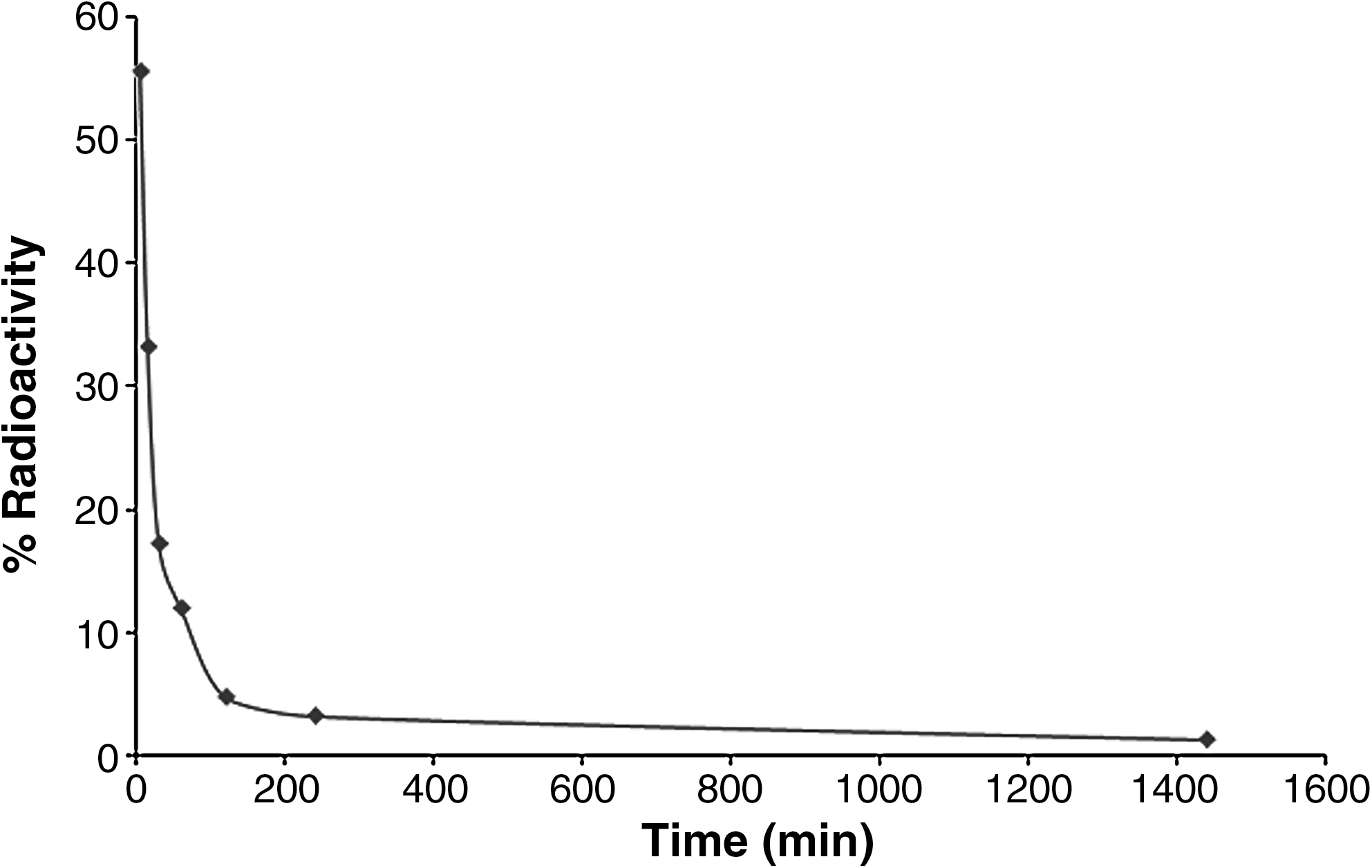
Blood kinetics study of 99mTc-DTPA-bis(His) in normal rabbit.
Scintigraphy in tumor-bearing nude mice
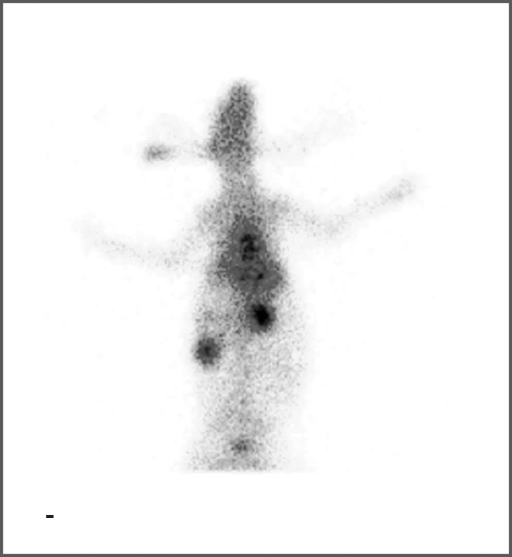
Tumor imaging was performed in mice bearing Ehrlich ascites tumor (EAT) tumor by administering 100 μL of the labeled conjugate (100 μg, 100 μci of activity). Images were taken by using a planar gamma camera equipped with a pinhole collimator. Images were obtained at different time intervals, starting from 15 minutes to 24 hours postinjection (Fig. 2).
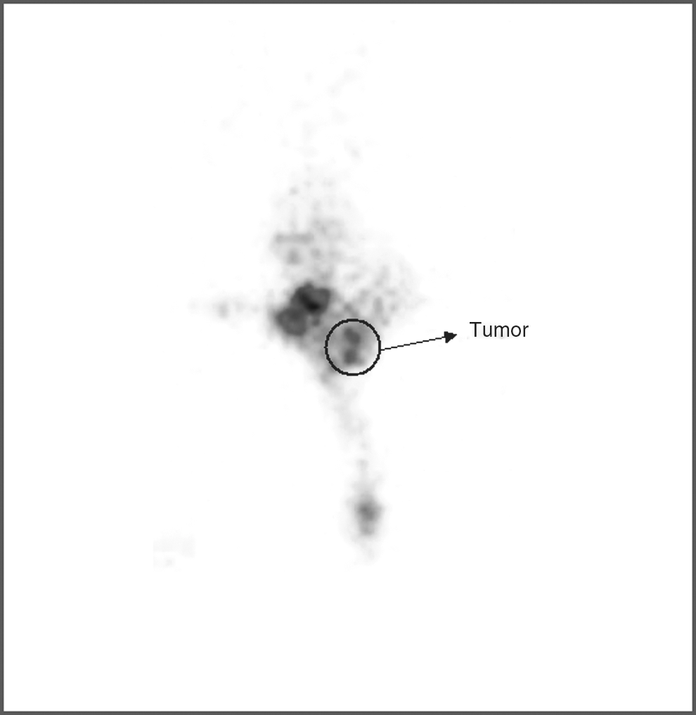
Whole-body γ-scintigraphy of 99mTc-DTPA-bis(His) in mice bearing Ehrlich ascites tumor.
Biodistribution study in mice
Balb/c mice with EAT grafted tumor in the hind leg was used for the tissue distribution studies. Animal handling and experimentation was carried out as per the guidelines of the Institutional Animal Ethics Committee. A dose equal to 10 μCi of labeled test compound was injected in each mice through the tail vein. The mice were sacrificed at different time intervals, and blood was collected, then different tissue and organs were dissected and analyzed. The radioactivity was measured in a gamma-counter. The actual amount of radioactivity administered to each animal was calculated by subtracting the activity left in the tail from the activity injected. Radioactivity accumulated in each organ was expressed as percentage administered dose per gram of tissue. Total volume of the blood was calculated as 7% of body weight. Biodistribution study of 99mTc-DTPA-bis(His) in mice bearing EAT tumor is shown in Table 2.
EAT, Ehrlich ascites tumor; T/M, tumor-to-muscle ratio.
Cell culture
Monolayer cultures of human glioma cell line U87-MG was maintained at 37°C in a humidified CO2 incubator (5% CO2, 95% air) in Dulbecco's modified Eagle's medium (DMEM; Sigma), supplemented with 10% fetal calf serum (FCS; Biological Industries, Israel), 50 U/mL of penicillin, 50 μg/mL of streptomycin sulfate, and 2 μg/mL of nystatin. Cells were routinely subcultured twice a week, using 0.05% Trypsin (Sigma) in 0.02% ethylene diamine tetraacetic acid (EDTA).
Receptor-binding studies
Exponentially growing cells (0.1 × 10⁶ cells/PD) (U87-MG) were plated at a uniform cell density and incubated overnight. Monolayer culture (U87-MG) of the cell lines were washed twice for 2 minutes with ice-cold binding buffer (25 mM HEPES, 10 mM MgCl2, and 1% bovine serum albumin BSA). The cell-line culture were then incubated for 40 minutes with labeled compounds (1–8 nM) in the absence and presence of the 100-fold excess unlabeled compounds for the estimation of total binding and nonspecific binding, respectively. Specific binding was obtained by subtracting nonspecific binding from total binding. At the end of each experiment, the cells were washed with ice-cold binding buffer three times for 3 minutes. The cells were lysed with 200 μL of lysis buffer. The cell-associated radioactivity was determined by gamma scintillation counting. Scatchard plot analysis was done by using EQUILIBRATE software from GraphPad (GraphPad Software, Inc., La Jolla, CA) (Table 2).
Results and Discussion
Synthesis and spectroscopic study of the complex
DTPA is an eminent chelate known to form stable complexes with metal ions, such as In, Y, Gd, and Eu. It can also be easily and efficiently labeled with 99mTc, with high radiochemical purity and stability. The 99mTc-DTPA complex is a classic nuclear-imaging agent and is used as a renal-imaging radiopharmaceutical in clinical practice. DTPA is also conjugated with biomolecules (such as antibodies, peptides, and steroids) to give target-specific pharmaceuticals that can display high tissue and organ selectivity. In an attempt to synthesize target-specific radiopharmaceuticals, novel DTPA-bis(amide)conjugate was synthesized in high yield, starting from DTPA. DTPA was first converted into DTPA-bis(anhydride), which further reacts with L-histidine in the presence of triethylamine in DMF to give the corresponding DTPA-bis(His) conjugate. The newly synthesized compound was characterized by spectroscopic techniques (IR, NMR, and mass spectroscopy). IR spectra at 1550 and 1680 cm−1 corresponds to the νN-H stretching, and νC = O stretching thereby indicated the formation of the amide bond, whereas in the 13C-NMR, two peaks at 178.33 and 172.99 ppm confirmed the presence of two types of the carboxylic acid group. In 13C-NMR, a spectrum peak at 170.49 ppm corresponded to a carbonyl group of the amide bond, which confirms the formation of the final conjugates.
Radiolabeling and blood kinetics study of the complex
Since technetium has many physicochemical properties, a variety of 99mTc-containing radiopharmaceuticals has become available for clinical use. In combination with favorable physical characteristics (γ-decay, 140 keV) and a convenient half-life (6 hours) which allow complex synthesis and prolonged imaging, 99mTc has become a popular nuclide, being used in over 80% of all routine diagnostic nuclear medicine procedures. Newly synthesized 99mTc- DTPA-bis(His) is also a very attractive radiopharmaceutical for target-specific scintigraphy, since it has several advantages, including good stability both in vitro and in vivo, low cost, and gamma-ray emissions, which are very important for clinical application of radiopharmaceuticals. The 99mTc-DTPA-bis(His) conjugate was prepared by the reduction of pertechnetate with stannous chloride, as reported in the literature. 28 –30 Radiocomplexation was completed in 10–15 minutes with a radiochemical purity of more than 96%, as monitored by ITLC-SG, using different solvent conditions. The complex was stable in dilute saline solution and in serum under physiologic conditions. In serum, neither transchelation of radioactive metal ions nor metal-ion transfer to serum proteins was observed. The blood-clearance study of 99mTc-DTPA-bis(His) showed a very rapid clearance of radioactivity from the blood circulation. Approximately 65%–75% of activity was cleared within 1 hour and more than 90% in 4 hour, which may be due to the hydrophilic nature of the radiotracers, which are responsible for their fast kinetics (Fig. 1), the biological half-life was found to be t1/2 (fast) 45 ± 0.041 minutes and t1/2 (slow) 6.5 hours ± 0.039 minutes, respectively.
In vitro cellular uptake assay
In vitro cellular uptake assay of 99mTc-DTPA-bis(His) was performed on human glioma and a nasopharyngeal carcinoma cell line (U87-MG and KB). 99mTc-DTPA was taken as the negative control and 99mTc-histidine as the positive control. Both 99mTc-DTPA-bis(His) and 99mTc-histidine significantly localized in the cancer cells, when compared with 99mTc-DTPA. Maximum cellular uptake of 99mTc-DTPA-bis(histidine) was observed at 2.5 hours, in comparison to 99mTc-DTPA.
Receptor-binding studies
Scatchard plot analysis revealed affinity of the compounds on tumor cell lines (U87-MG). K D was found to be 1.08 nM (Fig 3).
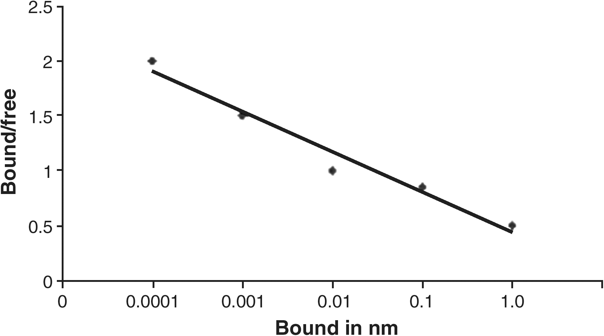
Scatchard plot of the specific binding data to the ratio of bound-to-free (B/F) for U87-MG cell lines.
Scintigraphy in EAT tumor-bearing mice
Tumor imaging was performed in mice bearing EAT tumor. Imaging of animals was carried out at different time intervals after administering the labeled compound i.v. through the dorsal ear vein. 99mTc-DTPA-bis(His) conjugate showed significant accumulation at the tumor site. γ-imaging in mice showed accumulation of activity in tumor at 30 minutes, which reached to maximum at 90 minutes and remained almost stable for 4 hours (Fig. 2).
Biobistribution studies
Biodistribution of the radiolabeled complex is an important phenomenon to study because it gives an idea about its metabolic excretory pathway and in vivo distribution of the radiocomplexed imaging agents. The percentage distribution of labeled conjugate in various organs of mice is shown as percentage of injected dose per gram organ or tissue at different time intervals. Initially, the radioconjugate was localized in the brain, heart, and liver, but with the passage of time, the activity in kidney was amplified, while in the intestine there was negligible increase in activity. This suggests that the major route of excretion of activity is through the kidneys. Also, with the passage of time, there was an increase in the accumulation of activity in the urinary bladder. Besides this, there was a retention of radioactivity in the liver for a considerable period, indicating that metabolism of amino acids takes place in the liver, but the excretion of radioconjugate and metabolites is mainly through the kidneys. Accumulation of radiocomplex in the liver may be because of the high-protein-binding nature of the radioconjugate. Biodistribution of DTPA-bis(His) in the normal rabbit is shown in (Fig. 4). Biodistribution of the compound in mice bearing EAT tumor showed remarkable localization at the tumor site. The high tumor accumulation of 1.47 ± 0.025 at 1 hour and 0.85 ± 0.001 at 4 hour were observed (Table 2).
Whole-body γ-scintigraphy of 99mTc-DTPA-bis(His) in normal rabbits.
Conclusions
The work represented in this article demonstrates the facile synthesis of amino-acid-based radioconjugate and its labeling with 99mTc. The radiotracer can be produced in high radiochemical yield and high radiochemical purity from stable precursors and is potentially valuable agent for imaging brain and other tumors with SPECT. Further, biodistribution study of the compound showed rapid, persistent accumulation of radioactivity in tumors with an excellent signal-to-background ratio. Other tissue studies, including those on muscle, lungs, heart, and liver, showed a relatively low uptake of radioactivity.
Footnotes
Acknowledgments
The authors thank Doctor R.P.Tripathi (Director, INMAS and DRDO) for providing us with the facilities and fellowship during the course of our research work. This work was supported under INM-306.
Disclosure Statement
No competing financial interests exist.