




Abstract
Introduction:
The use of radiolabeled molecules allows the study of in vivo biodistribution, target organs, and kinetic profile after systemic administration by 1) radioactive organ counting and 2) quantitative autoradiographic analysis of whole-body slices (WBA). However, these techniques are time- and animal consuming for several times studied. So, in vivo scintigraphic imaging should appear of interest for a first screening of compounds, as it is able to rapidly demonstrate tumoral uptake and kinetics by serial examinations in the same mice.
Materials and Methods:
In this study, the tumoral distribution and kinetics of six molecules considered as potential melanoma tracers radiolabeled with 125I were analyzed by gamma-scintigraphy comparatively to the results obtained by WBA. Tumoral uptake has been quantified and expressed by: 1) tumor-to-background ratios and 2) standardized tumoral uptake (STU) in percent injected dose per gram, with tumor weight being extrapolated from the measurement of the two diameters.
Results:
Results from STU analysis showed good agreement (correlation coefficient = 0.92) with those of WBA, and the same classification of compounds (on the basis of their melanoma affinity) was obtained, with two compounds (of six) being rejected.
Conclusions:
[125I] scintigraphic imaging appeared as a relevant, easy-going method for a first pharmacologic selection in mice.
Introduction
The development of radiopharmaceuticals for both a targeted imaging and therapy in oncology requires accurate preclinical animal studies for target identification and validation, drug-delivery quantitation in tumor, and pharmacokinetics determination. In preclinical studies, new drugs can be precisely compared with standard therapies or can be screened among series of analogs for further development on the basis of performance in mice bearing subcutaneous tumors. For all these studies, radiolabeled molecules constitute a powerful tool. 1 Radionuclide preclinical studies require radiolabeling procedures of the candidate drug that must not change the integrity of the structure, as well as their biologic properties. Drugs containing atoms that have multiple isotopes, such as iodine, in their structure appeared to us as very attractive, since the multiple isotopes of iodine [125I for flexibility of labeling in preclinical whole-body autoradiography (WBA), planar and single-photon emission computed tomography (SPECT) studies, 123I for extension into human SPECT, 124I for positron emission tomography (PET) imaging and 131I for radionuclide therapeutic approaches] provide a convenient bridge from animal models up into humans.
For many years, preclinical studies in small animal models, using radionuclide techniques, relied on tissue radioactivity counting and autoradiography in animal slices. WBA is an effective technique that allows an absolute quantitation of radiolabeled compound uptake in all the organs of the body, but is very time- and animal consuming. 2 Autoradiography could be considered as having limitations, since it requires the sacrificing of a large number of animals, which could be at the origin of significant interindividual variations and postmortem-induced artefacts. Indeed, such techniques preclude the possibility of serial studies in the same animals over time. Considering costs and timelines associated with the validation of preclinical studies, there is a real need for additional fast methods capable of rapid high-throughput screening, thus providing information on which rational scientific “go/no go” decision could be based. 1,3–4 Recent advances in instrumentation and methodology in radionuclide in vivo imaging modalities provide both a sensitivity and a resolution that allows preclinical “pharmacoimaging” studies in vivo in small animals. 5 –7 Scintigraphic imaging offers a noninvasive, functional access to a specific biologic target in vivo, with a high sensitivity for deep-tissue imaging (i.e., the ability to detect subnanomolar concentrations of radiotracer in the volume of interest) and signal-tissue quantitation.
Validation of tumor as a target was applied in our group for the development of innovative vectorization strategies toward a melanoma-associated molecular target (i.e., melanin) for application to diagnosis and therapy. 8 –10 Aromatic and heteroaromatic analogs of N-(2-diethylaminoethyl)-4-iodobenzamide have been recently developed as potential candidates for both targeted imaging and radionuclide therapy of melanoma. 11 The prerequisites for radiopharmaceuticals as imaging and therapeutic agents include favorable pharmacokinetics with a rapid, high tumor uptake, long tumor retention, rapid clearance from nontarget organs affording high tumor-to-healthy-organ ratios, and rapid whole-body clearance. 12
The aim of this study was to demonstrate the interest of “nuclear pharmacology” with scintigraphic technology for a rapid screening of such a series of iodobenzamide analog candidates, on the basis of melanoma affinity and pharmacokinetic tumor parameters determined in vivo in melanoma-bearing mice. With such a view, the tumoral distribution and kinetics of six molecules considered as potential melanoma tracers radiolabeled with 125I were analyzed by gamma-scintigraphy comparatively to the results obtained by WBA technology. Results showed how, for a first study focused on tumoral behavior, that gamma-scintigraphy appears as a relevant, rapid, and easy-going technique, as compared to WBA.
Materials and Methods
Chemistry
The following radioiodinated compounds evaluated in this study (Fig. 1) were synthetized and radiolabeled, according to the literature: [125I]N-(2-diethylaminoethyl)-1,4-dihydro-6-iodo-4-oxoquinoline-3-carboxamide hydrochloride salt ([125I]ICF01011), 11 [125I]N-(2-diethylaminoethyl)-6-iodoquinoxaline-2-carboxamide dihydrochloride salt ([125I]ICF01012), 11 [125I]N-(4-dipropylaminobutyl)-6-iodoquinoline-2-carboxamide dihydrochloride salt ([125I]ICF01014), 13 [125I]N-(2-diethylaminoethyl)-7-iodophenazine-1-carboxamide dihydrochloride salt ([125I]ICF01016), 13 [125I]N-(2-diethylaminoethyl)-9,10-dihydro-7-iodo-9-oxoacridine-4-carboxamide hydrochloride salt ([125I]ICF01035), 14 and [125I]N-(2-diethyl-aminoethyl)-7-iodo-9-(4-methanesulphonamido-2-methoxyanilino)acridine-4-carboxamide dihydrochloride salt ([125I]ICF01047). 14
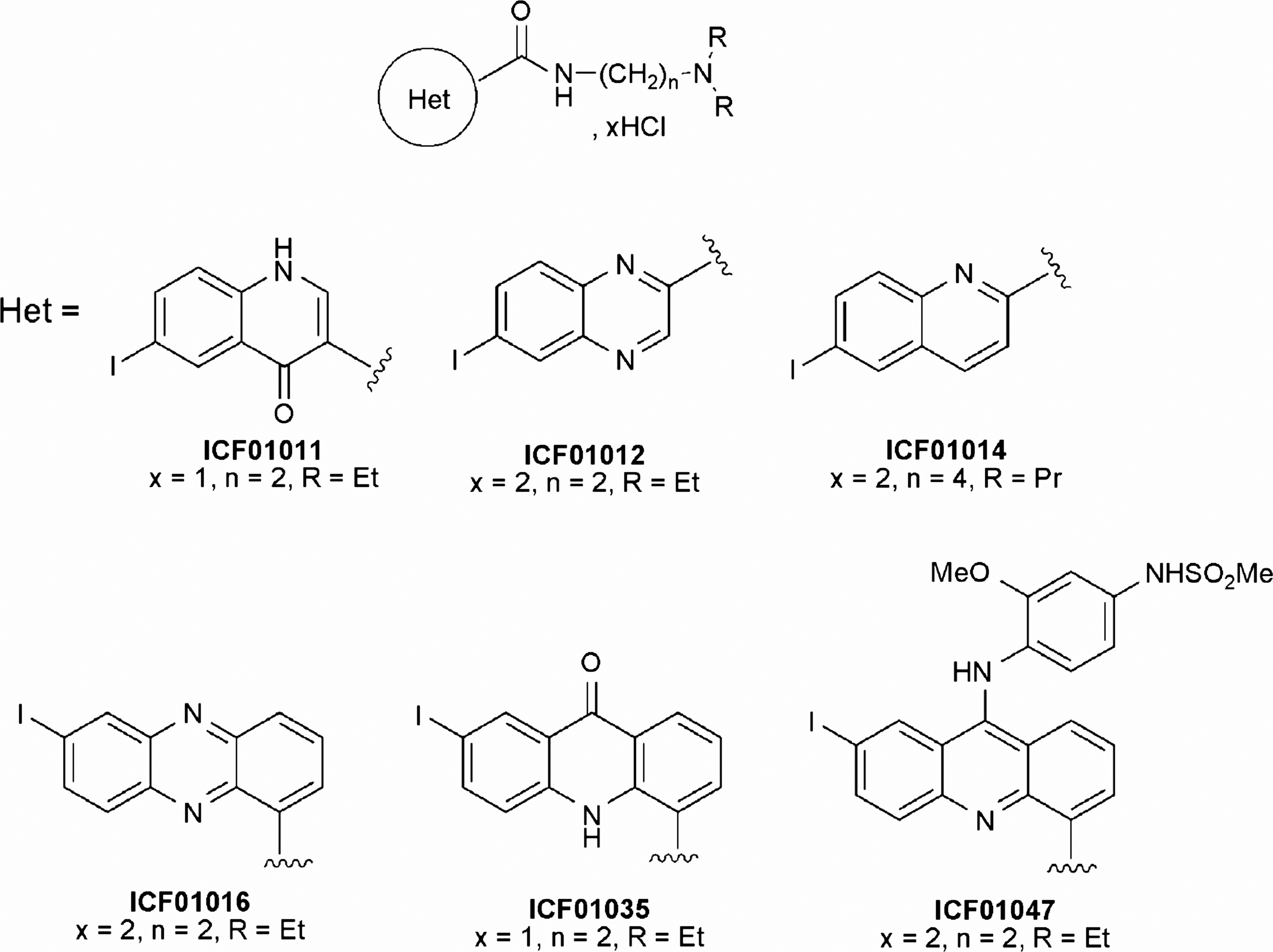
Chemical structures.
For each radiotracer, the actual injected dose to each animal was determined by measuring the syringe activity before and after injection, using both an activimeter (Capintec, Pittsburgh, PA) and scintigraphic acquisition.
Cells and animals
Murine melanoma cells (B16-F0) were obtained from ATCC (American Type Culture Collection, Manassas, VA) (no. CRL-6322). Stock cell cultures were maintained as monolayers in MEM Glutamax (Invitrogen, Cergy-Pontoise, France; Eagle's minimum essential medium with glutamine supplemented with 10% fetal calf serum, vitamins, sodium pyruvate, nonessential aminoacides, and gentamycin) and passaged by trypsination. The cells were grown in a humidified 37°C incubator containing 5% CO2. Early passages were frozen and stored in liquid nitrogen. For transplantation, an aliquot was grown in a monolayer culture.
Male C57BL/6J mice were obtained from Charles River (L'arbresle, France). They were handled and cared in accord with the guidelines for the Care and Use of Laboratory Animals (National Research Council, 1996) and European Directive 86/809/EEC. They were maintained at 21°C with a 12-hours light-dark cycle. They were fed with a breeding diet and received water ad libitum. Protocols were performed under the authorization of the French “Direction des Services Vétérinaires” (Authorization no. C63-113-10) and conducted under the supervision of authorized investigators in accord with the institution's recommendations for the use of laboratory animals.
Mice were inoculated subcuteanously (s.c.) in the left flank with 3 × 10⁵ cells in 0.1 mL of phosphate-buffered saline (PBS). Ten (10) days later, the tumors became palpable, with a percentage of tumor take of 98%–100%. Mice were then randomly divided into an WBA group and a scintigraphic-imaging group.
WBA
Each radioiodinated compound was administered intravenously (i.v.) (0.1 μmol, 0.74–0.92 MBq/animal). At selected time points (1, 3, 6, 24, and 72 hours), the animals (n = 2–3 animals per time point and per compound) were sacrificed by CO2 inhalation, immediately frozen in liquid nitrogen, and embedded in carboxy methyl cellulose (CMC). The frozen animals were cryosectionned at −22°C into 40-μm slices, which were dehydrated for 48 hours in a cryochamber and analyzed by using the technique already described. 15 Whole-body slices (8 slices per mouse and per time point) were then exposed for 1000 minutes in a digital autoradiographic analyzer (AMBIS 4000 detector; Scanalytics, CSPI, San Diego, CA). Surfacic activity (net cpm/mm2) was quantified in regions of interest (ROIs) delineated over tumoral pattern. All activity values were corrected for radioactive decay, and the overall mean intratumoral uptake was determined at each time point, and for all slices, as percent injected dose per (%ID/g), as previously described and validated. 16
Gamma-camera
Imaging was performed by using a gamma-camera dedicated to small animals (γIMAGER®; Biospace Mesures, Paris, France). The gamma-camera consists of a R 3292 Hamamatsu position-sensitive photomultiplier having a continuous 4-mm-thick × 120-mm diameter CsI(Na) crystal leading to a 10-cm field of view. For [125I] imaging, the camera was equipped with a parallel-hole collimator 1.8/0.2/20 (hole diameter/septum thickness/height in mm). All the acquisitions were performed with a 15% window centered on the 35-keV peak of [125I].
[125I] gamma-imaging of phantom
For the evaluation of camera sensitivity and response to activity variations in the range of activity injected, a phantom consisting of a 10-mm-thick Plexiglas plate, with holes of 0.8 cm in diameter (internal diameter), was used. The holes were filled with [125I]NaI solutions of known activity concentration (seven activities ranging from 15 kBq to 3.8 MBq, with a 2-dilution factor). Phantom was scanned five times (5 minutes for each acquisition). For each radioactive hole, the reproducibility of quantitative analysis was assessed by calculating the coefficient of variation (CV =100 × standard deviation/mean), and a mean reproducibility factor was determined for the range of 15 kBq–3.8 MBq investigated.
In vivo [125I] gamma-imaging of melanoma-bearing mice
The animals were anesthetized by intraperitoneal (i.p.) injection (200 μl/20g mouse) of a mixture (4:1) of ketamine (Imalgene 500,® Rhone Mérieux, Lyon, France) and xylazine (Rompun 2%,® Bayer, Puteaux, France).
For each of the radiolabeled compounds assessed, B16F0-melanoma-bearing mice (2–3 animals per compound) were submitted to serial scintigraphic imaging at 1, 3, 6, 24, 48, and 168 hours after injection (3.7 MBq/animal). A 10-minute duration image was acquired on anesthetized mice placed in the anterior position over the parallel-hole collimator 1.8/0.2/20 (hole diameter/septum thickness/height in mm) of the γ-IMAGER. Reproducibility in animal positioning for the serial images was achieved by using a graduated reference grid.
Quantitative analysis of scintigraphic scans was performed by using the GAMMAVISION+® software (Biospace Mesures, Paris, France). For each animal and for each acquisition, ROIs were drawn by the same experienced investigator around: 1) B16 melanoma by using a subjective visual boundary method in which attempts were made to draw the ROI boundary as close as possible to the presumed tumor boundary; 2) controlateral paw background; and 3) whole body. For each ROI, total activity, minimum and maximum pixel values, ROI size (in mm2), average activity per mm2, and standard deviation were obtained.
After correction for physical decay, whole-body and intratumoral distribution were determined at each time point. Whole-body distribution was expressed as %ID, and intratumoral distribution was assessed by determining: 1) tumor-to-background ratio and 2) standardized tumor uptake (STU) in %ID/g: For expressing tumoral uptake in terms of “standardized uptake value,” we used an extrapolation method of tumor mass from the Calculated Tumor Weight (CTW)
17
with L = length in mm; W = width in mm measured with a caliper; p = ponderation factor (determined from experimental measures from a large panel of tumoral samples in the same conditions).
Results
Phantom imaging: Assessment of reproducibility, sensitivity, and linearity of the gamma-camera response
For the range of activity tested (15 kBq–3.8 MBq), the reproducibility of activity measurement by ROI analysis was 99.05% ± 0.42%. The sensitivity of the gamma-camera for [125I] was 14.62 ± 2.88 cpm/kBq (560.47 ± 91.62 cpm/μCi). Figure 2 illustrates phantom activity measurements by ROI analysis versus actual activity, measured by gamma-counting: A linear response of the camera was observed, for the 15–3885 kBq (0.5–105 μCi)-activity range investigated, with a strong correlation coefficient (r 2 = 0.9987).
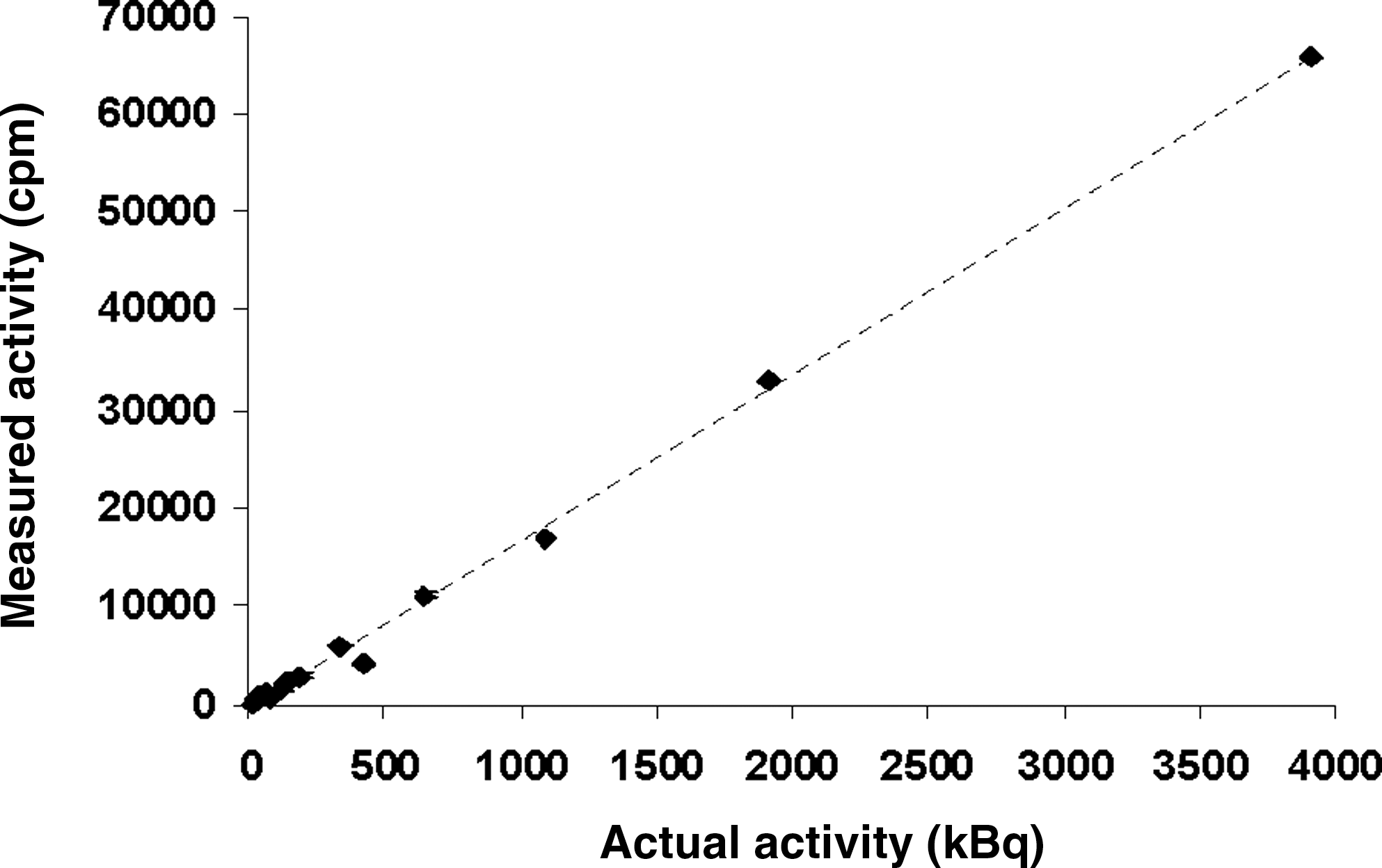
Phantom activity measurements: region of interest analysis of [125I] phantom scintigraphic imaging versus actual activity measured by gamma-counting, for the range of 15 kBq–3.8 MBq. A linear response of the camera was observed, with a correlation coefficient of 0.9987.
[125I] radiolabeled compound distribution and pharmacokinetics assessed by WBA
As illustrated in Figure 3, B16 melanoma accumulation was observed for five (i.e. [125I]ICF01012, [125I]ICF01014, [125I]ICF01016, [125I]ICF01011, and [125I]ICF01035) of the six compounds administered as early as 1 hour after injection. Four compounds (i.e., [125I]ICF01012, [125I]ICF01014, [125I]ICF01016, and [125I]ICF01035) evidenced tumoral accumulation levels higher than 10% ID/g from 1 to 72 hours postinjection (p.i.). The highest values obtained for [125I]ICF01012, [125I]ICF01014, [125I]ICF01016, and [125I]ICF01035 were 35 ± 7, 27.4 ± 9.4, 27.7 ± 6.1 and 27.4 ± 8.3%ID/g of melanoma, respectively. For [125I]ICF01012, [125I]ICF01014, and [125I]ICF01016, this highest tumoral accumulation was observed at 6 hours p.i. For [125I]ICF01035, highest tumoral uptake was maintained until 24 hours p.i., and tumoral retention was significantly higher at 72 hours p.i., respectively, to the other molecules (17.2 ± 2.5 %ID/g of melanoma for [125I]ICF01035; 12.4 ± 1.6 %ID/g for [125I]ICF01012; 10.9 ±3.3 %ID/g for [125I]ICF01014; and 6.3 ± 2.2 %ID/g for [125I]ICF01016). [125I]ICF01011 exhibited lower tumoral accumulation and retention, and respectively, to the other drugs (peak obtained at 6 hours p.i.; %ID/g = 5.8 ± 2). No significant tumoral uptake was observed for [125I]ICF01047 (peak obtained at 3 hours p.i.; %ID/g = 1.5 ± 0.1). As a consequence, ICF01011 and ICF01047 were rejected from the selection.
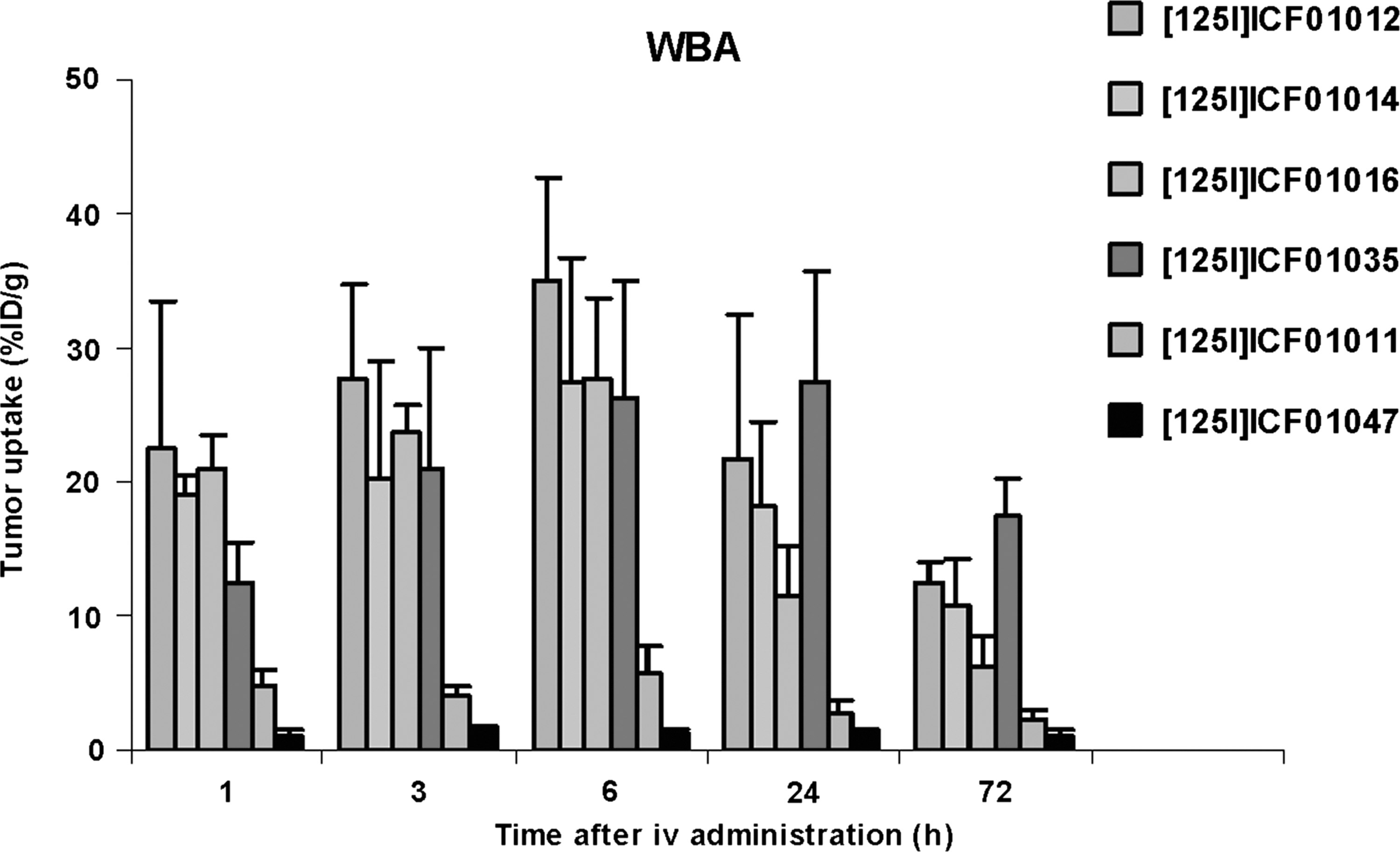
Tumoral accumulation of [125I]ICF01011, [125I]ICF01012, [125I]ICF01014, [125I]ICF01016, [125I]ICF01035, and [125I]ICF01047 determined by quantitative whole-body autoradiography at selected time points after intravenous administration to B16F0 melanoma-bearing mice (0.74–0.92 MBq/animal). Data are presented as mean ± standard deviation (2–3 animals/time point/compound).
[125I] radiolabeled compound distribution and pharmacokinetics assessed by two-dimensional (2D) planar scintigraphy
Qualitative analysis of scintigraphic scans
Qualitative analysis of scintigraphic scans evidenced radioactive patterns within digestive content, muscle, and thyroid (at later time points). Tumoral and eye accumulation was also observed, with the uptake level and time of retention, that may vary from one compound to another.
Figure 4 illustrates whole-body scintigraphy obtained in B16F0 melanoma bearing mice at various time points after i.v. administration of [125I]ICF01012 compound with high tumoral uptake for a long retention time. At an early stage after injection, localization of the radioactivity was observed in the tumor, digestive content, and muscle. At later stages, thyroid and eye accumulation was also detectable.

Representative serial [125I] imaging normalized with the same color (shown in black and white) scaling (at various time points, from 1 to 168 hours) after intravenous administration of [125I]ICF01012 to a B16F0-melanoma-bearing mouse (3.7 MBq/mouse and 10-minutes duration planar acquisition).
Quantitative analysis of scintigraphic scans
First, whole-body distribution was assessed as %ID as a way to evaluate retention and elimination of the six iodobenzamide derivatives (Fig. 5). Compound excretion showed a rapid kinetic for the first 24 hours p.i, and elimination was almost total after 72 hours (>90%), except for [125I]ICF01012 and [125I]ICF01035, with 20% and 40% ID still present, respectively (Fig. 5A). Tumoral uptake and retention were determined from ROI-based counts, according to two methods.

Whole-body accumulation of [125I]ICF01011, [125I]ICF01012, [125I]ICF01014, [125I]ICF01016, [125I]ICF01035, and [125I]ICF01047 determined by region of interest analysis of in vivo scintigraphic imaging (3.7 MBq/mouse and 10-minutes duration planar acquisition). (
Since the delineation of ROI was critical for radioactivity-uptake calculations, we looked at the relation between ROI-uptake surface and actual tumoral-surface caliper measurement. The correlation coefficient ranged from 0.73 to 0.93, for compounds with low and high tumor tropism, respectively. Considering the whole study (i.e., comparing all values obtained without any distinction of the compound administered), the overall correlation coefficient was 0.90.
Assessment of tumor-to-background ratio at each time point (Fig. 6) evidenced that background in the control flank was negligible, as evidenced by ratios in the range of 1.5–4 (for [125I]ICF01047 and [125I]ICF01016, respectively) at 1 hour p.i to ranges of 4.6–31.8 (for [125I]ICF01047 and [125I]ICF01012, respectively) at 72 hours pi. At 7 days p.i., no detectable signal could be evidenced in melanoma-bearing animals injected with [125I]ICF01011 and [125I]ICF01047. At this time, tumor-to-background ratios obtained for [125I]ICF01012, [125I]ICF01014, [125I]ICF01016, and [125I]ICF01035 were in the range of 18–43.

Quantitative analysis of scintigraphic imaging of B16F0-melanoma-bearing mice i.v. administered with [125I]ICF01011, [125I]ICF01012, [125I]ICF01014, [125I]ICF01016, [125I]ICF01035, and [125I]ICF01047 (3.7 MBq/mouse and 10-minutes duration planar acquisition. Results are presented as tumor-to-background ratio determined at each time point and for each compound. Data are presented as mean ± standard deviation (2–3 animals/time point/compound).
The content of radioactivity in the whole body, minus the tumoral uptake value presented in Figure 5B, showed that the elimination from nontarget organs was rapid for all compounds. After 24–72 hours, the activity was concentrated within tumor, more especially for [125I]ICF01012 and [125I]ICF01035.
Figure 7 shows STU values assessed at each time point. Four compounds ([125I]ICF01012, [125I]ICF01014, [125I]ICF01016, and [125I]ICF01035) of six evidenced STU higher than 10% ID/g and long-lasting tumoral uptake (up to 72 hours and also 7 days after administration). [125I]ICF01011 and [125I]ICF01047 accumulated within B16F0 melanoma at levels <5% ID/g and did not show any tumoral retention, as previously observed with WBA. As a consequence, [125I]ICF01011and [125I]ICF01047 were rejected from the selection.
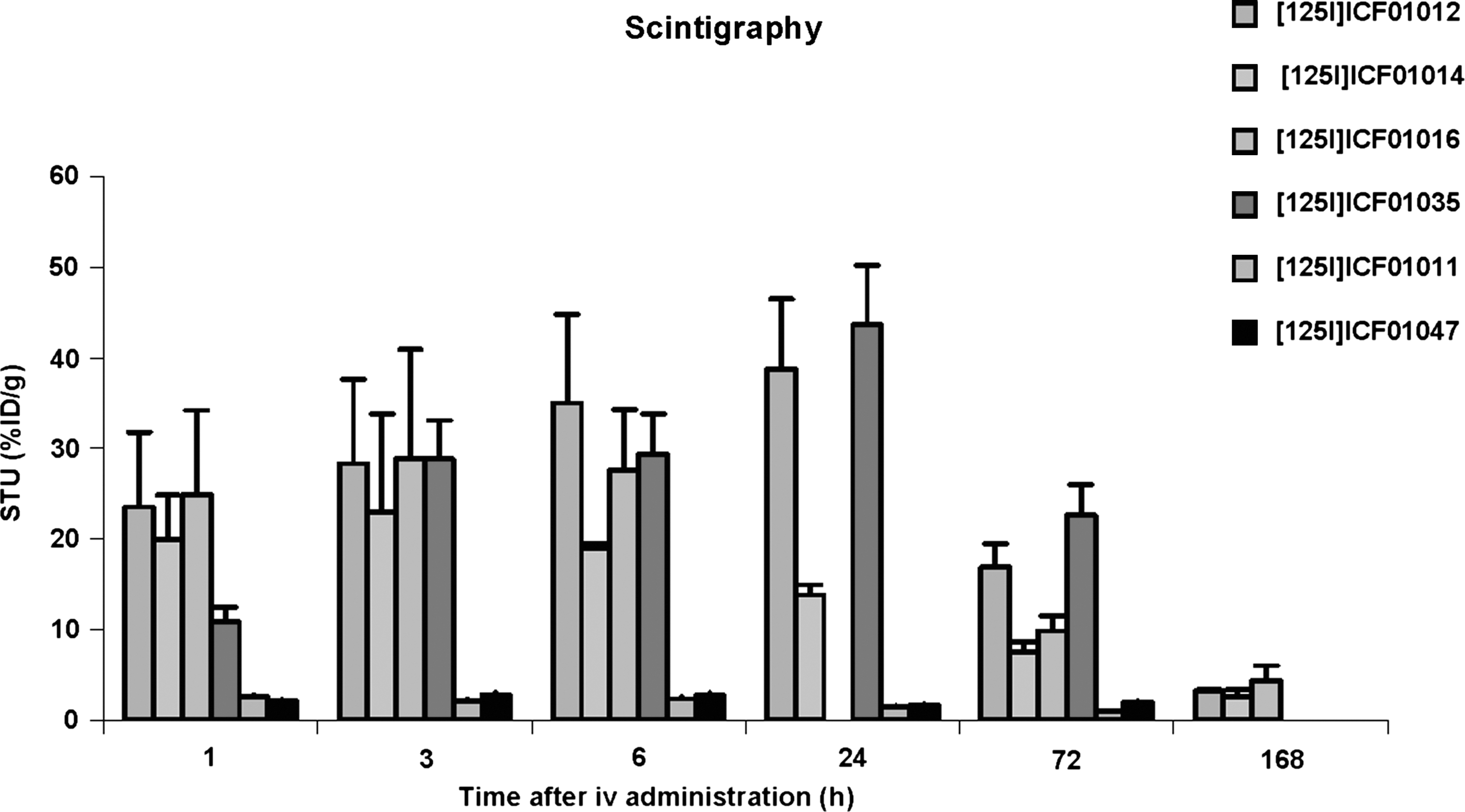
Quantitative analysis of scintigraphic imaging of B16F0 melanoma bearing mice i.v. administered with [125I]ICF01011, [125I]ICF01012, [125I]ICF01014, [125I]ICF01016, [125I]ICF01035, and [125I]ICF01047 (3.7 MBq/mouse and 10-minutes duration planar acquisition). Results are presented as standardized tumor uptake in percent injected dose per gram, determined at each time point and for each compound. Data are presented as mean ± standard deviation (2–3 animals/time point/compound).
Results from STU analysis showed good agreement (correlation coefficient = 0.92) with those of WBA (Fig. 8). Data obtained by WBA and scintigraphy were analyzed with a repeated-measures analysis of variance: No significant differences (p = 0.11) were observed between data obtained by using one or the other of the two methodologies.
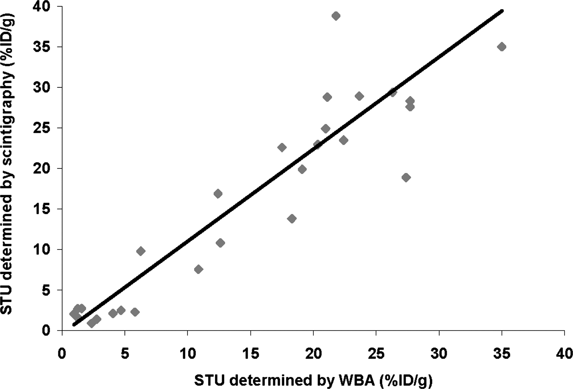
Agreements plots displaying tumoral uptake determined as standardized tumor uptake (STU) parameter in percent injected dose per gram from region of interest analysis of in vivo scintigraphies (y-axis) versus tumoral uptake determined from whole-body autoradiography (WBA) (x-axis). Results from STU analysis showed good agreement (correlation coefficient = 0.92) with those of WBA.
Discussion
The major aim of this work was to evaluate in vivo [125I] gamma-imaging for a first screening of compounds as potential melanoma radiopharmaceuticals, in view of imaging and radionuclide therapy applications. The primary finding in this study was that, in spite of the drawbacks in the radionuclide's physical properties (i.e., a low proportion of gamma-emitting rays and a low energy), in vivo scintigraphic imaging with iodine-125 can be easily applicable to s.c. melanoma-bearing mice, using a small-animal gamma-camera. Gamma-rays with low energies, such as those emitted by iodine-125 (35 keV), are known to be less penetrating than those emitted by technetium-99m (140 keV) or iodine-123 (159 keV), routinely used for diagnostic imaging in humans. Nevertheless, 35-keV rays emitted by 125I have an average path length of about 1 cm in soft tissue, which allows a sufficient fraction of emitted radiation flux to escape from the mouse for external imaging. 18 From all the 72 scintigraphic scans qualitatively analyzed in this study, whole-body, tumor, gastrointestinal tract, thyroid, bladder, and eyes were clearly identifiable in the 2D planar images. Thyroid patterns demonstrated some dehalogenation in vivo that was at the origin of an increased activity in nonspecific tissue (i.e., digestive organs). Faster blood clearance in vivo combined with tumoral accumulation and retention resulted in good tumor-to-background contrast images as early as 1 hour p.i. (tumor to background ratio around 2). At the later stages of the study, (day 7 p.i.), tumor-to-background ratios of 18–43 proved a high specific tumoral retention.
Quantitative analysis of in vivo scintigraphic scans, in terms of whole-body distribution, appeared as particularly useful to evaluate compound elimination in serial imaging in the same living animals. Intratumoral distribution was assessed in vivo by both tumor-to-background ratio and STU. Tumor-to-background ratio analysis allowed, in vivo, the evaluation of tumoral retention by taking into account whole-body elimination, and represents a relevant parameter for predicting clinical imaging. STU analysis allowed the evaluation of tumoral specific accumulation in terms of %ID/g, with a good correlation with data obtained from the reference method (i.e., whole-body quantitative autoradiography). Nevertheless, such in vivo screening imaging strategies should not exclude WBA analysis (in case of positive selection of compound) that remains the gold standard for absolute quantitation of radiopharmaceutical distribution in organs and allows a more sensitive detection of sites with low levels of radioactivity accumulation.
Planar imaging has, of course, limitations, because it represents a 3D distribution of radiopharmaceutical in a 2D display. It is well known that in vivo radioactivity-uptake calculations can also be affected by soft-tissue attenuation of photons that need to travel through the body to be detected. 19 –21 The good agreement obtained between in vivo and ex vivo determinations of tumoral uptake could be attributed to the model itself, namely the positioning of the s.c. tumor. The small size of animal reduces the magnitude of attenuation and scatter in the body and, therefore, reduces the relative magnitude of the error. Moreover, melanoma were s.c. situated in the flank of the animals; so, tumoral tissue protudes, and consequently, a part of it is covered with skin only, which contributes little to the background activity.
We should highlight that this screening study of compounds for their affinity for melanoma by 1) in vivo 125I scintigraphy, using a small-animal gamma-camera, and 2) WBA, led to the selection and exclusion of identical compounds.
This study, performed with a melanoma model, demonstrated that intratumoral uptake and retention of benzamide-derivative candidates for targeted imaging and/or radionuclide therapy can be accurately monitored noninvasively in the same animals by in vivo planar scintigraphic imaging. Such studies are able to provide both preliminary biodistribution and pharmacokinetics parameters useful for a rapid screening. Planar imaging is, by far, the simplest method for quickly draw conclusions about drug distribution, elimination, tumoral targeting, and retention by serial examination of the same living animals. Such 125I imaging protocols for screening molecule candidates for tumor targeting and retention could be, of course, applicable to other s.c. implanted-tumoral models in mice. It requires standardized experimental procedures, more especially for tumor implant on the flank of the animals.
Conclusions
The most important contribution that imaging can make to drug development is helping to distinguish between a drug that fails and a drug that should be selected for the proof of concept. This should be useful for reducing screening procedures from being time- and animal consuming.
Moreover, the recent development of dedicated small-animal computed tomography (CT) systems should also be helpful to assess, in vivo, the accurate tumor volume by CT ROI analysis for application to the determination of more precised activity accumulation. An additional study is under way in our lab to evaluate such a methodology.
Footnotes
Acknowledgments
The authors thank the French Agence Nationale de la Recherche and the Ligue Regionale contre le Cancer for their financial support.
Disclosure Statement
No competing financial interests exist.