
Abstract
Many tumor immunotherapy efforts are focused on upregulating the expression of 4-1BB/4-1BBL by transferring genes into immune cells or tumor cells. In this study, we sought to study whether 4-1BBL, expressed in normal tissue cells, inhibits the growth and metastasis of tumor. The expressing plasmid, p4-1BBL, was constructed and was used to treat established melanoma in situ model and metastasis tumor model mice, respectively, with injecting directly into the skeletal muscle of the inoculation site and systemically administering plasmid with the hydrodynamics-based gene-delivery approach. Administration of p4-1BBL resulted in high-level expression in the muscle and liver. Treatment of tumor-bearing mice with 4-1BBL plasmid DNA significantly suppressed the growth and metastasis of melanoma without significant toxicity, compared with mice treated with control-plasmid DNA. This treatment with plasmid p4-1BBL in vivo induced an infiltration of CD8+ T-cells into the tumor milieu and an increase of levels of interleukin-2 and interferon-gamma. Our study suggests that 4-1BBL expressed in normal tissue cells has significant antitumor activity and may have therapeutic potential as an antitumor agent in the clinic. Directly intramuscular injection peritumor and hydrodynamic intravenous injection may provide promising immune gene-therapy approaches for different tumor clinical stages.
Introduction
Melanoma is one of the malignant tumors with high propensity to recurrence and metastasis. Most clinical recurrences occur in the primary tumor with thickness bigger than 4 mm, 1 and the most frequent site of metastases is the lung. 2 Because the majority of deaths from melanoma are due to recurrent and metastatic disease, improvement in the probability of local control and inhibitions of metastasis of melanoma are expected to become effective treatments, leading to the prolongation of the lifespan. Among the treatment schemes, immunotherapy strategy has proven to be a standard for the treatment of patients with melanoma, 3,4 because melanoma has proven to have a low sensitivity to chemotherapy and radiotherapy. 5 –7
With the progression of malignant melanoma, the antitumor immune response of the body is always low. 8,9 Low-level expression of some costimulatory molecules is an important mechanism for tumor to evade from the attack of immunity system and increasing the expression of these molecules can enhance the antitumor immune response. Among the costimulatory molecules, 4-1BBL is a well-characterized costimulatory molecule expressed by APC, including B-cells, macrophages, and DCs. 10 4-1BBL/4-1BB signaling has profound effects on T-cells, including activation of both CD4+ and CD8+ T-cells, increased levels of cytokine secretion, enhanced clonal expansion, and increased survival. 11 –15 Recent studies have noted strong antitumor effects mediated by 4-1BB stimulation, using agonistic anti-4-1BB antibodies and adenoviruses expressing 4-1BBL. 16 –18
In this study, we constructed an expression-plasmid-encoding mouse 4-1BBL gene and transferred 4-1BBL gene into mice by using the direct intramuscular (i.m.) injection peritumor and hydrodynamic intravenous (i.v.) injection, 19,20 which demonstrated high expression level of plasmid in normal muscle cells and liver cells in vivo. The function of expressed 4-1BBL was determined by measuring levels of function of lymphocytes in mouse, melanoma in situ, and pulmonary metastasis was significantly inhibited by using recombinant 4-1BBL. Our data suggest that 4-1BBL expressed in normal tissue cells has significant antitumor activity and may have therapeutic potential as an antitumor agent in the clinical setting. Direct i.m. injection peritumor and hydrodynamic i.v. injection may provide promising immune gene therapy approaches for different tumor clinical stages.
Materials and Methods
Mice and cells
Male C57BL/6 mice (4–6 weeks old) were purchased from the Center of Experimental Animals of Chinese Academy of Medical Science (Beijing, China) and housed in specific pathogen-free conditions. All studies involving mice were approved by the institute's Animal Care and Use Committee. The murine melanoma B16F1 cell line was purchased from the China Center for Type Culture Collection (Wuhan, China) and was cultured in complete Dulbecco's modified Eagle's medium (DMEM), supplemented with 10% fetal calf serum (FCS), 10 mM of HEPES, 2 mM of L-glutamine, 100 μg/mL of penicillin, and 100 U/mL of streptomycin.
Plasmids
The eukaryotic expression plasmid, p4-1BBL, was constructed previously. 21 The plasmid, pcDNA3.1, was purchased from Invitrogen Life Technologies (Carlsbad, CA). Plasmid DNA used for gene therapy was prepared by the alkaline-lysis method and purified by polyethylene glycol, followed by selective compaction with spermine (Sigma-Aldrich, St. Louis, MO), as previously described. 22 The endotoxin levels were less than 3 pg/μg of plasmid DNA, as determined by the Limulus amebocyte lysate assay (BioWhittaker, Walkersville, MD). All plasmid preparations for injections were resuspended in sterile 0.9% saline.
Gene transfection
In the in situ tumor model mice, gene transfection was done by direct local injection. Briefly, naked plasmid was injected directly into the muscle (via i.m. injection) of the inoculation site. In the metastasis tumor model mice, the plasmid was injected into the tail vein (via i.v. injection) within 5 seconds with the hydrodynamics-based gene-delivery approach. The expression of plasmid was detected with conventional reverse-transcription polymerase chain reaction (PCR), real-time PCR, Western-blot, and immunohistochemical staining.
Conventional reverse-transcription PCR and real-time PCR
Total RNAs were isolated by using TRIzol reagent (Invitrogen Life Technologies) from tissues after treatment with p4-1BBL. After being treated with DNase I (Promega), equal amounts of RNAs were reverse-transcribed and the cDNAs were amplified by the PCR with gene-specific primers at 94°C for denaturing, 54°C for annealing, and 72°C for extension. β-actin mRNA was used as an internal control and coamplified. Previous experiments determined 29 cycles to be optimal. Specific primers used were: 4-1BBL, sense 5′-GAGTCAAAGCTTATGGACCAGCACAC-3′, antisense primer, 5′-GCGCTGAATTCTCATTCCCATGGGTT-3′; β-actin, sense 5′-ATGGGTCAGAAGGACTCCTATG-3′, antisense 5′-ATCTCCTGCTCGAAGTCTAGAG-3′. The amplified products were analyzed by electrophoresis on 1.5% agarose gels and stained with ethidium bromide. Quantitative real-time PCR for inter leukin-2 (IL-2) and inter feron-gamma. (IFN-γ) genes was done by using primers and procedures previously described. 23,24 The results were expressed as the expression level of each gene relative to that of housekeeping gene, β-actin.
Western blotting
Seventy-two (72) hours after transfection, tissue samples were lysed or homogenized for Western blot, as previously described, 25 and then incubated with 0.2 μg/mL of antimouse 4-1BBL mAb (TKS-1), provided as a gift by Dr. Hideo Yagita (Juntendo University of Japan). After washing, membranes were incubated with peroxidase-labeled antigoat IgG Ab (Amersham Pharmacia Biotech, Piscataway, NJ), and the bound antibodies were detected by using the ECL Western Blotting Detection System (Amersham Biosciences, Uppsala, Sweden), according to the manufacturer's instructions.
Immunohistochemical staining
Tissues from treated mice were surgically excised, fixed for 12–24 hours in 4% formamint, embedded in paraffin, and sectioned. Sections were stained with hematoxylin and eosin (H&E). For indirect immunostaining, sections were incubated overnight at 4°C with a mouse anti-CD8 mAb diluted at 1/100. Biotinylated Abs to mouse IgG were used as secondary Abs, followed by peroxidase-conjugated streptavidin in the third step. To analyze the expression 4-1BBL, the 4-1BBL mAb (TKS-1) was used for immunohistochemical staining. 26
Tumor in situ model and treatment protocol
For experiments on in situ tumor, C57BL/6 mice were inoculated with B16F1 melanoma cells by the injection of 1 × 10⁵ cells in 100 μL of phosphate-buffered saline (PBS) into the right hind thigh muscle. Three (3) days after inoculation, the naked expression plasmids were injected i.m., every other day six times, into the inoculation site at the 100-μg dose in 100 μL of sterile saline. Mice of the control group received an equal volume of saline or an equal amount of pcDNA3.1 plasmid. Tumor size was measured by using calipers fitted with a Vernier scale, when tumor could be palpated. The tumor diameter was calculated by using the formula; (a × b)/2, with a as the larger diameter and b as the smaller diameter. Mice were sacrificed and tumors were dissected and weighed on the indicated days after inoculation or for ethical reasons when animals showed severe distress. The average inhibition rate percentage = ((average tumor weight of pcDNA3.1 plasmid control group − average tumor weight of treated group)/average tumor weight of pcDNA3.1 plasmid control group) × 100%.
Experimental tumor metastasis model and treatment protocol
To establish the pulmonary metastasis model, C57BL/6 mice were injected i.v. with 1 × 10⁵ B16F1 melanoma cells suspended in 100 μL of PBS. Metastatic foci appeared on lungs as discrete black pigmented foci that were easily distinguishable from normal lung tissue. For the treatment of mice with 4-1BBL, naked plasmid p4-1BBL dissolved in 2 mL of saline was injected i.v. via the tail vein within 5 seconds on day 3 after tumor injection, 27,28 every 3 days four times. Mice of the control group received an i.v. injection of an equal volume of pcDNA3.1 and saline every 3 days. The number of pulmonary metastatic foci in the treatment and control groups was counted in a double-blind fashion on day 28 after tumor inoculation. Metastatic foci, which were too consolidated and numerous to count, were assigned an arbitrary value of ≥600.
ELISA
The levels of IFN-γ and IL-2 in serum were assessed by enzyme-linked immunoadsorbent assay (ELISA), using murine IL-2 or IFN-γ ELISA kits (eBioscience, San Diego, CA), according to the manufacturer's protocol.
Cytotoxicity assay
Standard 51Cr-release assays were performed. B16F1 melanoma as target cells, spleen lymphocytes from tumor bearing mice were individually treated with p4-1BBL or pcDNA3.1 used as effector cells. The procedure was previously described. 29 Specific lysis was determined as follows: percent specific release = 100 × (experimental cpm −spontaneous release)/(maximum release − spontaneous release). Spontaneous release was ≤20% of maximum release in all experiments.
Statistics
Results were expressed as mean values ± standard deviation, and the differences were determined by the analysis of variance (ANOVA) test, except for the survival rate, which was determined by Wilcoxon's rank test. A p-value less than 0.05 (p < 0.05) was used for statistical significance.
Results
Transgene expression in normal muscle cells and liver cells
We identified the expression of p4-1BBL in muscle-treated B16F1 tumor-bearing mice by i.m. injection with p4-1BBL by RT-PCR, Western blot, and immunohistochemical staining (Fig. 1A). We found 4-1BBL was expressed on the surface of normal muscle cell membrane (Fig. 1B). The result showed that direct i.m. injection peritumor was a convenient, effective gene-transfer method.
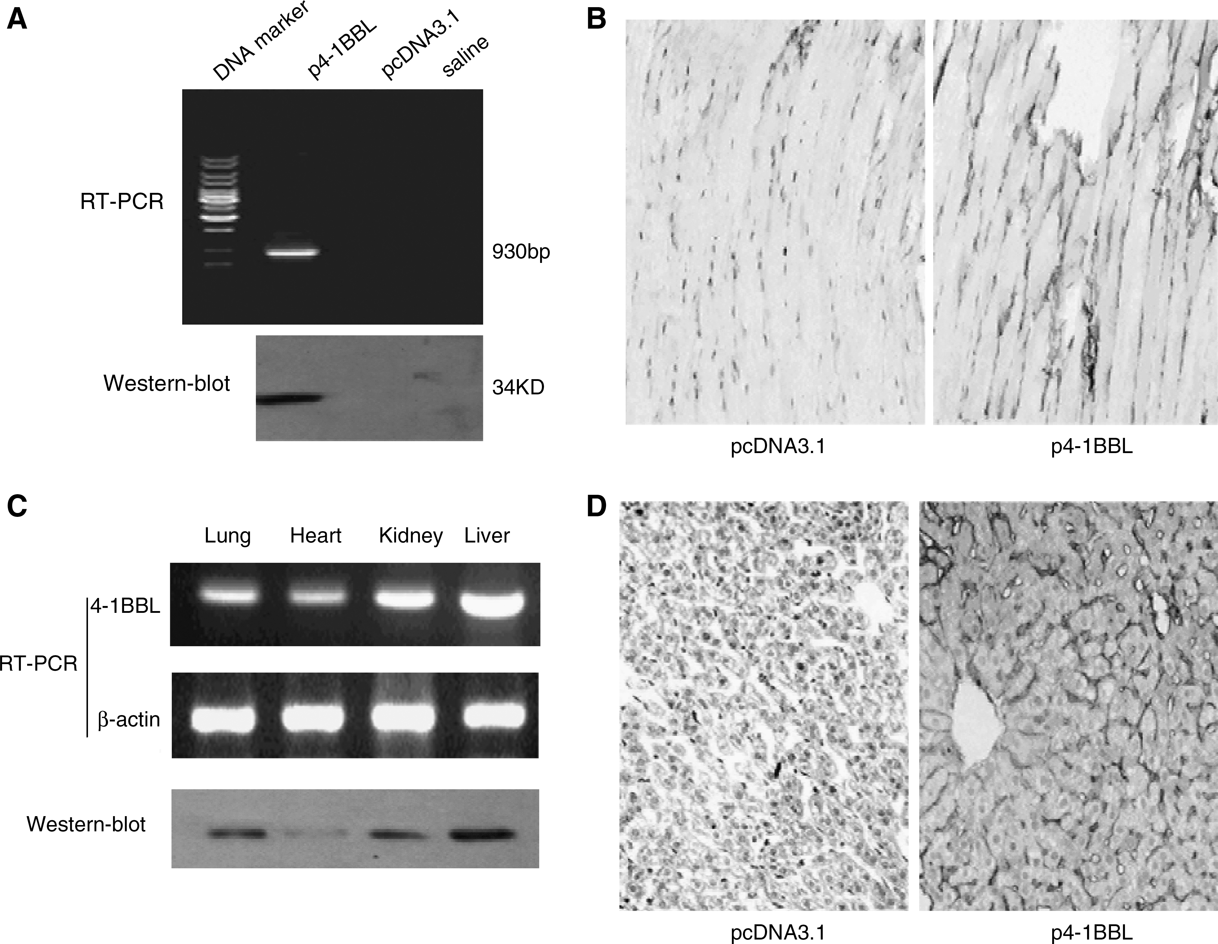
Expression of p4-1BBL in normal tissue cells. (
Studies demonstrated that the hydrodynamics-based gene transfection achieved an efficient expression of targeted genes predominantly in the liver, but much less in the kidney and spleen, and the plasma. 27,28 Previous studies selected targeted genes encoding the secretory proteins. In our study, we examined whether 4-1BBL, a cellular membrane protein, could be expressed by normal tissue cells in vivo after plasmid transferred via this gene delivery. The result showed that the expressed product of p4-1BBL was confirmed by RT-PCR and Western blot analysis (Fig. 1C); we further verified the 4-1BBL expression on the surface of mouse liver cells with immunohistochemical staining (Fig. 1D). Rapid tail-vein injection of a large volume of plasmid DNA solution into a mouse results in a high level of transgene expression in the liver, which was applicable to membrane protein molecules.
Suppression of tumor in situ growth by 4-1BBL expressed by normal muscle cells
On the basis of 4-1BBL expression in the muscle and liver, we next examined whether 4-1BBL would inhibit tumor growth in vivo. B16F1 melanoma cells were s.c. implanted into C57/BL6 mice, and p4-1BBL was repeatedly administered with an interval of 2 days; the increase in tumor volume was inhibited by the expression of 4-1BBL, using the directly i.m.-injection gene-delivery technique. The volume of S.C. primary tumor in 4-1BBL-treated mice was inhibited to 45% on day 25, as compared to control mice given empty plasmid (n = 6; p < 0.05) (Fig. 2A and 2B).
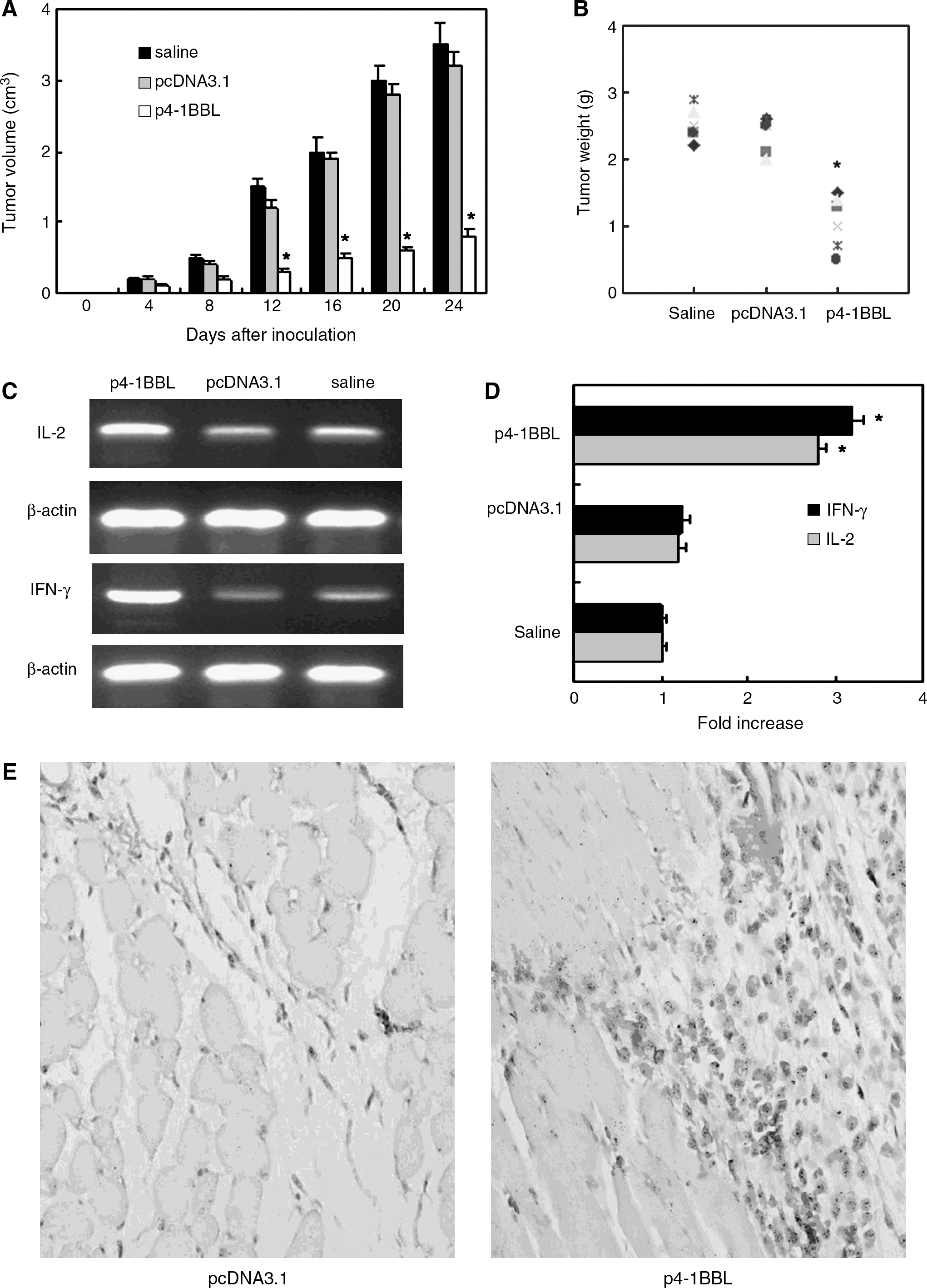
The regulatory effect of 4-1BBL expressed in muscle in tumor milieu and inhibition of growth of melanoma in situ. Mice inoculated with 1 × 10⁵ B16F1 melanoma cells in the right hind thigh muscle were treated, starting from day 3 after inoculation, with intramuscular injection of 100 μg of naked p4-1BBL or 100 μg of pcDNA3.1 or saline 6 times. (
Real-time PCR results showed that the transcription activity of IL-2 and IFN-γ genes were further increased in peritumor tissue, and the modulation of IL-2 and IFN-γ in the tumor microenvironment was obviously due to the presence of 4-1BBL (Fig. 2C and 2D). Immunohistochemical staining estimation exhibited that there was a larger number of CD8 T-lymphocytes in the tumor milieu with the p4-1BBL treatment group than in pcDNA3.1 group (Fig. 2E). These data indicate that the administration of 4-1BBL can modulate tumor microenvironment and make it biased to antitumor immunity.
According to experimental design, an equal dosage of plasmid p4-1BBL was used to treat the tumor in situ with the hydrodynamics-based gene-delivery technique. The result indicated that the volume of tumor in p4-1BBL i.v. injection via the tail vein was a little bigger than the p4-1BBL i.m.-injection group; the tumor inhibition rate percentage was 30% (image not shown). This experiment demonstrates that, to suppress tumor in situ growth, direct the i.m. injection gene-delivery technique is better than the hydrodynamics-based gene-delivery technique.
Inhibition of the progression of pulmonary metastatic melanoma by 4-1BBL expressed by normal liver cells
To evaluate whether the blockade of tumor metastasis by 4-1BBL was expressed by normal tissue cells in vivo, the mice challenged with B16F1 cells by tail-vein injection were treated with the transfection of naked p4-1BBL, which was performed by the hydrodynamics-based gene-delivery technique. Plasmids p4-1BBL and pcDNA3.1 were repeatedly delivered four times, and the mice were analyzed on day 25 (Fig. 3A). In control mice injected with the empty plasmid, the mean number of the surface metastases in the lung reached 550 ± 50 in control mice injected with p cDNA3.1; however, it was suppressed to 80 ± 30 in mice injected with p4-1BBL (Fig. 3B).
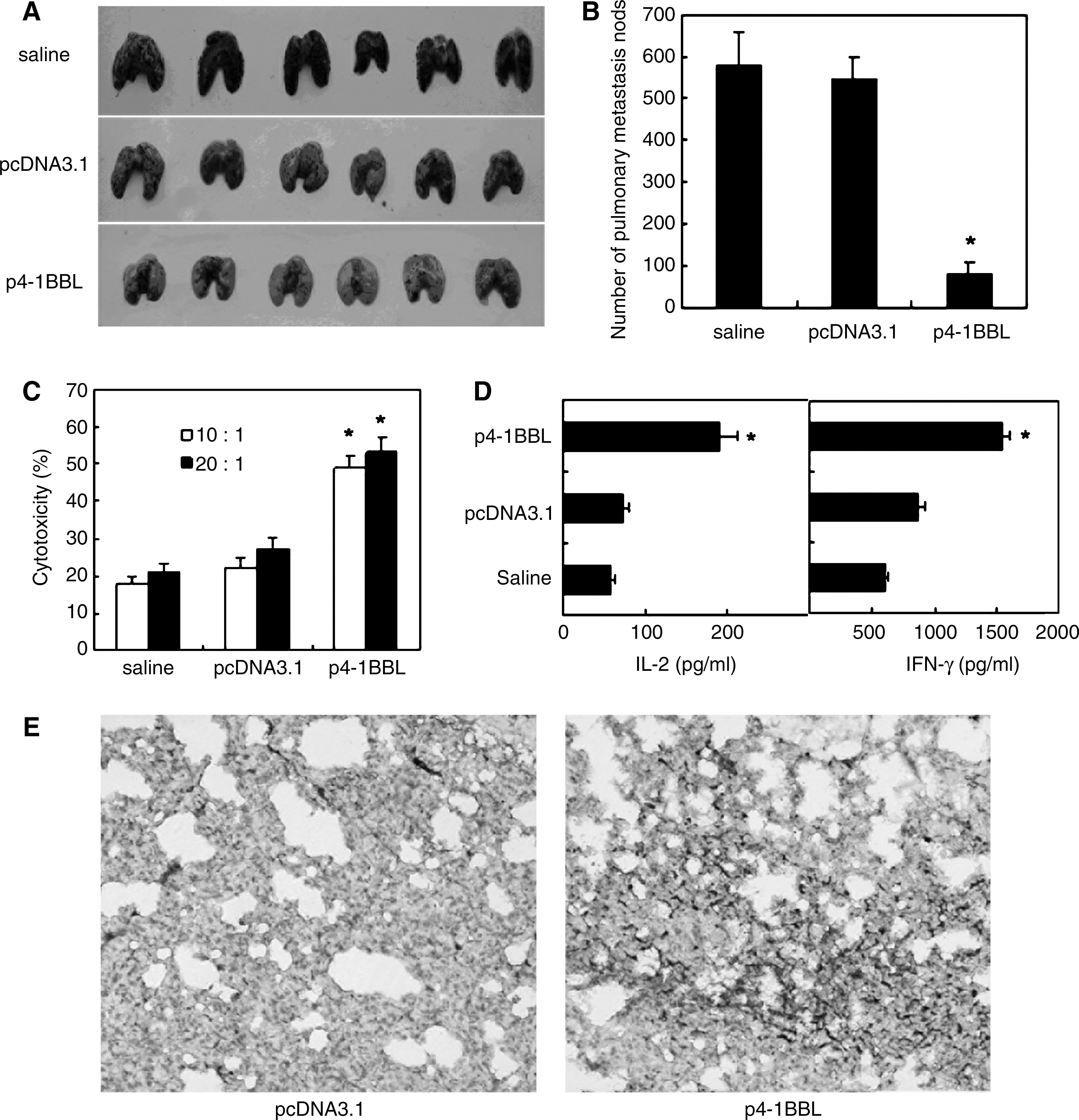
Suppression of melanoma pulmonary metastasis of 4-1BBL expressed in liver cells. First, 1 × 10⁵ B16F1 melanoma cells was inoculated into mice via the tail vein, then the mice bearing tumor were treated, starting from day 3 after inoculation, with intravenous injection of 100 μg of naked p4-1BBL or 100 μg of pcDNA3.1 dissolved in 2 mL of saline four times. (
To analyze the possible antitumor mechanism of 4-1BBL gene treatment, spleen lymphocytes were prepared from mice on day 25 after tumor inoculation and antitumor CTL activity was tested. As shown in Figure 3C, the cytotoxicity of spleen lymphocytes from the p4-1BBL treatment group to melanoma target cells was significantly augmented, compared with the pcDNA3.1 treatment group. To assess the immunologic reactivity in the tumor microenvironment in the process of treatment, we serially examined the distribution of CD8+ T-cells. Results showed that the number of CD8+ T-cells in the tumor microenvironment was larger in the p4-1BBL treatment group than in the control group (Fig. 3E). As Figure 3D shown, the expression levels of IL-2 and IFN-γ in serum from p4-1BBL-treated mice were significantly increased.
Our experiment also showed that an equal dosage of plasmid p4-1BBL direct i.m. injection gene delivery had only a tiny function to inhibit pulmonary metastasis (image not shown). These data indicated that the administration of the 4-1BBL gene, using the hydrodynamics-based gene-delivery technique, can modulate the tumor microenvironment and enforce antitumor immunity to inhibit tumor metastasis effectively.
Discussion
The prognosis for patients with advanced melanoma remains poor, despite the many advances in cancer treatment that have occurred over the last several decades; the 5-year survival rate for patients with distant metastases is less than 10%. 30 For these patients, surgery and radiation therapy are primarily used to palliate symptoms. Most patients with advanced melanoma receive systemic therapy. Single-agent cytotoxic chemotherapy with dacarbazine is the standard of care in community practice, but the response rate is generally low. 31 Biotherapy using high-dose IL-2 and IFN has been shown to induce durable responses; however, these immunotherapy strategies have very low tumor-response rates, usually in the order of 5%–10% of treated patients. 32 On the other hand, daily use of high-dose exogenous recombinant IL-2 or IFN has been developed to increase toxicity. We propose that the antitumor activity of adequately stimulated tumor antigen-specific cytotoxic T-lymphocyte (CTLS) is limited by local factors within the tumor milieu and the pharmacologic modulation of endogenously expressed IL-2 and IFN. To achieve this role, the targets turn to costimulatory molecules, which involve lymphocyte activation, proliferation, and differentiation.
Recent advances in the development of immune-based therapies in our understanding of the function of costimulatory molecules have revitalized enthusiasm in the development of immune therapies for cancer. Among these costimulatory molecules, 4-1BBL is well-characterized and is expressed by APC, including B-cells, macrophages, and dendritic cells (DCs). 4-1BBL is capable of enhancing the expression of B7 molecules on DCs to augment the DCs-mediated second signal (B7/CD28 signaling) for T-cell activation. In particular, 4-1BBL/4-1BB signaling, unlike B7/ CD28, is very important for the effector phase of T-cell response. The stimulation of 4-1BBL can prevent the apoptosis of CD8+ cells after several cell-division cycles.
Recently, the research in the aspect of tumor therapy of costimulatory molecules mostly uses the method of gene metastasis, which transfects tumor cells to express costimulatory molecules and observes tumorgenicity, 33 –35 and there are no reports about transferring costimulatory molecule genes to normal cells to be expressed, thus far. In this study, we transferred 4-1BBL plasmid DNA into the normal tissue cells by using the direct i.m. injection and hydrodynamic i.v. injection. Our data show that the 4-1BBL-expressing vector does not have to enter into specific cells, such as tumor or immune cells, and the expression of 4-1BBL in muscle and liver cells can efficiently exert its function in the experiments through acting with lymphocytes in tumor milieu and blood circulation. This study reveals that 4-1BBL expressed by normal tissue cells increases the level of IL-2 and IFN-γ in tumor milieu and peripheral blood and obviously improved the function of tumor-antigen-specific CTLS (Figs. 2 and 3).
The activated immunocytes by 4-1BB/4-1BBL signal, on one hand, can proliferate and secrete more IL-2 and IFN-γ, and on the other hand, can transform into tumor antigen-specific T-cells (CTLS) after tumor-antigen presenting; the latter reaches the tumor milieu and specifically kills tumor cells. 4-1BBL interacted with activated T-cells and improved the ability of antiapoptosis and survival of T-cells. IL-2 and IFN-γ are both important immune cytokines, which promote the antitumor function of the body. IL-2 can promote activated T-cell proliferation, and IFN-γ plays an important role in promoting the killing function of activated T-cells. 36 On the other hard, IL-2 and IFN-γ can promote the nonspecific killing action of other immunocytes (such as natural killer LAK, and DC cells), 37 and plays a role in removing the tumor cells. In addition, some studies showed 4-1BBL could improve the secretion of IL-8 and IL13 and had an obvious antitumor action. 38
Though antitumor immunology gene therapy is a promising strategy for cancer treatment, it generally requires highly efficient delivery systems. To date, success of this strategy has depended almost exclusively on the delivery of high titers of viral vectors, which can result in effective exogenous gene expression. However, their cytotoxicity and immunogenicity are a major concern for clinical applications. Recent advances in the delivery efficiency of naked DNA could potentially meet the requirement for both high transgene expression and minimal side-effects. Currently, most products expressed by naked DNA were soluble or secretory-type molecules; there are no reports about cytomembrane molecules expressed in normal tissue cells via naked DNA injection either by i.m. or i.v. In this study, we transferred naked DNA into mouse via i.m. or i.v. injection and compared two approaches of gene transfer to inhibit growth and metastasis, which are different clinic stages of melanoma progression. Our results indicated that 4-1BBL not only could be expressed on the surface of mouse muscle and liver cells (Fig. 1), but could also suppress tumor growth and metastasis by regulating immune-cell activities. Comparing two gene delivery approaches in treating with a tumor in situ model and tumor metastasis model, we found that direct plasmid DNA i.m. injection was mainly fit to treat tumor in situ, and the hydrodynamics-based gene delivery technique could inhibit tumor metastasis more effectively.
In addition, the liver and renal functions in our study were detected, and the data showed that transfection and expression 4-1BBL in normal muscle and liver cells had no impact on the function of the liver and renal system, indicating that there was no autoimmune response. Therefore, 4-1BBL expressed by normal tissue cells can be used to treat tumor with large security.
Conclusions
In summary, the therapeutic strategy with 4-1BBL expressed by normal tissue cells effectively inhibits the growth and metastasis of melanoma. Naked plasmid can be transferred into the body by using direct i.m. injection or hydrodynamic i.v. injection and can exert powerful immunoregulating function, providing provide promising immune gene-therapy approaches for different tumor clinical stages.
Footnotes
Acknowledgments
This work was supported by the National Natural Science Foundation of China (no. 30600735) and the National Development Program (973) for Key Basic Research (no. 2002CB513100).
Disclosure Statement
No competing financial interests exist.