
Abstract
The synthesis of eight ligands by using 2-amino benzimidazole with different mono/bis aldehydes is described herein. The final products were characterized by spectral techniques such as FT-IR, 1H NMR, and EI-Mass. The structure–activity relationships of the benzimidazole derivatives are also reported. Studies on the complexation of the ligands with 99mTc were optimized by using stannous tartrate as reducing agent under various reaction conditions. The radiochemical stability was ≥95% for all the complexes, and they were to be stable for 12–14 hours in serum. Most of the ligands showed fast blood clearance. Biodistribution studies of the 99mTc complexes of these ligands showed no significant uptake in the brain or in the heart, and the clearance was mainly through the hepatobiliary system. Among the eight compounds evaluated for their antiproliferative activity in vitro, L8 produced good activity against the cancer cell lines A549 and PC-3.
Introduction
Benzimidazole derivatives are key components in a large number of bioactive compounds of both natural and synthetic origin. Specifically, this nucleus is a constituent of Vitamin-B12. 1 The biological activities of the compounds containing this basic moiety have been well-documented. 2 Similarly, 2-substituted benzimidazoles and their derivatives are potent biologically active compounds, 3 that have a wide range of pharmacological activity and inhibitory properties. 4 –7
Several efforts have been made to develop radiopharmaceuticals by using 99mTc-labeled small-molecule complexes because of their superior medico-imaging characteristics (t1/2 = 6.02 hours, Ey = 141 keV) and the availability of radionuclides. One strategy is to explore novel complexes of small-sized, multifunctional ligands that produce specific bioactivity. Schiff bases are one of the examples which contain the azomethine (-C = N-) group and synthesized by the condensation of primary amines with carbonyl compounds. 8 –10
In recent years, considerable interest has been shown in the medicinal chemistry and pharmacokinetics of Schiff-base compounds derived from heterocyclic moieties, because of their ability to serve as anticonvulsant, anticancer, and antifungal agents. 11 –17 Schiff-base complexes have also been used as catalysts for many reactions and as biological models for understanding biological processes and the structure of biomolecules. Novel Schiff-base derivatives can also be used for interactions with DNA 15 as well as for inhibition of various tumors. 16
Considering the importance of benzimidazoles and azomethine compounds in biological systems, compounds containing both benzimidazole and -C = N- moieties were designed and synthesized, thus generating a new series of radiopharmaceuticals.These compounds were also screened for their potential efficacy, as part of an ongoing project in which the synthesis of some biologically active indole-based Schiff-base analogues were reported. 17
Materials and Methods
All the chemicals used in the present study were of analytical grade and were purchased from Sigma, Aldrich, and Merck Chemical. All the solvents were used after distillation. Thin layer chromatography (TLC) was run on silica gel–coated aluminum sheets (silica gel 60 F254; E Merck) and viewed under ultraviolet (UV) light. Infrared (IR) spectra were recorded on a FT-IR Perking Elmer spectrum BX spectrophotometer. Nuclear magnetic resonance (NMR) spectra were obtained with a Brucker NMR instrument (400 MHz). Electron impact mass spectra (EI-MS) were recorded on JEOL SX 102/DA-6000 spectrometer using m-nitrobenzyl alcohol as the matrix. Radio complexation and radiochemical purity were checked by instant strip chromatography (silica gel–impregnated paper chromatography) with ITLC-SG (Gelman Sciences).
Preparation of Schiff base
The Schiff bases were synthesized (as presented in the Chemical Scheme 1) by mixing an ethanolic solution of 25 mL of 2-amino benzimidazole (1.45 g, 0.01 mol) with 0.01 mol of aldehydes (glyoxal, methylglyoxal, benzaldehyde, salicylaldehyde, m-anisaldehyde, o-nitrobenzaldehyde, 3-indolecarboxaldehyde, and 5-indolecarboxaldehyde) in the same volume of ethanol. The mixture was refluxed for 2–6 hours. The precipitate was collected by filtration through a Buckner funnel, recrystalized from ethanol, and dried at room temperature giving 70%–85% yield. The complete spectral analyses of these compounds are given in Table 1.
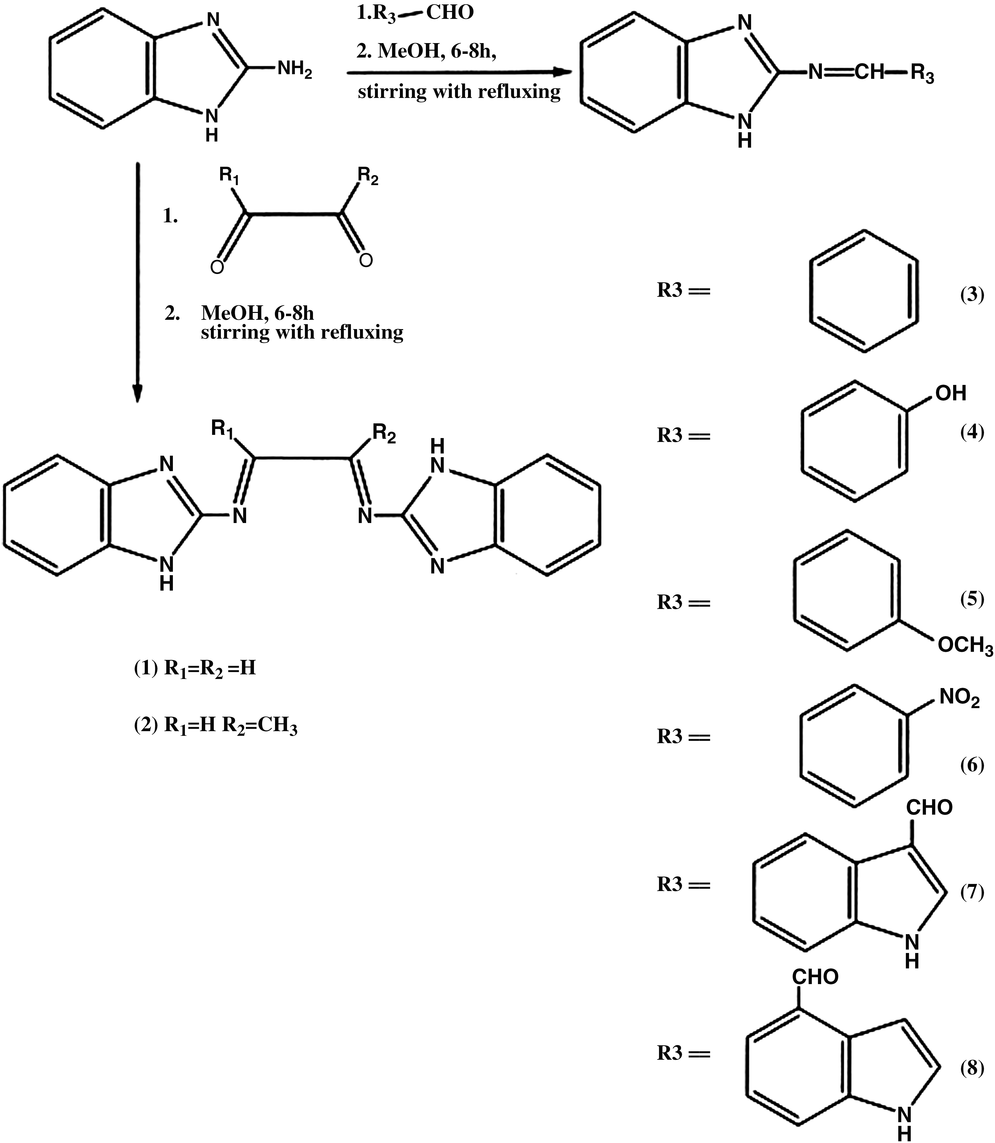
Chemical scheme for preparation of Schiff bases.
ppm, parts per million.
Quantitative structure—property relationship analysis
The quantitative structure–property relationship (QSPR) investigation was performed via the linear free energy relationship model proposed by Hansch and Leo. 18 Selection of parameters is the first and foremost step in any QSPR study. In the present study, electronic, hydrophobic, and steric factors were selected for a Schiff base and considered to be consistent. These included molar refractivity (MR), Van der Waals volume (VDW), Connolly accessible area, Connolly molecular area, Connolly solvent excluded area, dipole–dipole energy, partition coefficient, HOMO, and so forth. These parameters were calculated by MM2 studies with Chem 3D 6.0 software. Geometries of all compounds were completely optimized with the same software package.
A classical Hansch multivariate regression analysis using the least-square method was chosen to derive the QSPR equations for the data set. The level of significance of each coefficient was judged by statistical procedures such as F tests. Statistical analysis was carried out by using least squares through a stepwise selection and elimination procedure. For each equation, several indices of best fit were considered, namely, the regression coefficient r, the standard deviation s, and the measure of level of statistical significance F.
Using these equations, two models were found suitable to analyze the Schiff-base complexes.
QSPR model for Schiff base series (Model 1)
−log C = [3.01462(±0.5208)] −VDW [0.0103(±0.518)]
n = 10 l r l = 0.915 s = 0.192 F = 99.128
QSPR model for Schiff base series (Model 2)
−log C = [4.08827(±0.1368)] −MR [0.0186(±0.102)]
n = 10 l r l = 0.902 s = 0.178 F = 98.958
Anticancer activity
All the compounds were evaluated for their cytotoxic activity in vitro against two cancer cell lines, A549 (human lung carcinoma) and PC-3 (human prostate cancer) (Table 2).
Cell numbers given should be multiplied by 1014.
Routine culture and experimental studies were carried out in a humidified cell incubator maintained at 37°C and 5% CO2. Cell propagation for A549 was achieved in minimum essential medium Eagle with Earle's salts and with phenol red, 10% fetal bovine serum,
The exponentially growing cells were resuspended in drug-free medium or in the presence of 24 nM doxorubicin. The resistant cells require doxorubicin to maintain normal cell proliferation. Cells were seeded at a density of 106 cells/mL (in triplicate six-well plates) and allowed to attach for 24 hours before the addition of 10 μL of the drug at 0.5% concentration. After 72 hours of incubation, the cells were trypsinized and counted.
PC-3 cells were maintained in RPMI 1600 containing 2 mmol/L of
Cell-cycle analysis
Test substances were dissolved in DMSO and diluted to 10 mmol/L. A549 cells (5 × 10⁴ cells/mL, 5 mL) were cultured in five flasks for 24 hours with or without the addition of test compounds (for each test compound, one flask was used). The cells were harvested, washed with PBS, and centrifuged. The cells were then fixed by adding 4 mL of ethanol (70% ice-cold) and keeping at −20°C overnight until DNA staining. The fixed cells were treated with 100 mg/mL of RNase A in PBS for 1 hour, followed by staining with 50 mg/mL of propidium iodide in PBS in the dark. The DNA content of eukaryotic cells was then measured with flow cytometry.
Flow cytometric measurements were performed with a 15-mW solid-state argon-ion laser emitting at 488 nm. Fluorescence was collected using a 575/25 nm band-pass filter (orange–red fluorescence [FL1]) after linear amplification. For each measurement, data from 10,000 to 20,000 single-cell events were collected, while cell doublets and aggregates were gated out using a two-parameter histogram of FL1–area versus FL1–width. Cell-cycle histograms were analyzed such that only cell cycles with a low coefficient of variation of the G0/G1 peak (<5) and low reduced chi-square values (<3), an indication of how well the model describes the observed data, were considered. The proliferative potency of a chemical in this assay was indicated by the percentage of cells in the S(ynthesis)-phase of the cell cycle (Table 3).
Radiolabeling of the compounds with 99mTc
Radiolabeling of the compounds was performed by using a freshly eluted saline solution of sodium pertechnetate (74 MBq, 100 mL), which was reduced with the help of 60 μL of 1 × 10⁻² M SnCl2.2H2O (dissolved in 1 mL of nitrogen-purged 10% acetic acid). To this, 100 μL of 0.03 nM solution of the compounds in DMSO was added, and pH of the reaction mixture was adjusted to 6.5 with 0.1 M NaHCO3 solution. The reaction mixture was put in a shielded vial and incubated for 20–30 minutes at room temperature. Labeling efficiency of the 99mTc-based complex was determined with ITLC-SG strips with 100% acetone as the developing solvent and simultaneously in pyridine, acetic acid, and water in the ratio of 3:5:1.5 in to differentiate the labeled complex from the reduced and hydrolyzed colloids formed during the labeling procedure. Each ITLC strip was cut into 0.1-cm segments and counted in a γ counter.
In vitro serum stability assay
Fresh human serum was prepared by allowing blood collected from healthy volunteers to clot for 1 hour at 37°C in a humidified incubator maintained at 5% CO2 and 95% air. The sample was then centrifuged at 400 rpm, and the serum was filtered through a 0.22-μm syringe filter into sterile plastic culture tubes. The freshly prepared technetium radio complexes described above were incubated in freshly prepared human serum at physiological conditions, that is, at 37°C and at a concentration of 100 nM/mL. These were then analyzed with ITLC at different time intervals to detect any dissociation of the complexes. The percentage of free pertechnetate at any particular time point was estimated, using saline and acetone as the mobile phase.
Biodistribution studies
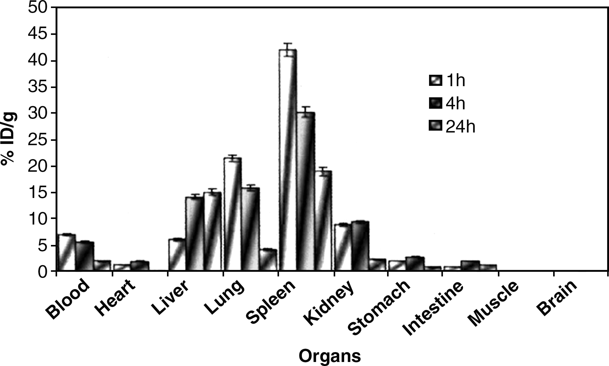
Balb mice (taken in triplicate sets) were used for the tissue distribution studies. Animal handling and experimentation were performed according to the guidelines of the Institutional Animal Ethics Committee. Equal doses of 10 μCi of the labeled test compounds were injected through the tail vein of each animal. At different time intervals, mice were sacrificed, their blood collected, and different tissues and organs dissected and analyzed. Of the eight analogues, L8 was the most efficient (Fig. 1). Radioactivity was measured in a γ counter. The actual amount of radioactivity administered to each animal was calculated by subtracting the activity left in the tail from the compound injected. Radioactivity accumulated in each organ was expressed as the percentage of administered dose per g of tissue. Total volume of blood was calculated as 7% of body weight.

Biodistribution of 99mTc–L8 in mice.
Blood kinetics studies
The blood clearance study was performed in albino New Zealand rabbits weighing approximately 2.5–3.0 kg, by administering 0.3 mL (10 MBq) of the 99mTc-labeled compounds via the ear vein. At different time intervals, 0.5 mL of blood samples were withdrawn from the dorsal vein of the other ear and radioactivity measured in a γ counter. The data from the experiment were expressed as the percentage of administered dose per g of tissue (organ; Fig. 2).
Blood kinetics of Schiff-base analogues.
Results and Discussion
Condensation reactions of 2-aminobenzimidazole with glyoxal, methylglyoxal, benzaldehyde, 2-nitrobenzaldehyde, 3-anisaldehyde, salicylaldehyde, indole-3-carboxylaldehyde, and indole-5-carboxaldehyde provided a high yield (70%–85%) of Schiff bases. These reactions were monitored with TLC, which showeds the single spot that was different from the original materials. The IR spectra of the compounds L1–L8 showed absorption bands in the range of 1600–50 cm−1. The (C = N) linkage, characteristic of the azomethine groups, appeared in all the ligands, indicating that condensation between the aldehyde and the amino group of 2-aminobenzimidazole had occurred. The 1 H NMR spectral data are reported along with possible arrangement of protons in final products. All the protons were in their expected regions, and comparison with the original materials confirmed the proposed stoichiometry and structure of these compounds. MS spectra of the Schiff bases showed the presence of their molecular ions.
All the analogues were analyzed against two cell lines, namely, A549 and PC-3. Most of the compounds were effective against A549, while only L2, L4, L5, and L8 were effective against PC-3.
Preliminary studies on complexation of novel synthesized compounds with 99mTc produced sufficiently stable complexes under physiological conditions. In vitro serum stability of the radiocomplexes is a necessary parameter for measuring the effectiveness of a chelating moiety for coordinating the radiometal. Generally, trans-chelation occurs between radiometal and serum proteins, particularly albumin. In vitro serum stability of the synthesized complexes falls initially but becomes constant thereafter. The initial fall in the labeling efficiency after addition of fresh serum could be attributed to the trans-chelation that would have occurrred in the serum as a result of the high affinity of plasma proteins for metal ions.
In vivo blood kinetics and biodistribution studies were performed for all eight complexes. Biodistribution is a very important phenomenon in the in vivo drug distribution and excretion pathway. These studies showed fast blood kinetics of the synthesized Schiff bases. In L8 a high amount of radioactivity accumulated in the liver and in the stomach, which increased with time, while there was negligible increase in activity in the intestine. This shows that the kidney is a major pathway of excretion (Fig. 2), while the brain and the heart retain very little amount of the drug.
The percentage distribution in the various organs of the mice is shown as the percentage of injected dose per organ or tissue at different time intervals. Long retention of the drug in the liver indicates that, most probably, the metabolism of the drug occurs in the liver but excretion of the drug and metabolites occurs through the lung and the spleen.
Conclusions
Preliminary studies on complexation of novel Schiff-base ligands with 99mTc produced sufficiently stable complexes under physiological conditions. The results are encouraging and merit further in vivo experiments for targeted imaging of human subjects. The therapeutic potential of these complexes can be further extended further by applying them in different animal models and cell lines.
Footnotes
Acknowledgments
We thank Dr. Rajendra Prasad Tripathi, director, of Institute of Nuclear Medicine & Allied Sciences, Delhi, India, for providing all necessary facilities and constant encouragement during the course of this study.
Disclosure Statement
No conflicts of interest exist for any of the authors.