
Abstract
The clinical use of cisplatin, a potent antineoplastic agent, is limited by its severe adverse effects. The present study was designed to evaluate the effects of resveratrol on cisplatin-induced cardiac injury. Resveratrol is a potent free radical scavenger. In the present study, we tested whether resveratrol would prevent cisplatin-induced cardiotoxicity in rats. Plasma-enzyme activities and histologic myocardial changes were examined. The anticancer role of resveratrol and/or cisplatin were measured by MTT. Our data showed that cisplatin led to cardiac-function deterioration, myocardial injury, increased lactate dehydrogenase, creatine kinase, malondialdehyde activities, and decreased activities of superoxide dismutase, glutathione, glutathione peroxidase, and catalase. Treatment with resveratrol effectively hindered the adverse effects of cisplatin in a dose-dependent manner, such as myocardial injury and impaired heart function. An in vitro cytotoxic study showed that resveratrol could increase the antineoplastic activity of cisplatin to A549 adenocarcinoma cells. All the above lines of evidence suggest that resveratrol protects cardiomyocytes from cisplatin-induced cardiotoxicity via the suppression of oxidative stress.
Introduction
Cisplatin (cis-diamminedichloroplatinum II; CDDP) is an antitumor agent against several types of cancer and has been widely used in chemotherapy, such as head and neck, testicular, ovarian, bladder, and small-cell lung cancers. However, it has dose-limiting side-effects, such as nephrotoxicity, 1 ototoxicity, 2 hepatotoxicity, 3 and gastrointestinal dysfunction. In spite of these toxicities, many survivors may experience acute or chronic cardiovascular complications that can impair their quality of life after cisplatin treatment. Cisplatin-induced cardiotoxicity includes atrial fibrillation, supraventricular tachycardia, left bundle-branch block, and myocardial infarction. 4,5 In addition, cardiac complications of cisplatin lead to chemotherapy dose reduction and delay, and in some cases, have necessitated discontinuation of treatment. The potential for cardiotoxicity should be recognized before treatment, and preventive measures should be taken.
Drugs that ameliorate cardiotoxic effects would allow us to exploit the full therapeutic potential of cisplatin, with a considerable impact on cancer therapy. Until now, pharmacologic and clinical attempts to reduce the cardiotoxicity of cisplatin have met with little success. Consequently, it is important to develop a therapy to decrease cisplatin-induced cardiotoxicity. The possible mechanisms of cisplatin-induced cardiotoxicity are mainly alterations in oxidant/antioxidant balance. 6 Thus, antioxidative agents could provide possible approaches to reduce toxicity induced from the clinical use of cisplatin.
Resveratrol (trans-3,4′,5-trihydroxystilbene) is a polyphenolic phytoalexin, found abundantly in various fruits, vegetables, and grapes. Intriguingly, several pharmacologic effects of resveratrol, including oestrogenic, antiplatelet, anticancer, and anti-inflammatory properties, have been demonstrated. 7 Additionally, resveratrol has been shown to have cardioprotective properties, partly because of its antioxidant, -apoptotic, and -arrhythmic effects. 8,9 Recently, there has been abundant epidemiologic and clinical evidence showing that resveratrol may act as an antioxidant, promote nitric oxide production, 10 increase H2O2 tolerance, 11 and inhibit cyclooxygenase-2 (COX-2). 12
Based on these findings, we hypothesized that resveratrol, along with its antioxidant effects, can potentially increase its therapeutic value in the prevention of patients with cisplatin-induced cardiotoxicity. In this study, we test the effects of resveratrol in vivo to test this hypothesis. Finally, using A549 adenocarcinoma cells, an in vitro test is used to determine whether resveratrol affects the antitumor activity of cisplatin.
Materials and Methods
Chemicals
Cisplatin (30 mg/6 mL, Code 061002) was obtained from Yunnan Gejiu Biological Pharmaceutical Co., Ltd. (Gejiu, China) Resveratrol [RES; botanical name: polygonum cuspidatum; part of plant used: root; manufacturing methods: maceration reflux; extract method: alcohol; test method: high-performance liquid chromatography (HPLC); color: pure white; and purity: 98%] was purchased from Feida Biotechnology Company (Xian, China). Resveratrol was suspended in isotonic Na chloride. Thiobarbituric acid (TBA) was obtained from Nanjing Jiancheng Biotechnology Company (Nanjing, China).
Animals and treatment
Fifty (50) healthy adult male Wistar rats (15 weeks old, 220–270 g in body weight) were used in this study. The animals were obtained from Shanghai SiLaike (SLAC) Laboratory Animal Company (Shanghai, China). The animals were housed individually in stainless-steel wire-bottomed cages in an air-conditioned room where the temperature was 22 ± 2°C with 50% ± 10% relative humidity and a 12-hours light-and-dark cycle. Rats were fed with the standard diet and water throughout the experimental period.
Experimental protocol
The rats were randomly divided into five groups, with each group containing 10 rats. Cisplatin was injected intraperitoneally (i.p.) to animals at the single dose of 7 mg/kg at day 6, except for the normal control and resveratrol groups. Normal control: Isotonic Na chloride was administered to animals by gavage for 10 days following a single i.p. injection of isotonic saline at day 6. Low-dose resveratrol+cisplatin (RES-L+DDP): Resveratrol was administered to animals by gavage at the dose of 5 mg/kg/day for 10 days. Middle-dose resveratrol+cisplatin (RES-M+DDP): Resveratrol was administered to animals by gavage at the dose of 15 mg/kg/day for 10 days. High-dose resveratrol+cisplatin (RES-H+DDP): Resveratrol was administered to animals by gavage at the dose of 45 mg/kg/day for 10 days. Cisplatin (DDP): Isotonic Na chloride was administered to animals by gavage for 10 days.
For the five groups, the systolic blood pressure (BP) was measured noninvasively in prewarmed rats by the tail-cuff plethysmographic method on days 5 and 10. Before treatment, the rats were trained to be accustomed to BP measurements.
On day 11, the rats were anesthetized i.p. with ketamine (0.75 mg/100 g of body weight) and acepromazine (0.075 mg/100 g of body weight) and heparinized. Blood was collected by cardiac puncture. A thoracotomy was then performed, and the heart was removed. Ventricular muscle was taken for light microscopy and the study of enzyme metabolism.
Sample collection
Blood was collected by cardiac puncture under ether anesthesia and allowed to clot for 30 minutes at room temperature. The serum was separated by centrifugation at 2500 rpm at 30°C for 15 minutes and used for the estimation of marker enzymes, lactate dehydrogenase (LDH) and creatine kinase (CK).
The hearts were dissected out immediately, washed with ice-cold saline, and 10% homogenates in phosphate buffer (50 mM, pH 7.4) were prepared. An aliquot was used for the estimation of malondialdehyde (MDA), glutathione (GSH), glutathione peroxidase (GSH-Px), and catalase (CAT). The homogenates were centrifuged at 7000 g for 10 minutes at 4°C, and the supernatants were used for the assays of superoxide dismutase (SOD). Similarly, hearts were also fixed in 10% buffered formalin for histologic studies.
Marker enzyme assays
The marker enzymes, LDH and CK, were assayed in the serum by using standard kits supplied from Nanjing Jiancheng Biotechnology Company. The results were expressed as IU/L for LDH and CK, MDA, SOD, GSH, GSH-Px, and CAT assays.
The amount of MDA formed was quantified by reaction with thiobarbituric acid-reactive substances (TBARS) and used as an index of lipid peroxidation LPO. The results were expressed as nmol of MDA/g of wet tissue, using the molar extinction coefficient of the chromophore (1.56 × 10⁻⁵/M/cm) and 1,1,3,3-tetraethoxypropane as the standard. Every sample was assayed in duplicate, and the assay coefficients of variation for MDA were less than 3%.
SOD, GSH, GSH-Px, and CAT assay kits were purchased from Nanjing Jiancheng Biotechnology Company. The activity of SOD was measured by the xanthine oxidase method. Tissue GSH concentration was measured by a kinetic assay, using a dithionitrobenzoic-acid recycling method, and was expressed as μmol/g protein. GSH-Px activity was based on the oxidation of GSH by GSH-Px coupled to the disappearance of NADPH by glutathione reductase measured at 37°C and 340 nm and were expressed as U/g protein. CAT activity was determined by measuring the decomposition of hydrogen peroxide at 240 nm and was expressed as k/g protein.
Histopathologic examinations
The heart tissues were fixed in 10% formalin, embedded in paraffin, sectioned at 5 μm, and stained with hematoxylin and eosin. The slides were coded and semiquantitative analysis of the sections was performed without knowledge of the treatment protocol. In these tissues, degeneration, necrosis, and other pathologic changes were evaluated.
Effect of resveratrol on the antitumor activity of cisplatin in vitro
The human A549 lung adenocarcinoma cells were plated in a 96-well plate with approximately 10,000 cells per well, 100 μL/well, each sample seeded three wells, and incubated in Dulbecco's modified Eagle's medium (DMEM), containing 10% fetal bovine serum (FBS), 100 units/mL of penicillin, and 100 mg/mL of streptomycin. Cultures were maintained at 37°C in a humidified 5% CO2 atmosphere.
After a 24-hours incubation period, the cells were exposed to the resveratrol solution (10 μM) and/or cisplatin (70 μM) for another 48-hours incubation. Cytotoxicity was analysed by using the MTT method. Briefly, after 72 hours of incubation, 20-μL of MTT (5 mg/mL; Sigma, St. Louis, MO) was added into each well, and the plates were reincubated for 2 hours. The supernatant was removed carefully and 100 μL of dimethyl sulfoxide (DMSO) was added into each well to dissolve the formazan crystals. The plates were shaken for 5 minutes to dissolve the crystals completely. The absorbance at a wavelength of 490 nm (A490) of cells in each well was measured by a microliter enzyme-linked immunosorbent.
Statistical analysis
Data are presented as mean ± standard error of the mean of at least three independent experiments. The paired t-test was used to compare the data within groups, and analysis of variance was used to compare the data between groups. Differences were considered to be significant at p < 0.05.
Results
Cardiac function
After the cisplatin treatment, the rats were sick and weak and their fur became scruffy.
The time course of systolic BP and heart rate (HR) changes in the five groups on days 5 and 10 are shown in Table 1. Systolic BP and HR on day 5 of the four groups continually decreased with the administration of cisplatin, suggesting a severe cardiac dysfunction of the cisplatin-treated rats. Resveratrol treatment induced an increase of systolic BP and HR, when compared with the cisplatin group, while only the high-dose resveratrol+cisplatin group increased significantly (p < 0.05). In the high-dose resveratrol+cisplatin group, systolic BP and HR had no significant difference, as compared with the control (p > 0.05).
p < 0.05 versus normal control; # p < 0.05 versus DDP control.
RES, resveratrol; DDP, cisplatin.
Resveratrol prevented cisplatin-induced LDH and CK release
As shown in Table 2, a significant rise in LDH and CK activities together confirmed the development of heart impairment caused by cisplatin. A careful examination and comparison of the serum-marker enzymes, LDH and CK, for all treated doses of resveratrol (5, 15, and 45 mg/kg) revealed a decrease in their levels in a dose-dependent manner. Treatments with resveratrol to cisplatin-challenged animals decreased significantly (p < 0.05) the activities of these enzymes elevated by cisplatin, though not to normal.
p < 0.05 versus normal control; # p < 0.05 versus DDP control.
NA, not applicable; RES, resveratrol; DDP, cisplatin.
Effects of resveratrol on antioxidant enzymes
As shown in Tables 3 and 4, the activity of MDA in the cisplatin control group was significantly elevated, compared with the normal group (p < 0.05). Animals treated with resveratrol (45, 15, and 5 mg/kg), followed by cisplatin injection, significantly decreased the activity of MDA (p < 0.05). Significant decline of SOD in myocardial (p < 0.05) was observed in the cisplatin control group, as compared to the normal group, and this decline was reversed by pretreatment with resveratrol.
p < 0.05 versus normal control; # p < 0.05 versus DDP control.
NA, not applicable; RES, resveratrol; DDP, cisplatin.
p < 0.05 versus normal control; # p < 0.05 versus DDP control.
RES, resveratrol; DDP, cisplatin.
The content of GSH was lower in the cisplatin control, compared with the control, group (p < 0.05), while having significantly lower GSH-Px concentration and CAT activities. Resveratrol administration significantly increased the activity of GSH, GSH-Px, and CAT (p < 0.05). However, administration of high-dose resveratrol restored the plasma levels of GSH, GSH-Px, and CAT in cisplatin-treated rats to almost normal values (p < 0.05).
Protective effect of resveratrol on cisplatin-induced histopathologic injury
Histopathologic examination of the myocardium of normal animals showed clear integrity of myocardial cell membrane (Fig. 1A), and no inflammatory cell infiltration was seen. Rats treated with 7 mg/kg of cisplatin showed marked multifocal cadiocyte coagulation necrosis, karyopycnosis with nuclear hyperchromasia, equally interstitial edema, vacuole, myocardial fiber necrosis, and moderate infiltration of lymphocytes and macrophages (Fig. 1B). The changes were pronounced against most of the endocardium and in papillary muscles. Resveratrol revealed a decrease of cardiotoxicity in a dose-dependent manner, as shown in Figure 1C, 1D, and 1E.
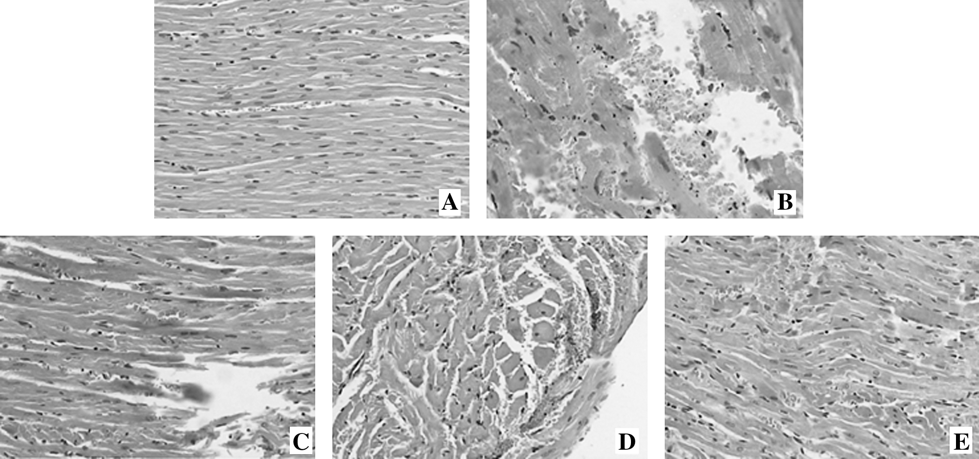
Histopathology. (
Antitumor activity
As shown in Figure 2, the cytotoxicity of cisplatin was shown by the finding that nearly 50% of A549 cells were killed by cisplatin at a concentration of 70 μM. Resveratrol (10 μM) showed some level (≈15%) of cytotoxicity to the adenocarcinoma cells. The presence of resveratrol could enhance the antitumor activity of cisplatin. This is supported by our finding that the cells' survival rate in the wells treated with resveratrol+cisplatin was higher than cisplatin alone.

Effect of resveratrol (RES) on the antineoplatic potency of cisplatin (DDP) to human lung carcinoma cells (A549). The cells were incubated with RES (10 μM) and/or DDP (70 μM) for 48 hours. Values are expressed as means ±standard error of the mean (n = 5 each groups). *p < 0.05, compared with control.
Discussion
Cisplatin is a platinum-based chemotherapy drug used to treat various types of cancers, including sarcomas, some carcinomas (e.g., small-cell lung cancer, and ovarian cancer), lymphomas, and germ-cell tumors. Additionally, cisplatin has been shown to exhibit cardiot-, 13 hepato-, 3 and nephrotoxicity. 1 In the present study, cardiac damage, such as cytoplasmic vacuolization and myofibrillar disorganization, was observed after cisplatin treatment with a single dose of 7 mg/kg. Severe cardiac dysfunction was manifested by the reduction of BP and HR in our study. Consistent with the change of serum-enzyme activities caused by cisplatin in other studies, our results confirmed that the myocardial damage increased the levels of LDH, CK, MDA, and decreased the activities of SOD, GSH, GSH-Px, and CAT in the animals treated with cisplatin. These results are in accord with the findings that free oxygen radical damage plays an important role in cisplatin-induced cardiotoxicity. 1,14 Development of therapies to prevent the generation of free radicals may influence the progression of oxidative damage induced by cisplatin. With this aim, many chemoprotective agents may be used, such as plant extracts, which can act as antioxidants and counteract the damage induced by antitumor drugs.
As an effective antioxidant, resveratrol, a natural antioxidant found in grapes and wines, has attracted great attention in the therapy and prevention of oxidative stress-induced cardiovascular diseases. 15,16 A study demonstrated that resveratrol is inversely related to mortality from coronary heart disease. 17 In addition, several investigators have reported that cisplatin-induced oxidative stress is responsible for damages observed in heart, kidney, and liver. However, resveratrol exerted protective properties on the heart, 18 kidney, 19 and liver. 20 In the study, we investigated the protective effects of resveratrol against cisplatin-induced cardiotoxicity in vivo. Resveratrol (5 mg/kg) treatment could elicit a protective effect against cisplatin-induced cardiotoxicity, embodied with lower cytoplasmic vacuolization in cardiomyocytes and better cardiac function in the resveratrol+cisplatin, compared with the cisplatin, group. With the increasing of resveratrol, 15 and 45 mg/kg of resveratrol show a better protective effect against cisplatin-induced cardiotoxicity. The protective effect has also been shown by the fact that treatment with resveratrol significantly attenuated the promotion of LDH, CK, and MDA activities induced and SOD, GSH, GSH-Px, and CAT activities inhibited by cisplatin exposure. Resveratrol exhibits a strong antioxidant capability. Resveratrol treatment effectively prevented increased production of intracellular reactive oxygen species (iROS) and inflammatory markers (interleukin-1-alpha (IL-1-α), IL6, IL8, and ELAM-1) in trabecular meshwork cells. 21 In addition to antioxidant properties, resveratrol has been demonstrated to modulate the activity of antioxidant enzymes, such as COX-1, endothelial nitric-oxide synthase, peroxidase, CAT, and superoxide. 22,23 In other words, we demonstrate that the protective effect of resveratrol is paralleled by the increasing of myocardial antioxidant potency.
A perfect cardioprotective agent for cisplatin chemotherapy must not compromise the antitumor activity of cisplatin. According to our preliminary data, the cytotoxic study demonstrated that resveratrol did not attenuate the antitumor activity of cisplatin in A549 cells, whereas treatment of resveratrol exhibited cytotoxicity to the carcinoma cells. Obviously, resveratrol has antitumor activity. In this regard, some studies have implied that resveratrol is linked to cancer prevention and therapy by its attenuating oxidative-induced DNA damage in C6 glioma cells 24 and shows cytotoxicity to several tumor cells, including breast, pancreatic, and endometrial cancer. 25,26 The potential cytotoxicity of resveratrol to tumor cells made it more promising to investigate resveratrol as the adjunct in cisplatin chemotherapy. 27
Conclusions
In conclusion, our study has shown that resveratrol elicits a typically protective effect on cisplatin-induced myocardial damage and cardiac dysfunction by scavenging free radicals in rats; however, resveratrol could increase the antitumor activity of cisplatin. These results indicate that cisplatin, with its cardioprotective capability, merits further evaluation as an adjunct during the chemotherapy of cisplatin. However, one limitation of our study was that the effect of cisplatin treatment and resveratrol administration may not reflect the overall effects of resveratrol administration in patients. Consequently, our data should be discussed cautiously in terms of their effects in patients.
Footnotes
Acknowledgments
This study was supported, in part, by the Department of Pathology, Tumor Hospital of Harbin Medical University (Harbin, China). The authors thank the Tumor Research Institute of the Tumor Hospital of Harbin Medical University for providing technical assistance and animal breeding.
Disclosure Statement
No competing financial interests exist.