




Abstract
The use of radiolabeled antibodies that are able to target primary tumors as well as metastatic tumor sites with minimal reactivity to normal tissues is a promising approach for treating pancreatic cancer. In this study, the integrin αvß5 is studied as a target for the diagnosis of and potential therapy for human pancreatic cancer by using the radiolabeled murine monoclonal antibody (mAb) 14C5. Biopsy specimens from human pancreatic tumors were examined for the expression of the integrin αvß5. The pancreatic tumor cell line Capan-1 was used to test the in vitro targeting potency of mAb 14C5 labeled with 125/131-iodine and 111-indium. Internalization, retention, and metabolism were investigated in cellular radioimmunoassays. Biodistribution and tumor-targeting characteristics were studied in Capan-1 xenografts. All tumor sections were positive for the integrin αvß5, with an extensive positive staining of the stroma. Saturation binding experiments showed high affinity with comparable K ds. In vitro internalization experiments showed a longer intracellular retention of 111In-p-benzyl isothiocyanate-1,4,7,10-tetraazacyclododecane-1,4,7,10-tetraacetic acid (p-SCN-Bz-DOTA)-14C5 in comparison to 125I-14C5 and 111In-p-isothiocyanatobenzyl diethylenetriaminepentaacetic acid (p-SCN-Bz-DTPA)-14C5. In vivo radioisotope tumor uptake was maximum at 48–72 hours, with the uptake of 111In-p-SCN-Bz-DOTA-14C5 (35.84 ± 8.64 percentage of injected dose per g [%ID/g]) being 3.9- and 2.2-folds higher than 131I-14C5 (12.16 ± 1.03%ID/g) and 111In-p-SCN-Bz-DTPA-14C5 (14.30 ± 3.76%ID/g), respectively. Planar gamma imaging with mAb 14C5 indicated clear localization of the pancreatic tumors versus minimal normal tissue uptake. mAb 14C5 is a promising new antibody for targeting the integrin αvß5 for the diagnosis of and potential therapy for pancreatic cancer.
Introduction
Despite recent advances in diagnostic modalities and therapeutic strategies, the prognosis of pancreatic cancer remains poor. With an incidence of 58,100 per year and a mortality rate almost identical to this cancer's incidence, it is the fifth leading cause of death in Europe. The 5-year survival rate for patients with this malignancy is less than 5%. 1 Although much research has been undertaken in the past 50 years, conventional treatment approaches, such as surgery, radiation, chemotherapy, or combinations of these, have little impact on the course of this aggressive neoplasm. 2 One of the reasons for the poor prognosis of pancreatic carcinoma is the difficulty of obtaining an early diagnosis. Metastatic spread, which would have already occurred in half of the patients at the time of diagnosis, is one of the major problems in the treatment of this aggressive malignancy. 2
One potential approach is the use of radiolabeled antibodies that are able to target primary tumors as well as metastatic tumor sites with minimal affinity for normal tissues. 3 Different monoclonal antibodies (mAb) have been used to target tumor antigens with high expression on malignant pancreatic cancer cells, including ferritin, 4 HER-2/neu, 5 MUC1, 6,7 α6 ß4, 8 VEGF, and EGFR. 9 Some of these mAbs have produced encouraging results, but none of them is yet commercially approved for pancreatic cancer therapy.
Recently, a promising new antibody targeting the integrin αvß5 was identifiedfor diagnosing human breast, colon, squamous-cell, and lung cancers. 10 –15 By FACScan analysis, the integrin αvß5 was shown to be overexpressed on the tumor surfaces of several cancer types, including pancreatic cancer. 13,15 Immunohistologic staining of human lung, colon, and breast tumor tissues with mAb 14C5 showed antigen expression predominantly in the stroma surrounding the tumor cells and on the stromal fibroblasts (squamous-cell carcinoma, 5 of 5; lung adenocarcinoma, 3 of 3; large cell carcinoma, 1 of 1; colon adenocarcinoma, 19 of 20) and was only localized on the tumor cells themselves in some cases (squamous-cell carcinoma, 2 of 5; lung adenocarcinoma, 1 of 3; large-cell carcinoma, 0 of 1; colon adenocarcinoma 10 of 20). 15 Except for a weak reactivity within the myoepithelial cells in biopsies of breast tumor and in tubular cells of the kidney, the antibody was not reactive against normal tissues. 11,12,15
Integrins are not only key mediators in normal development, as recent evidence indicates that integrin-mediated tumor–stroma interactions play an active and crucial role in directing the malignant phenotype in different cancer types, including pancreatic cancer. 16 A hallmark in pancreatic adenocarcinoma is the presence of desmoplasia, which is defined as proliferation of fibrotic tissue with an altered extracellular matrix (ECM) conductive to tumor growth and metastasis. 17 This desmoplastic reaction is associated with an abnormal vasculature as well as abundant connective tissue in which matrix proteins, such as collagen, fibronectin, and vitronectin, interact with cell surface integrin receptors to provide survival signals for pancreatic stellate cells (PSCs) and adenocarcinoma cells. 17 These PSCs or stromal fibroblasts express the integrin αvß5 18 and play a pivotal role in the development of fibrosis in adenocarcinoma of the pancreas and chronic pancreatitis. 18,19 Targeting these PSCs by antifibrosis drugs has been suggested as a therapeutic strategy for pancreatic fibrosis. 20
In vitro experiments conducted by Grzesiak et al. 16 revealed integrin αvß5 overexpression on all examined pancreatic tumor cells (13 of 13 for αv and 10 of 10 for ß5) and previous experiments demonstrated that mAb 14C5 showed a higher affinity for different cancer cells, including pancreatic cancer, 13 compared with a commercially available anti-αvß5 mAb (P1F6). 15 In vivo, integrin αvß5 expression on human carcinoma cells was correlated with a higher potential to metastasize 21 and coinjection of PSCs with pancreatic tumor cells has led to a markedly increased growth rate in nude mice. 22 Immunohistochemical studies revealed the presence of αv on centroacinar cells of the normal pancreas, 23 and an increased heterogenous presence of αv was shown in pancreatic carcinoma. 24
Even though pancreatic cancer differs markedly for normal pancreas through an altered ECM composition, activated PSCs, and changed distribution of integrins on the cell surface, the role of integrins, particularly integrin αvß5, in the development of pancreatic carcinoma needs further delineation.
Burvenich et al. 13 showed that binding of 125I-14C5 to its antigen resulted in the internalization of the complex in human lung and colon carcinoma cells. It is well-established that catabolism of iodine–tyrosine-labeled antibodies results in the generation of iodotyrosine within the lysosomes, which leaves the cell rapidly. 25 Conversely, indium chelator–labeled antibodies are metabolized to leave a chelator–amino acid fragment, which is typically not released from the cell and is trapped for further degradation. 26 –29 This effect can produce different residence times for two differently labeled forms of the same antibody. A longer retention time of a radioisotope within tumor cells is advantageous not only for imaging purposes but also for cancer RIT where a long duration of exposure is desired. Another benefit of specifically targeting tumors with a mAb coupled with 111-indium is the scouting potential before administration of therapeutic doses of ß-emitting radiometals such as 90-ytrium and 177-luthetium. Performing radioimmunoscintigraphy with trace-labeled mAbs as a scouting procedure before RIT confirms the tumor-targeting properties of the mAb, and quantification of the biodistribution allows estimation of dose delivery to tumors and normal tissues. 30
In this study, the potency of integrin αvß5 as a target for the diagnosis and potential treatment of pancreatic cancer with mAb 14C5 was tested. Cellular radioimmunoassays with human pancreatic carcinoma cells (Capan-1) were used to compare the retention of radioactivity, targeted to tumor cells using mAb 14C5, with iodine and 111-indium. Studies were performed to evaluate the radiolabeling efficiency of iodine and 111In (with p-isothiocyanatobenzyl diethylenetriaminepentaacetic acid [p-SCN-Bz-DTPA] and p-benzyl isothiocyanate-1,4,7,10-tetraazacyclododecane-1,4,7,10-tetraacetic acid [p-SCN-Bz-DOTA] as chelators) and their stability, in vitro binding capacity, and immunoreactivity. Comparative in vivo studies were performed with iodine-, indium-p-SCN-Bz-DTPA- and indium-p-SCN-Bz-DOTA-labeled 14C5 in Capan-1 tumor–bearing mice. At various timepoints, planar γ imaging scans were conducted with 123I-14C5.
Materials and Methods
Monoclonal antibodies
All antibodies used are of murine origin. The mAb 14C5 (anti-integrin αvß5 immunoglobulin G1 [IgG1]) was produced and purified as described previously. 11,12 Control antibody MAB002 (IgG1 isotype control) was purchased from R&D Systems and anti-CD31 antibody (a blood-vessel marker) was purchased from Millipore. mAb LMH-3 and mAb F19 (antifibroblast activation protein [anti-FAP] mAb) were produced at the Biological Development Facility, Ludwig Institute for Cancer Research, Melbourne, Australia.
Cell lines
Capan-1, a human pancreatic cancer-cell line, was a kind gift of J&J Pharmaceutical Research and Development. Capan-1 cells were cultured in DMEM (Cambrex) containing 2 mM
Immunohistochemistry
Biopsy specimens from human pancreatic tumors (n = 4) and normal pancreatic tissues (n = 3) (Austin Health, Ethics Committee number H2005/02169) were examined for the expression of the integrin αvß5. Sections (5 μm) from fresh, frozen tissues were cut and fixed in acetone for 1 minute at 4°C. For immunohistochemical detection, an avidin–biotin system (DakoCytomation) was applied using a 1 μg/mL dilution of mAb 14C5 as previously described. 15 As a fibroblast marker, mAb F19 31 was used at a concentration of 5 μg/mL. mAb LMH-3 32 was included as an IgG1 isotype control, and anti-CD31 Ab was used as a blood-vessel marker. As a negative control, the primary antibody was omitted.
Radiolabeling
Preparation of iodine-labeled 14C5
Iodination of mAbs 14C5 and MAB002 was performed using the Iodo-Gen method as described previously. 10,13,14
Preparation of 111In-labeled 14C5
The bifunctional chelators p-SCN-Bz-DTPA and p-SCN-Bz-DOTA were purchased from Macrocyclics. mAb 14C5 was conjugated with both chelators under strict metal-free conditions, using a modification of the procedure described by Perk et al. 33 The mAb was transferred in metal-free 0.1 M NaHCO3 (pH 8 for DTPA, pH 9 for DOTA) via Centricon YM-30 (5500 rpm, 3 × 30 minutes) at a 15 mg/mL concentration. Conjugation of the chelator to the antibody was performed at a 50:1 concentration. DTPA conjugation was performed at room temperature for 10 minutes, and conjugation with DOTA was allowed to proceed overnight at 4°C. Buffer exchange into 0.1 M ammonium acetate buffer (pH 5.5) and removal of unbound chelator was performed using ultrafiltration with Centricon YM-30 (Centrifugal Filter Device YM 30; Millipore).
Indium [111In] chloride (0.05 M HCl) was purchased from Tyco Healthcare NV. The pH of indium was adjusted to 5.5 by adding metal-free sodium acetate (pH 5.5). Typically, labeling reactions were performed using 50 μg of mAb with 3.7 MBq of radionuclide to obtain a specific activity of 74 MBq/mg of mAb. DTPA conjugates were incubated for 10 minutes at RT, whereas DOTA conjugates were incubated for 60 minutes at 45°C. The reaction was quenched by adding a 100-fold excess of EDTA. Removal of free 111In was performed using ultrafiltration with Centricon YM-30 (5500 rpm, 3 × 30 minutes).
The molar ratios of p-SCN-Bz-DTPA and p-SCN-Bz-DOTA were determined following a method described by Meares et al. 34 In short, conjugates were labeled with a known excess of indium acetate spiked with 111In. After labeling, the solution was challenged with EDTA and subsequently spotted for instant thin layer chromatography (ITLC). ITLC data were used to calculate the chelator-to-mAb molar ratio.
Quality control
All conjugates were analyzed by ITLC for radiochemical purity and by high-performance liquid chromatograph (HPLC) and sodium dodecyl sulfate (SDS)–polyacrylamide gel electrophoresis followed by phosphor imaging analysis for integrity. ITLC analysis of radiolabeled mAb was performed on silica gel-impregnated glass fiber sheets (Gelman Sciences). For iodine, citric acid (20 mM, pH 5.0) was used as an eluant. For 111In-labeled antibodies, samples were incubated for 5 minutes in an EDTA solution (20 mM) and subsequently spotted for ITLC, using 0.9% NaCl as mobile phase.
The radioimmunoconjugates were monitored with HPLC using a Shodex KW 802.5 column (7.8 × 300 mm; Thomson Instrument Company). Elution was done with 0.1 M potassium phosphate buffer (pH 7.4) at a flow rate of 0.8 mL/minutes. Gel electrophoresis was performed on a Mini-Protean III cell system (BioRad Laboratories) using 10% SDS gels under nonreducing conditions and analyzed via phosphor imaging (Cyclone Storage Phosphor System; Perkin Elmer).
Immunoreactivity and in vitro stability
The immunoreactivity of the iodine- and 111In-labeled mAb preparations was assessed using Capan-1 cells by the method of Lindmo et al. 35 Cells were diluted in PBS (0.5% BSA) at concentrations ranging from 1.6 × 10⁵ to 2.5 × 10⁶ cells/mL and 500 μL of these suspensions were incubated with a fixed amount of labeled mAb (250 μL, 39 ng/mL) for 2 hours at 4°C under agitation. To control the in vitro stability, 125I-14C5, 111In-p-SCN-Bz-DTPA-14C5, and 111In-p-SCN-Bz-DOTA-14C5 were incubated at 4°C in PBS with 0.5% BSA or at 37°C in PBS:mouse serum (50:50). Samples were taken periodically for ITLC analysis up to 168 hours, using citric acid or ammonium acetate as an eluant.
Saturation-binding assay
The affinities of both conjugates were analyzed by a saturation-binding assay using Capan-1 cells. Specific activities of 37 MBq/mg protein were typically used. Twelve duplicate test samples containing increasing amounts of 125I-14C5, 111In-p-SCN-Bz-DTPA-14C5, 111In-p-SCN-Bz-DOTA-14C5, or 125I-MAB002 and 0.5 × 10⁶ Capan-1 cells in a total volume of 1 mL of cell medium were incubated at 4°C for 2 hours. The supernatant was removed by centrifugation (15 seconds, 8000 rpm, RT) and cells were washed twice with ice-cold PBS. Nonspecific binding was estimated in the presence of 167 nM (i.e., approximately 1000-fold excess of K d value of mAb 14C5) unlabeled mAb. Nonlinear regression and K d values were determined using GraphPad Prism Software 5.0.
Internalization experiments
The degree of internalization was investigated in a radioimmunoassay with Capan-1 cells as described previously. 13 Petri dishes (10 cm2) with confluent Capan-1 cells were incubated with 37 kBq 125I-14C5 and 37 kBq 111In-p-SCN-Bz-DTPA-14C5 or 111In-p-SCN-Bz-DOTA-14C5 (with a total of 0.2 μg mAb 14C5 per dish) at 4°C for 2 hours. Excess unbound mAb was removed and the cells were incubated for various time periods at 37°C to allow internalization or at 4°C to inhibit internalization. The labeled antibodies bound to the cell surface were stripped by washing the cells twice with ice-cold low-pH buffer (pH 2.25) for 1 minute. Cell surface-bound and intracellular activities were determined by measuring the activity of the acid washes and cell-associated radioactivity after treatment with low-pH medium. The % activity shown in the graphs represents the amount of radioactivity as a percentage of the total radioactivity available in the Petri dish at each timepoint (activity released in the medium + cell surface-bound activity + internalized activity). The radioactivity released in the supernatant and bound to the cell fraction was analyzed by passing the supernatant over a PD-10 column to determine the percentage of free isotopes and antibody-coupled radioactivity. The different fractions were counted with a γ counter (Cobra II; Canberra). The fractions containing antibody or antibody fragments were pooled, concentrated with YM-30, and analyzed with SDS–polyacrylamide gel electrophoresis and Cyclone phosphor imaging. For the analysis of the internalized radioactivity, cells were treated with a 2% RBS solution to disrupt the cell membranes, and after centrifugation, cell lysate was passed over a PD-10 column, as described previously.
Biodistribution and pharmacokinetic studies
Athymic mice (nu/nu; female, 5 weeks; Charles River) were used for in vivo biodistribution. Capan-1 cells (1 × 10⁶) were injected subcutaneously, and when tumors reached a size of 0.5–1.0 g, biodistribution studies were performed. All experiments were approved by the local ethics committee for animal experiments (ECD08/18).
For biodistribution studies, mice were injected in the tail vein with 0.074 MBq 131I-14C5 (37 MBq/mg) or 0.148 MBq 111In-conjugated 14C5 (74 MBq/mg). Typically, groups of 3 mice were sacrificed at 1, 24, 48, 72, and 168 hours after injection. To show the specificity of mAb 14C5, a biodistribution with 131I-labeled nonspecific MAB002 was performed at preset timepoints (1, 24, 48, and 168 hours). Tumors and organs were removed immediately, blotted dry, and weighed. Tumor, organ, and blood activities were counted in a γ counter. Standards prepared from the injection solution were counted at each timepoint with the tissues and tumors. Tissue radioactivity concentrations are expressed as %ID/g of organ. Differences in blood clearance between the different isotopes were calculated from the blood samples. The biologic half lives were calculated and the data were analyzed by a two-phase exponential curve fit.
Statistical analysis
Experimental data were analyzed with GraphPad Prism Software. Data are expressed as mean ± standard deviation and evaluated for statistical significance with a nonparametric Mann-Whitney U test.
Results
Immunohistochemistry
Figure 1 shows the immunohistochemical investigation of human pancreatic carcinoma tissues and their surrounding stroma. All tumor sections (n = 4) were moderately to poorly differentiated ductal adenocarcinomas and all were positive for the integrin αvß5. An extensive positive staining of the stroma surrounding the tumor cells (4 of 4) was seen with no staining of tumor cells. A comparison with the fibroblast marker mAb F19 (Fig. 1B) and a blood-vessel staining antibody (anti-CD31; Fig. 1C) confirmed that 14C5 was not localized in the blood vessels but on the fibroblasts surrounding the tumor. No acute pancreatic inflammation was seen, but all sections showed areas of chronic pancreatitis. Intense staining of the pancreatic-cancer stroma was seen with minimal or no staining in the areas of chronic pancreatitis fibrous tissue. No staining was seen for the IgG1 isotype control (Fig. 1D) and the secondary antibody control (data not shown). Normal tissues of the pancreas (n = 3) were analyzed for expression of the antigen 14C5 and no specific staining with mAb 14C5 was detected (data not shown). In one section, a very weak focal staining of the pancreatic-duct cells and stroma was seen but considered nonspecific.
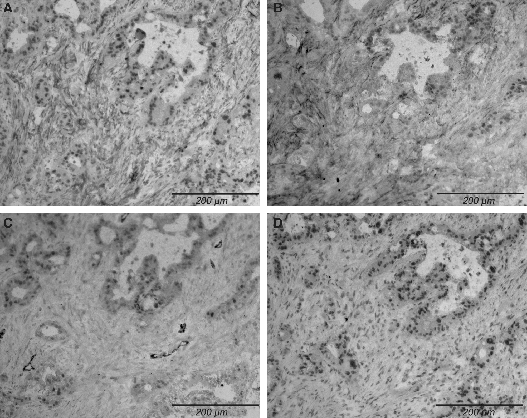
Immunohistochemistry on frozen tissue of human pancreatic carcinoma ( × 100). (
Radiolabeling, stability, and immunoreactivity
Premodification of mAb 14C5 resulted in 1.5 p-SCN-Bz-DTPA and 0.6 p-SCN-Bz-DOTA moieties per mAb. Subsequent labeling with 111In resulted in an overall labeling yield of >82% for 111In-p-SCN-Bz-DTPA and >60% for 111In-p-SCN-Bz-DOTA. A radiochemical purity of at least 98% was noted for both products. Labeling of mAbs 14C5 and MAB002 with iodine resulted in an overall labeling yield of 90.4% ± 2.7% (n = 8) for 14C5 and 77.8% ± 3.5% (n = 3) for MAB002 and radiochemical purity was >98% for the purified products. The specific activities were 37 MBq/mg for 125/131I-mAbs and 74 MBq/mg mAb for 111In-14C5.
All radioimmunoconjugates were stable for 7 days at 4°C in 0.5% BSA. In serum at 37°C, 125I- and 111In-DOTA-14C5 were stable for at least 7 days (<5% of the radiolabel was released during this incubation), but a faster radiometal release was seen for 111In-p-SCN-Bz-DTPA-14C5 (17% of radiolabel release at day 7). Radiolabeled mAb 14C5 was found to have an immunoreactivity of >78% for all immunoconjugates, even after storage for 48 hours in mouse serum at 37°C. At day 7, a decrease in immunoreactivity was seen for 111In-p-SCN-Bz-DTPA-14C5 to 38%, whereas no decline was seen for iodine-14C5 and 111In-p-SCN-Bz-DOTA-14C5.
Saturation-binding assay in Capan-1 cells
To determine the binding capacity of the radioimmunoconjugates, the dissociation constants (K d) of 125I-14C5, 111In-p-SCN-Bz-DTPA-14C5, and 111In-p-SCN-Bz-DOTA-14C5 were estimated for Capan-1 cells by a saturation-binding assay. 125I-14C5 showed specific binding with a dissociation constant of 0.11 ± 0.01 nM. No major influence of p-SCN-Bz-DTPA or -DOTA conjugation was seen on the binding capacity of 14C5 with a K d of 0.24 ± 0.02 and 0.11 ± 0.03 nM, respectively (n = 3). The saturation binding curves obtained were characteristic of high-affinity specific binding of an antibody to its antigen. No specific binding was seen for 125I-MAB002.
Internalization experiments
The degree of internalization was investigated in a radioimmunoassay with Capan-1 cells. Cells were loaded with 125I- and 111In-labeled mAb 14C5 at 4°C for 2 hours. After removing unbound mAb 14C5 and adding fresh medium, the cells were incubated for various timepoints at 37°C to allow internalization or at 4°C to prevent internalization. Figure 2 shows the distribution of radioactivity in Capan-1 cells following incubation with 125I-14C5 (Fig. 2A, n = 6), 111In-p-SCN-Bz-DTPA-14C5 (Fig. 2B, n = 3), or 111In-p-SCN-Bz-DOTA-14C5 (Fig. 2C, n = 3) at 37°C, corrected for the acid-resistant fraction observed at 4°C. At the start of the internalization assay (t = 0 minute) and at 4°C, the acid-resistant cell-associated fraction was 10%. Because the energy-dependent internalization process cannot occur at 4°C, the average 10% internalized fraction observed during incubation at 4°C was considered to be a result of handling of the cells during the internalization assay and not because of internalization of mAb 14C5. Therefore, the cell-associated fraction observed at 37°C was corrected for this cell-associated radioactivity observed at 4°C at each timepoint and the difference was considered the internalized fraction. A rapid internalization was seen for all radioimmunoconjugates, with a significantly faster and higher internalization for 111In-p-SCN-Bz-DOTA-14C5. At 24 hours, 125I-14C5 showed a significantly lower internalized fraction of 6.93% ± 0.88%, compared with 49.44% ± 0.75% for 111In-p-SCN-Bz-DOTA-14C5 and 36.66% ± 1.42% for 111In-p-SCN-Bz-DTPA-14C5 (p < 0.05, nonparametric Mann-Whitney U test). The activity released in the medium was higher for both 125I-14C5 (64.40% ± 0.10%) and 111In-p-SCN-Bz-DTPA-14C5 (40.83% ± 1.62%), compared with 111In-p-SCN-Bz-DOTA-14C5 (19.13% ± 0.45%; p < 0.05, nonparametric Mann-Whitney U test). Analysis of the media revealed that the radioactivity of 111In-p-SCN-Bz-DTPA-14C5 and 111In-p-SCN-Bz-DOTA-14C5 was primarily present in a high–molecular-weight form, with no appreciable amounts of free 111In, 111In-p-SCN-Bz-DTPA, or 111In-p-SCN-Bz-DOTA (<6%), whereas the released radioactivity derived from 125I-14C5 was primarily present in the degraded form (>77%). Tumor cell lysates showed no free 125I, consistent with the rapid release of free iodine from the lysosomes. In contrast, both free 111In/111In-p-SCN-Bz-DTPA/111In-p-SCN-Bz-DOTA and intact 111In-p-SCN-Bz-DTPA-14C5 and 111In-p-SCN-Bz-DOTA-14C5 were observed in the tumor cell lysate.

Distribution of radioactivity in Capan-1 cells following incubation at 37°C with 125I-14C5 (
Biodistribution in Capan-1 tumor-bearing mice
For biodistribution studies, approximately 2 μg (37 MBq/mg) antibody per mouse was used. The biodistribution with nonspecific 131I-MAB002 in Capan-1–bearing mice showed a significantly lower uptake in tumor tissue at all timepoints (p < 0.05, nonparametric Mann-Whitney U test; Fig. 3A) and a significantly longer retention in the blood compared with 131I-14C5 (5.07 ± 0.84%ID/g and 2.24 ± 0.03%ID/g at 168 hours, respectively). At all timepoints, tumor uptake with 131I-14C5 was higher than that with 131I-MAB002, with the highest uptake of 12.16 ± 1.84%ID/g at 48 hours p.i. Tumor-to-blood ratios of 131I-14C5 exceeded 1 at 24 hours and remained high at all timepoints. At 48 hours, the time of highest tumor uptake, ratios of 131I-MAB002 were 6.6-fold lower than those of 131I-14C5. Tumor-to-blood ratios of 131I-14C5 were significantly higher compared with those of 131I-MAB002 for all timepoints (p < 0.05; Fig. 3B).
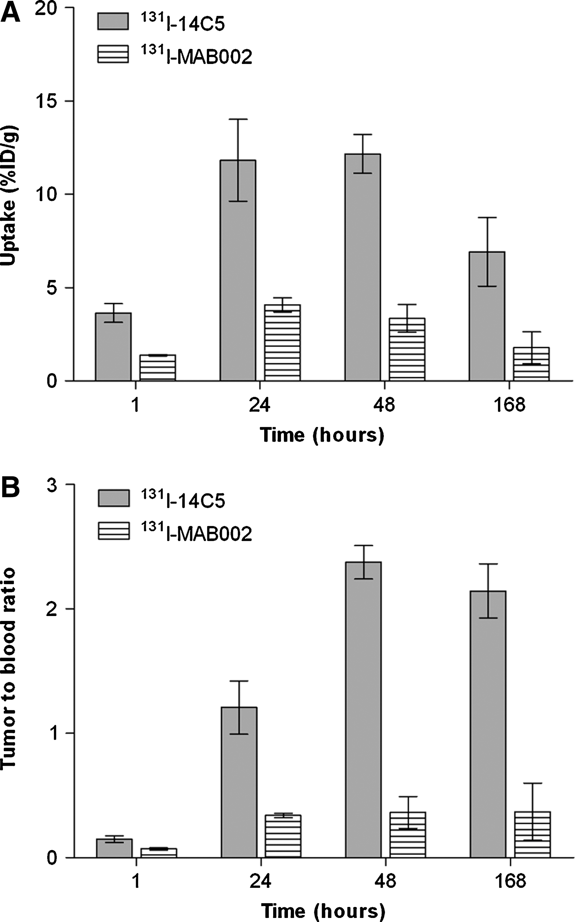
Antigen specificity of in vivo tumor binding. (
Table 1 summarizes the uptake of radiolabeled mAb 14C5 in the tumor and the important organs. The %ID/g of 131I-14C5 in Capan-1 xenografts peaked at 24–48 hours (11.22–12.16%ID/g), whereas tumor uptake for 111In-conjugated 14C5 was highest at 48–72 hours after antibody injection (with 12.93–14.30%ID/g for p-SCN-Bz-DTPA and 24.68–35.84 for p-SCN-Bz-DOTA), with the uptake of 111In-p-SCN-Bz-DOTA-14C5 being 2.2- and 3.9-fold greater than that of 111In-p-SCN-Bz-DTPA-14C5 and 131I-14C5, respectively, and reaching statistical significance (p < 0.05, Mann-Whitney U nonparametric test). Tumor-to-blood ratios for 111In-labeled-14C5 peaked at 11.54 ± 7.53 (p-SCN-Bz-DTPA) and 15.09 ± 4.16 (p-SCN-Bz-DOTA) at 168 hours.
Values are given in percent of injected dose per g (%ID/g); n = 3. Data are presented as mean ± standard deviation.
For iodine, other organs did not show specific uptake but showed a progressive washout of radioactivity, which mimicked the blood clearance. For both 111In-p-SCN-Bz-DTPA-14C5 and 111In-p-SCN-Bz-DOTA-14C5, the liver showed a high uptake of 8.85–13.88%ID/g and 11.30–14.02%ID/g, respectively. Kidney and spleen uptakes were also increased, but other organs showed no uptake of the indium-labeled 14C5. Bone uptake for both radiometals was <2.49%ID/g, uptake in muscle was <1%ID/g, and uptake in pancreas, intestines, and stomach was <2%ID/g. Uptake in the lungs was comparable for all three conjugates, with a high uptake at early timepoints, but with a declining uptake over time.
The half-life of each radioimmunoconjugate was estimated from the %ID/g of blood obtained over the timepoints sampled. Assuming an exponential two-phase decay in a two-compartmental model, the T 1/2α for 131I-, 111In-p-SCN-Bz-DTPA-, and 111In-p-SCN-Bz-DOTA-14C5 was 51.93, 55.38, and 24.94 minutes and mean T 1/2 ß was 2109, 868, and 1166 minutes, respectively.
To evaluate the influence of antibody concentration on liver, kidney, and spleen uptake, biodistributions with 50 and 100 μg (both 1.5 MBq) of 111In conjugates were carried out (Table 2). No influence was seen for p-SCN-Bz-DTPA, but for p-SCN-Bz-DOTA significant differences were noted, with a decrease in the liver from 12.18 ± 4.40%ID/g (2 μg) to 3.80 ± 0.66%ID/g (100 μg) and in the spleen from 8.03 ± 1.84%ID/g (2 μg) to 4.50 ± 0.80%ID/g (100 μg), but with an increased uptake in the kidneys from 6.39 ± 2.25%ID/g to 13.60 ± 1.70%ID/g (p < 0.05, Mann-Whitney U nonparametric test).
%ID/g, n = 3; Data are presented as mean ± standard deviation.
Significant differences between uptake of 2 μg and 100 μg of 111In-p-SCN-Bz-DOTA-14C5; p < 0.05 (Mann-Whitney U nonparametric test).
n.d., not done.
Planar γ camera imaging
Localization of radiolabeled mAb 14C5 in Swiss nu/nu mice bearing a Capan-1 tumor is presented in Figure 4, in which panel A shows 123I-14C5, panel B shows 111In-p-SCN-Bz-DTPA-14C5, and panel C shows 111In-p-SCN-Bz-DOTA-14C5. For 123I-14C5, no images were acquired after 48 hours because activity levels were too low for detection. For all radioimmunoconjugates, no tumor localization was evident at 3 hours after injection, with the image showing blood-pool activity and increased activity in the central portion of the scan corresponding to the cardiac and liver areas. At 24 hours, defined localization of mAb 14C5 to the Capan-1 tumor was seen. Although higher uptake in the liver area was seen for 111In-conjugated 14C5 (Fig. 4B, C), consistent with biodistribution data, the Capan-1 tumor was clearly defined. Radioactivity was localized in the Capan-1 tumor, and via region of interest analysis, the tumor:contralateral-side ratios were determined. For all isotope conjugates, ratios exceeded 2 at 24 hours, with the highest ratio of 4.09:1 for 123I-14C5 (48 hours), 3.77:1 for 111In-p-SCN-Bz-DTPA-14C5, and 6.46:1 for111In-p-SCN-Bz-DOTA-14C5 (both 96 hours).
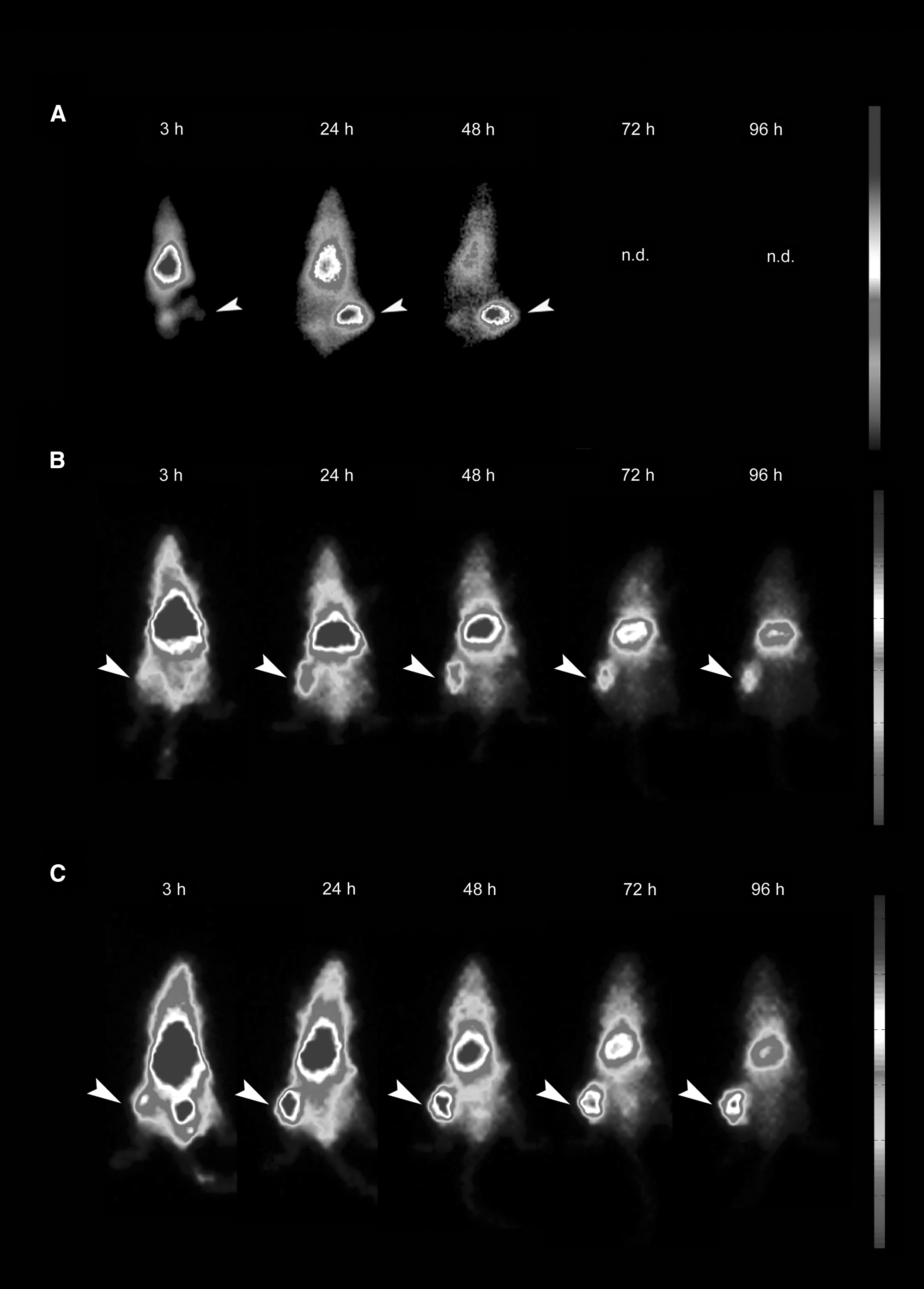
Whole body planar images of 22.2 MBq 123I-14C5 (
Discussion
Pancreatic cancer has one of the poorest prognoses of all cancers, with no available curative treatment except surgical resection if possible. 2 14C5 is a mAb that binds to the integrin αvß, 15 an antigen that is overexpressed on a variety of tumor cells, including pancreatic Capan-1 cells. This study investigated the use of mAb 14C5 to target radioisotopes specifically to pancreatic cancer. Labeling stability, in vitro binding capacity and internalization, and in vivo biodistribution of the mAb 14C5 were investigated, comparing iodine-, 111In-p-SCN-Bz-DTPA-, and 111In-p-SCN-Bz-DOTA-labeled mAb in a Capan-1 xenografted Swiss nu/nu mouse model.
Although the expression of integrin αvß has been investigated extensively in breast-, colon-, and lung-cancer tissues, 11,12,15 its expression had not been examined in human pancreatic-cancer tissues. By immunohistochemistry, specific stromal uptake of mAb 14C5 was demonstrated in all tumor sections. An intensive staining was seen in the stroma surrounding the pancreatic tumor cells, but little Ag 14C5 was present on the tumor cells, similar to the data provided by Burvenich et al. 15 Cancer-associated fibroblasts (CAFs) play an emerging role in the initiation and progression of cancer. These activated fibroblasts can enhance growth and metastasis of cancer cells by altering the ECM within the tumor stroma and changing the release and availability of growth factors. 36 The cell-surface serine protease known as FAP is a CAF-specific marker, and targeting FAP with sibrotuzumab in a phase I dose-escalation study showed specific uptake in the tumor sites without apparent side-effects. 37 Coexpression of the anti-FAP F19 antibody with mAb 14C5 in human pancreatic tumor sections shows that targeting the integrin αvß on activated fibroblasts or PSCs with mAb 14C5 might serve as a new approach for cancer therapy. As described by Mahadevan and Von Hoff, 17 activated fibroblasts could play an important role in the oncogenic process of pancreatic cancer, not only by altering the microenvironment of the tumor but also by altering the resistance to radiation and chemotherapy. 38 By not only targeting tumor cells but also CAFs, radioimmunotherapy with mAb 14C5 in pancreatic tumors could be a promising new approach for pancreatic cancer therapy.
The mAb 14C5 is a murine mAb that could potentially lead to immunogenic reactions when used in patients. Nowadays, chimerization and humanization techniques can be used to reduce immunogenicity 39,40 and even fully human antibodies can be generated. 41 Efforts for genetically engineering mAb 14C5 to a chimeric and humanized mAb are currently undertaken.
mAb 14C5 could be efficiently labeled with iodine and 111In in high yields and with high stability. In vitro experiments showed a small effect of conjugation with p-SCN-Bz-DTPA compared with p-SCN-Bz-DOTA on the affinity and immunoreactivity of mAb 14C5, with a twofold lower affinity for 111In-p-SCN-Bz-DTPA-14C5 compared with 125I- and 111In-p-SCN-Bz-DOTA-14C5 and a decrease in immunoreactivity from 78% to 38% after 7 days at 37°C in mouse serum. Comparison with other mAbs used for targeting pancreatic carcinoma showed that mAb 14C5 has comparable affinity with hPAM4 42 and a 2- to 20-fold higher affinity to pancreatic tumor cells compared with mAb AMB8LK (antiferritin), mAb DU-PAN 1, and mAb B20.4.1 and G6 (anti-VEGF). 3,43,44
Internalization of the 14C5–receptor complex was demonstrated in pancreatic tumor cells for all radioimmunoconjugates, but delayed retention times for 111In-p-SCN-Bz-DTPA-14C5 and 111In-p-SCN-Bz-DOTA-14C5 compared with 125I-14C5 were shown, consistent with literature suggesting retention of 111In-p-SCN-Bz-DTPA-lysine or 111In-p-SCN-Bz-DOTA-lysine catabolites by tumor lysates. 25 –28 The prolonged intracellular retention of 111In-labeled 14C5 leads to a long-lasting association between the radionuclide and the tumor cells, which would be beneficial not only for imaging but also for radioimmunotherapy of pancreatic cancer. Substituting 111-indium with the ß-emitter 90-ytrium or 177-lutetium could lead to an effective treatment of both bulky pancreatic tumors (90Y with a ß−-energy range of 12.0 mm) as well as minimal residual disease (177Lu with a ß−-energy range of 1.5 mm) with mAb 14C5. Nowadays, excellent results in imaging are also obtained with 124-iodine and 89-zirconium 33 and conjugation of these PET isotopes to mAb 14C5 could be promising in making a valuable PET tracer for pancreatic cancer.
In accordance with Press et al., 29 the DTPA-conjugated antibody was more prone to passive dissociation. Because a higher number of p-SCN-Bz-DTPA molecules are coupled to 14C5 mAb compared with p-SCN-Bz-DOTA (1.5 for DTPA and 0.6 for DOTA), a critical lysine nearby the antigen-binding site could be occupied for the p-SCN-Bz-DTPA-conjugated 14C5 complex, leading to a decreased affinity and immunoreactivity. The lower stability of the 111In-p-SCN-Bz-DTPA-14C5 mAb-Ag complex leads to a higher passive dissociation of 111In-p-SCN-Bz-DTPA-14C5 and this consequently influences the internalization and trapping of the DTPA-conjugated mAb. The faster degradation of 125I-14C5 and dissociation of 111In-p-SCN-Bz-DTPA-14C5 could lead to a faster in vivo clearance and subsequently to a lower tumor uptake. Biodistributions with Capan-1 xenografts were conducted to compare the in vivo characteristics of the radioimmunoconjugates.
In the biodistribution, excellent and specific tumor uptake was observed for all mAb 14C5 conjugates compared with a nonspecific antibody (MAB002). A lower iodine-14C5 uptake compared with 111-indium-14C5 was seen in the Capan-1 xenografts, which has also been demonstrated in other studies, 45,46 and this, together with differences in tumor retention of the radiolabel, most likely represents dissimilarities in cellular processing as seen in the internalization study. The differences seen in the stability and in vitro experiments were also reflected in the in vivo results, with a significantly higher tumor uptake of 111In-p-SCN-Bz-DOTA-14C5 compared with 131I- and 111In-p-SCN-Bz-DTPA-14C5 (p < 0.05 at 48 and 72 hours, respectively). Sabbah et al. 4 reported comparable tumor (12.8–23.6%ID/g) and blood (12.7–14.0%ID/g) values for an 111In-p-SCN-Bz-DTPA-conjugated antiferritin mAb in Capan-1 tumor–bearing mice. However, tumor uptake for the p-SCN-Bz-DOTA-mAb was lower (11.2–14.1%ID/g), in contrast to the uptake of 111In-p-SCN-Bz-DOTA-14C5 seen in the present experiments (24.7–35.8%ID/g). Also a significantly higher liver uptake (18.4–19.7%ID/g) was noted for the p-SCN-Bz-DOTA-conjugated antiferritin mAb compared with the p-SCN-Bz-DTPA-coupled mAb. Many different researchers 47 –50 have reported stable conjugations of mAbs with p-SCN-Bz-DTPA. However, it was not possible to establish this stability with p-SCN-Bz-DTPA-14C5.
In the present study, no differences in liver uptake were seen between both 111In-conjugates in a low antibody concentration. Similar uptakes in liver and spleen have been published by several researchers, 7,48,49 and these results are consistent with known sequestration of radiometals in the reticuloendothelial organs. The higher uptake of 111In-labeled 14C5 in the kidneys, compared with 131I-14C5, indicates differences in metabolism of radiometal conjugates as shown by different researchers. 18,45,46,51 Increasing the amount of 14C5 mAb reduced the liver and spleen uptake but elevated the kidney uptake for 111In-p-SCN-Bz-DOTA-14C5, whereas no differences were seen for 111In-p-SCN-Bz-DTPA-14C5. As postulated by Lub-de Hooge et al., 52 the lower stability of 111In-p-SCN-Bz-DTPA-14C5 could cause transchelation to transferrin, leading to faster blood clearance and higher uptake in the liver. Knogler et al. 53 showed a decrease in liver uptake of p-SCN-Bz-DOTA-conjugated chCE7 antibody, with a decreasing ratio of chelator. The 111In-p-SCN-Bz-DTPA-14C5 mixture has a higher number of chelator molecules compared with 111In-p-SCN-Bz-DOTA-14C5, which could result in the high liver uptake at 100 μg with 111In-p-SCN-Bz-DTPA-14C5. Preadministration of unlabeled mAb 14C5 did reduce the uptake of 111In-p-SCN-Bz-DOTA-14C5 in the liver but not in the kidneys or spleen (data not shown). As shown by Morris et al. 54 and Pandit-Taskar et al., 55 clearance of 111In-mAb in serum and liver was dependent on antibody mass, with liver uptake being inversely proportional with mAb concentration. The reason for the increased uptake in the kidneys with increasing amounts of 111In-p-SCN-Bz-DOTA-14C5 is unclear and should be further examined.
Uptake of radiolabeled 14C5 mAb in other organs reflects the blood flow and catabolization of the radiolabeled conjugates and is not a consequence of specific targeting attributable to the presence of 14C5 antigen expression.
Data from the biodistributions were confirmed in the planar γimaging, with 111In-p-SCN-Bz-DOTA-14C5 showing the highest specific tumor uptake. Because of the longer half-life of 111-indium compared with 123-iodine, images could be taken even after 48 hours p.i., with the highest tumor-to-background ratio being 6.46:1 for 111In-p-SCN-Bz-DOTA-14C5 at 168 hours p.i. The relatively high uptake of 111In in the normal liver and the difficult anatomic location of the pancreatic lesions could hamper the diagnosis of pancreatic cancer. But, in patients, Pai-Scherf et al. 56 showed that even small liver metastasis gave a clear positive uptake against the 111In-uptake of the normal liver. Future preclinical and clinical trials could elucidate the potential application of 111In-conjugated 14C5 in the diagnosis of pancreatic adenocarcinoma. Nevertheless, radioimmunotherapy with p-SCN-Bz-DOTA-conjugated 14C5 could be very promising, considering the relative radioresistance of normal liver cells. Based on data from the present study, with a high and specific uptake of 111In-p-SCN-Bz-DTPA-14C5 and 111In-p-SCN-Bz-DOTA-14C5 in the pancreatic tumor and low uptake in normal tissues, 111In-conjugated 14C5 and 111In-p-SCN-Bz-DOTA-14C5 in particular might be an interesting mAb for scouting pancreatic tumors prior to RIT with 90Y or 177Lu. Because combinations of gemcitabine, the standard of care for advanced pancreatic cancer, with radionuclide therapy has shown very promising results, 57,58 and the possibility of therapy with gemcitabine and 14C5 mAb will be explored further.
Conclusions and Future Perspectives
This study shows that mAb 14C5 is a promising new antibody for targeting the integrin αvß5 in pancreatic cancer cells, not only in vitro but also in tumor-bearing xenografts. The superior affinity of mAb 14C5 to human pancreatic tumor cells was demonstrated in a cell-binding assay and in vivo biodistribution. An IgG1 isotype control antibody showed no specific targeting on human pancreatic tumor sections and pancreatic tumor cells in vitro as well as in vivo. Staining of human pancreatic tumor tissues showed intense staining of the tumor stroma and minimal to no staining of surrounding chronic inflammation areas. Radioimmunotherapy in which the CAFs are targeted with radiolabeled mAb 14C5 could be a promising new approach for therapy of pancreatic cancer, especially in combination with gemcitabine, a radiosensitizing chemotherapeutic targeting tumor cells.
Internalization experiments reveal a prolonged retention and association of 111In to the tumor cells compared with 125I-14C5, confirming reports from other investigators. 25 –29 Passive dissociation of 111In-p-SCN-Bz-DTPA-14C5 as a result of lower stability of the mAb-Ag complex leads to a lower in vivo uptake in Capan-1 tumors compared with 111In-p-SCN-Bz-DOTA-14C5. In the biodistributions, the superior internalization of 111In-p-SCN-Bz-DOTA-14C5 is reflected in a significantly higher uptake of the radiometal in Capan-1 tumors compared with 131I-14C5 and 111In-p-SCN-Bz-DTPA-14C5. The large liver uptake for the 111-indium-conjugated mAb 14C5 could hamper imaging of small primary pancreatic tumors. Therefore, it is recommended to use single photon emission computed tomography/computed tomography (SPECT/CT) scans in cases where tumors are located in the head to distinguish between hepatic uptake or tumor uptake and a prolonged clearance time after injection of 111In-p-SCN-Bz-DOTA-14C5 could improve tumor-to-liver ratios. Changing the chelator to C-functionalized trans-cyclohexyl diethylenetriaminepentaacetic acid (CHX-A"-DTPA), a stable chelator for 111In labeling as described by Tolmachev et al., 59 could lower the hepatic background uptake, improving the imaging potential of 111In-p-SCN-Bz-DOTA-14C5. For now, awaiting improved and more stabile chelator conjugations, mAb 14C5 and 131I-14C5 in particular are promising new tools in the detection of pancreatic cancer. Because of the low penetration of mAbs in solid tumors, 60 smaller IgG fragments such as F(ab′)2 are promising tools in increasing the tumor penetration grade and hence radionuclide uptake. Burvenich et al. 10 showed excellent tumor uptake of the bivalent F(ab′)2 14C5 fragment in a mouse A549 lung-tumor model, with improved tumor-to-background contrast images. The use of F(ab′)2 14C5 fragments in the diagnosis and treatment of pancreatic cancer should be explored.
For radioimmunotherapy, it is possible that targeting pancreatic cancer in patients with subacute or acute pancreatitis is hampered by the inflammation area frequently noted adjacent to pancreatic tumors, but further research has to be conducted to reveal the expression of αvß5 in these types of inflammation. As described by Sultana et al., 61 careful selection of patients prior to RIT with a pretherapy dose of mAb 14C5 could assist in the selection of patients with high enough tumor uptake before administration of therapeutic dosages. Supplementary immunologic research of pancreatic metastases could elucidate the possible application of mAb 14C5 in RIT in disseminated pancreatic adenocarcinoma.
To reduce radiotoxic damage to penetrating normal pancreatic glands or adjacent normal tissue, 14C5 variants conjugated with toxins or Auger electron emitters are interesting options. Because of the internalizing nature of mAb 14C5, nuclear localizing sequences could promote nuclear translocation and enhance the radiotoxicity of mAb 14C5 labeled with Auger electron emitters 62,63 targeting stromal fibroblast in the tumor and sparing surrounding normal tissue. Further investigation on the expression of the integrin αvß5 and its possible role as a marker for CAFs is necessary to understand fully the therapeutic and diagnostic potential of mAb 14C5.
Footnotes
Acknowledgments
The data and human normal and pancreatic tissues used in this project were provided by the Victorian Cancer Biobank with appropriate ethics approval. The Victorian Cancer Biobank is supported by the Victorian Government. The authors thank Dr. Peter Crowley, Director, and Dr. Siddhartha Deb, Senior Pathology Registrar, Department of Anatomical Pathology, Austin Health, for scoring the pancreatic tissues.
Disclosure Statement
No conflict of interest exists for any of the authors.