
Abstract
Objective:
Buthus martensi Karsch (BmK) CT, a kind of scorpion toxin peptide, was found to inhibit glioma cell proliferation in previous researches. 131I-BmK CT may have more inhibition effect and could be used as a glioma cell-targeted therapy and imaging agent. The purpose of this study was to investigate whether 131I-BmK CT could specifically conjugate with C6 glioma cell and induce glioma cell inhibition in vitro.
Methods:
After cloning, expression, and purification, BmK CT was labeled with 131I by indirect labeling (Bolton–Hunter method). The cell conjugation experiment was performed to investigate the connection between the reciprocal of cell conjugation rate and the reciprocal of cell count. 3-(4,5-dimethylthiazol-2-yl)-2,5-diphenyl-tetrazolium bromide (MTT) method and flow cytometry were used to detect the inhibition effect of BmK CT and 131I-BmK CT on glioma cell proliferation.
Results:
131I-BmK CT was successfully prepared with the overall yield of 34.5%. The cell conjugation experiment indicated that 131I-BmK CT could specifically conjugate with C6 cells. MTT tests indicated that both BmK CT and 131I-BmK CT could inhibit C6 growth. The ability of 131I-BmK CT to inhibit cell growth is superior to that of BmK CT. The inhibitory rate (IR) of glioma cells was 60.5% (p < 0.01) at the concentration of 2 μg/mL with BmK CT. And the IR was 71.2% (p < 0.01) at the radioactivity concentration of 50 μCi/mL (concentration was much lower than 2 μg/mL) with 131I-BmK CT. BmK CT could block the C6 glioma cell cycle in the G0/G1 stage. 131I-BmK CT blocked the cell cycle in the S stage (the proportions of C6 in the S, G0/G1, and G2/M phases were 24.5% ± 0.4% vs. 44.0% ± 2.3%, 63.9% ± 0.6% vs. 51.8% ± 1.6%, and 11.6% ± 1.0% vs. 4.3 ± 0.7% [p < 0.05], respectively, at an initial radioactivity concentration of 50 μCi/mL).
Conclusions:
On the basis of cytology experiments, it was found that 131I-BmK CT could specifically conjugate with C6 glioma cell and inhibit cell growth. Hence, it may be used as a glioma-targeted agent.
Introduction
Gliomas are currently the most common and lethal type of primary brain tumor, accounting for 40%–50% of all brain tumors. In the last 20 years, standard treatments (a combination of surgery, radiotherapy, and chemotherapy) for glioma patients have not produced a significant improvement in survival. 1 Most gliomas' tumor progression and recurrence occur within 2 cm of tumor margin. 2 There is a need for new approaches to provide a higher probability of tumor control with less normal brain tissue damage. One novel approach is to target the brain tumor cells via a unique receptor found on their surface. Chlorotoxin (Cltx), also called TM-601, is a 36-amino acid peptide derived from the scorpion Leiurus quinquestriatus, which has been found to specifically bind to malignant brain tumors, but not to normal brain tissue. 3 Cltx has been widely used as a blocker of small-conductance Cl− channels. 3 Glioma cells have been reported to express a glioma-specific chloride ion channel that is sensitive to Cltx. 4,5 In in vivo targeting and biodistribution experiments, 125I- and 131I-labeled Cltx selectively accumulated in the brain of tumor-bearing mice. 6 –8 A phase I clinical trial of intracavitary 131I-TM-601 in adult patients with recurrent high-grade glioma had been performed to determine the biodistribution, toxicity, and therapeutic effect of Cltx. 9 A full-length cDNA sequence encoding the precursor of a venom peptide, Buthus martensi Karsch (BmK) CT, with homology to Cltx has been isolated from a cDNA library made from the venom glands of the Chinese scorpion BmK. The sequence of BmK CT is similar (68% identities) to that of Cltx. 10,11 It has been shown that BmK CT is a short-chain glioma chloride channel blocker and can induce cell death of cultured malignant glioma cells in vitro. 11,12 Previous studies show that BmK CT can specifically bind to gliomas cells, which will undoubtedly shed light on further diagnostic and therapeutic research on this interesting peptide. The purpose of this study was to investigate whether 131I-BmK CT could specifically conjugate with C6 glioma cell and induce glioma cell growth inhibition in vitro.
Materials and Methods
Materials
EcoRI, XhoI, Tris-HCl, IPTG, reduced glutathione, N-succinimidyl-3-(4-hydroxyphenyl)-propionate (Bolton–Hunter [BH] reagent), chloramine-T, sodium metabisulfite, potassium iodide, phosphate solution, N,N-dimethylformamide, benzene, boric acid, glycin, MTT, dimethyl sulfoxide (DMSO), and propidium iodide were purchased from Sigma Aldrich. pGEM-T Easy vector was purchased from Promega. T4DNA was purchased from Invitrogen. Sephadex G-25 was purchased from Pharmacia. Sodium 131I-iodide was purchased from Shanghai GMS Pharmaceutical Company. Culture medium (Ham's F12K) was purchased from AppliChem. C6 rat glioma cell was purchased from the Shanghai Institute of Biochemistry and Cell Biology.
Cloning, expression, and purification of scorpion toxin BmK CT
According to the mutated cDNA sequence of BmK CT, two primers were designed to synthesize BmK CT:
Primer 1: 5′-ACTGAATTCATCGAAGGTCGTTGTGGGCCTTGCTTTACAACGGATGCTAATATGGCAAGGAAATGTAGGGAATGTTGC
Primer 2: 5′-AGCTCGAGTTATATACGGTTACACAGACATTGTGGGCCAAAGCATTTTCCAATACCTCCGCAACATTCCCTACATTT
The BmK CT gene was obtained by using a polymerase chain reaction (PCR) method. The PCR product was subcloned into a pGEM-T Easy vector and this cDNA sequence was determined by Ying Jun Biotechnology Company. The PCR product was digested with EcoRI and XhoI. And then, the ligation reactions were performed between the digested product and the vector pGEX-4T-1 by using ligase T4DNA to obtain the expression recombinant plasmid pGEX-4T-1-BmK CT. The plasmids were then transformed into Escherichia coli BL21 (DE3) by using the heat-shock method. The induction of expression was carried out at 16°C for about 3 hours with 0.4 mM IPTG. Cells were harvested by centrifugation at A 600 = 0.8–1.0 and then lysed by ultrasound and again centrifuged. The supernatant was applied to GST volume. Elution buffer used was 10 mM reduced glutathione in 50 mM Tris-HCl (pH 8.0). After elution, the obtained liquid was kept at 4°C for 16 hours and then freeze-dried. After weighted freeze drying, the obtained powder was dissolved and then enzymolysed for 18 hours with XhoI at 24°C. After high-performance liquid chromatography (HPLC) purification of the enzymolysis output, elution liquid of wave crest was gathered for mass spectrographic analysis.
Radiolabeling procedure
BH reagent was labeled with iodine-131 (I-131) by the chloramine-T method and purified by solvent extraction. Briefly, to 1 μL of a fresh BH solution in benzene (0.5 mg/mL), blow dried with nitrogen, 5 μL chloramine T (5 mg/mL in phosphate buffer) and 3.7–37 MBq (0.1–1.0 mCi) of sodium 131I-iodide were added. Iodination was carried out by incubation for ∼15 seconds, and then 5 μL of 12 mg/mL sodium metabisulfite in phosphate buffer (0.25 mol/L, pH 7.5) and 10 μL of 10 mg/mL potassium iodide in phosphate buffer (0.05 mol/L, pH 7.5) were added. To avoid ester hydrolysis, the radiolabeled BH was immediately extracted with 5 μL N,N-dimethylformamide and 200 μL benzene. The radiochemical purity was checked using thin layer chromatography. The organic solvent was then evaporated using a dry nitrogen stream before adding the BmK CT.
For the encapsulation of 131I-BH, 10 μg of 1 mg/mL BmK CT in boric acid buffer (0.125 mol/L, pH 8.4) was added to the dry 131I-BH reagent in a glass tube. After 30 minutes, 150 μL of 0.2 mol/mL glycin was added to link with the redundant 131I-BH. The 131I-BH-BmK CT solution was eluted by sephadex G-25, medium grade, which was equilibrated with boric acid buffer (0.125 mol/L, pH 8.4). One milliliter (1 mL) of liquid was collected in every tube. A total of 30 tubes were collected. The radioactivity of each tube was measured by activity meter to draw the elution graph.
Experiment of cell conjugation
C6 glioma cell suspension was prepared, with 107 cells/mL as the initial concentration. Five cell suspension concentration gradients were diluted (one half, 1.5 mL in each tube). To each test tube, 50 μCi 131I-BmK (500 μCi/mL) was added. After 1.5 hours, radioactive counting of the sediment after centrifugation was performed. The connection between the reciprocal of cell conjugation rate and the reciprocal of cell count was investigated.
Cell counting and MTT assay
In brief, 5000 cells/well were seeded into flat-bottomed 96-well plates. After 8 hours, 100 μL BmK CT of different concentrations, sodium iodide I-131 solution, and 131I-BmK CT of different radioactivities were added for 48 hours to achieve synchronous growth arrest, respectively. The initial concentration of BmK CT was 0.2 mg/mL. The initial radioactivity of I-131 solution and 131I-BmK CT was 50 μCi/mL. Eight diluted solutions of different concentrations and six diluted solutions of different radioactivities were used. Forty-eight (48) hours after the experimental stimulation, MTT was added (0.5 μg/mL) and incubated for 4 hours, followed by addition of 150 μL DMSO to dissolve the formed formazan crystals. Optical density (OD) values for each well were measured at 490 nm using an enzyme-linked immunosorbent assay meter. Inhibitory rate (IR) was calculated according to the following formula: IR = [1 − (mean OD of treated group − mean OD of blank group)/(mean OD of control group − mean OD of blank group)] × 100%.
Flow cytometric analysis of the cell cycle
Cells (1 × 10⁵ per mL) were seeded into a culture bottle. After 6–8 hours, BmK CT of different concentrations or 131I-BmK CT of different radioactivities was added to achieve synchronous growth arrest, respectively. The end concentration of BmK CT was 0.2, 0.02, and 0.002 mg/mL. The end radioactivity of 131I-BmK CT was 50, 5, and 0.5 μCi/mL. In the control group, culture medium was added. The cells were incubated for 72 hours. Finally, samples were processed to obtain simple cell suspensions (target cell number was 1 × 10⁶ per mL). Then they were stained with propidium iodide (final concentration, 50 μg/mL) for 30 minutes. Finally, the cell suspensions were analyzed for the cell cycle using a nylon monofilament mesh screen with 44-μm openings and by flow cytometry.
Results
Cloning, expression, and purification of scorpion toxin BmK CT
The recombinant plasmid pGEX-4T-1-BmK CT was transformed into a competent cell (DH12S). Several positive clones were identified using restriction enzymes and gel electrophoresis. Among them, 14 positive clones were selected at random for sequence analysis, and 2 of them were found to have the correct sequences. After the pGEX-4T-1-BmK CT plasmids were transformed into E. coli BL21 (DE3), a single correct colony was selected. Recombinant plasmid was obtained by double restriction enzyme digestion (EcoRI and XhoI). Enzyme electrophoretogram showed an electrophoretic band at 120 bp, which was similar to gene encoding mature peptide of BmK CT. After induction of expression, sodium dodecyl sulfate–polyacrylamide gel electrophoresis analysis showed a band of 10 kDa in the recombinant plasmid lane. The increased level of band density after induction indicated that the expression of fusion protein extremely increased after induction. Fusion protein was applied to GST column and separated after 10 minutes at 26°C. After purification, mass spectrographic analysis showed that the molecular weight of fusion protein (M W 31,197) was the same as the theorically expected value (the molecular weight of BmK CT plus the molecular weight of GST equals 31,197). After weighted freeze drying, the obtained powder was dissolved and enzymolysed for 18 hours with XhoI at 24°C. After HPLC purification of the enzymolysis output, mass spectrographic analysis indicated that the molecular weight of the obtained protein was 3748, similar to the value 3749 in theory (Fig. 1).
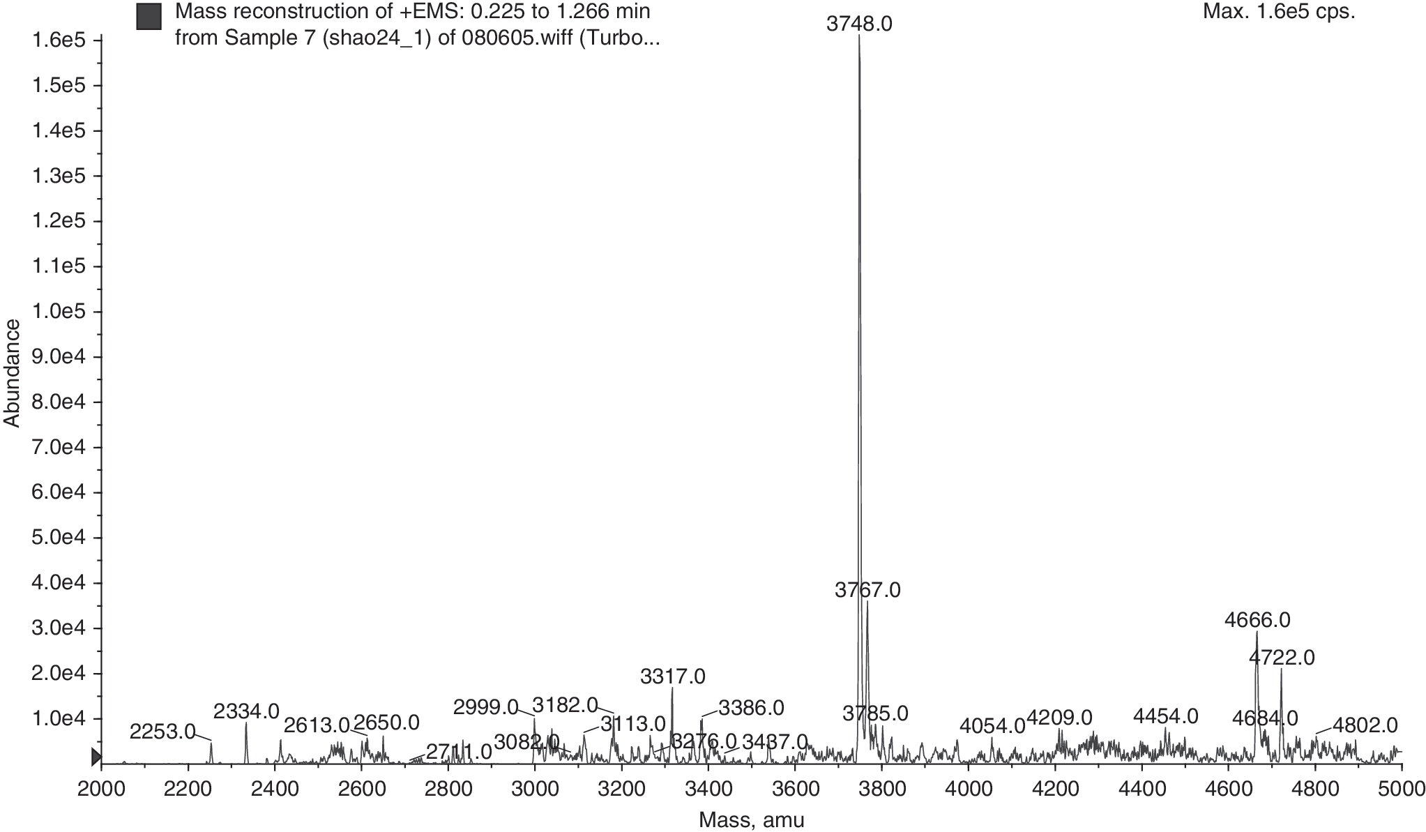
Mass spectrographic analysis of obtained protein: the molecular weight of obtained protein was 3748, similar to the value 3749 in theory.
Radiolabeling procedure and cell conjugation experiment
The radioactivity of each of the 30 collected tubes was measured by activity meter to draw the elution graph (Fig. 2). 131I-BmK CT was successfully prepared by indirect labeling (BH method) with the overall yield of 34.5% (mean rate: 30.2% ± 2.8%; range: 25.6%–34.5%). The mean radiochemical purity of 131I-BmK CT was 94.0% ± 0.8%. 131I-BmK CT has excellent stability in vitro, with the radiochemical purity being higher than 92% within a period of 48 hours. The cell conjugation experiment indicated that the reciprocal of cell conjugation rate is of linear correlation with the reciprocal of cell count (R = 0.85).

Indirect labeling (BH method): The radioactivity of each of the 30 collected tubes was measured by activity meter. In the radioactivity graph, there are two peaks. The first peak is for 131I-BH-BmK CT and the second small peak is for free iodine. BH, Bolton–Hunter; BmK, Buthus martensi Karsch.
MTT assay of BmK CT and 131I-BmK CT to glioma
MTT tests indicated that both BmK CT and 131I-BmK CT could inhibit C6 glioma cell growth (Fig. 3). The IR of glioma cell reached up to 90.5% (compared with the control group; p < 0.01) at the concentration of 0.2 mg/mL with BmK CT. The IR of glioma cell was 60.5% at the concentration of 2 μg/mL with BmK CT. And the IR of glioma cell was 71.2% (p < 0.01) at the radioactivity concentration of 50 μCi/mL (concentration is much lower than 2 μg/mL) with 131I-BmK CT. In addition, the IRs were 56.2% and 31.5% at the radioactivity concentration of 50 and 5 μCi/mL with sodium iodide I-131 solution, respectively.
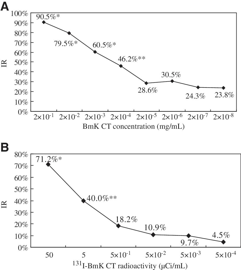
Detection of the IRs of BmK CT and 131I-BmK CT to C6 glioma cells by MTT method. (
Flow cytometric analysis of the cell cycle
As shown in Table 1, the inhibition treatment with BmK CT at initial concentration (0.2 mg/mL) increased the proportion of C6 glioma cells in the G0/G1 phase (79.3% ± 1.1% vs. 88.9% ± 1.5%, p < 0.05) and decreased the proportion of C6 glioma cells in the S phase (15.7% ± 2.1% vs. 6.8% ± 1.6%, p < 0.05). This result suggested that BmK CT could block the C6 glioma cell cycle in the G0/G1 stage.
Compared with the control group (p < 0.05).
BmK, Buthus martensi Karsch.
In contrast to BmK CT, however, 131I-BmK CT blocked the cell cycle in the S stage (Table 2). The inhibition treatment with 131I-BmK CT at the initial radioactivity concentration (50 μCi/mL) increased the proportion of C6 glioma cells in the S phase (24.5% ± 0.4% vs. 44.0% ± 2.3%, p < 0.05) and decreased the proportion of C6 glioma cells in the G0/G1 phase (63.9% ± 0.6% vs. 51.8% ± 1.6%, p < 0.05) and the G2/M phase (11.6% ± 1.0% vs. 4.3% ± 0.7%, p < 0.05). The effects of BmK CT and 131I-BmK CT on the cell cycle of glioma cells could also be observed from the flow cytometry results (Fig. 4).
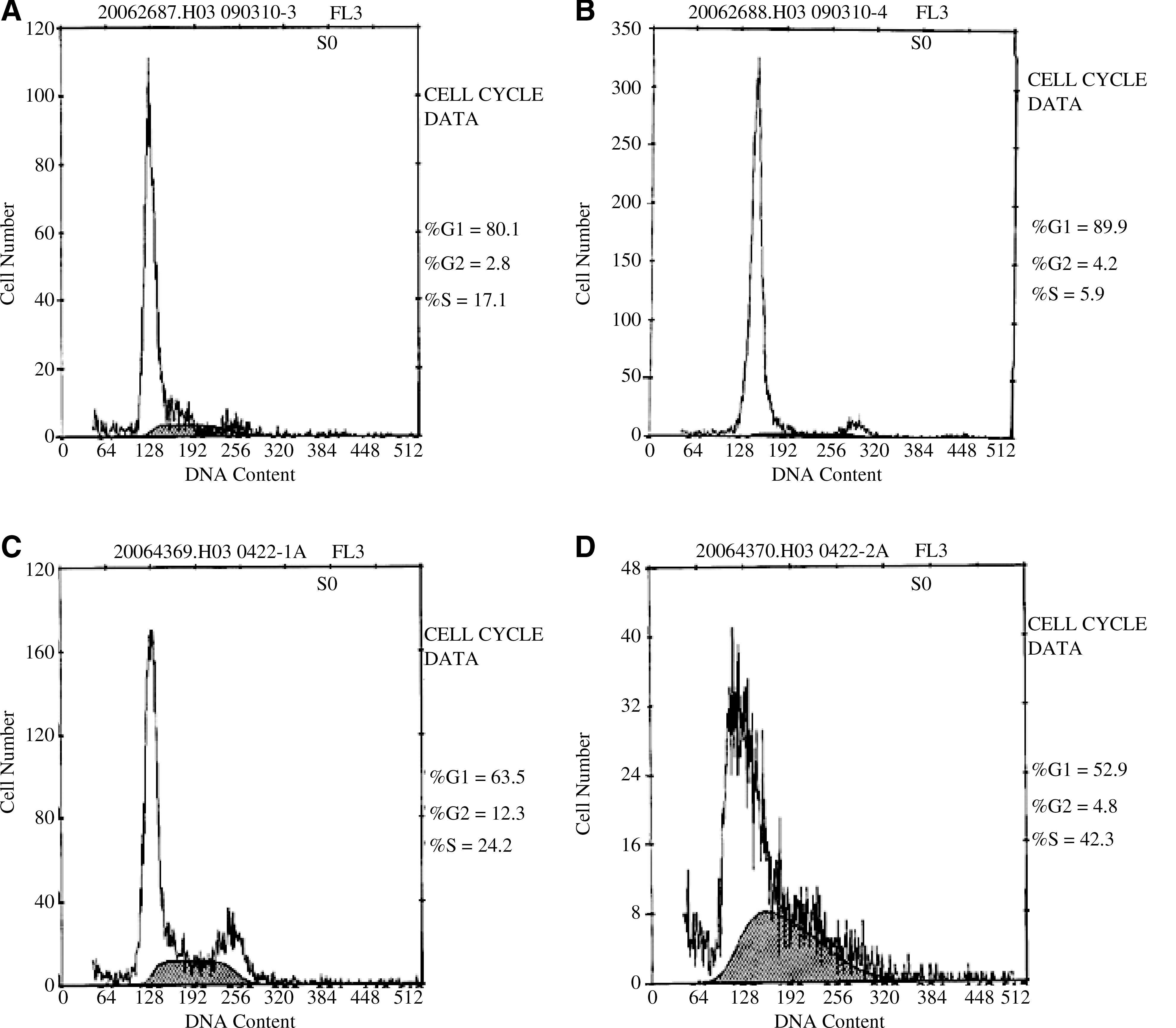
Flow cytometry results showing the influence of BmK CT and 131I-BmK CT on the cell cycle of C6 rat glioma cells. (
Compared with the control group (p < 0.05).
BmK, Buthus martensi Karsch.
Discussion
Gliomas are currently the most common and lethal type of primary brain tumor and their treatments have limitations. Postsurgery treatment of glioma using targeted internal radiotherapy is very appealing. Targeting of glioma has been successful using monoclonal antibody against components in the tumor area. Anti-tenascin BC-2 and 81C6 have been radiolabeled with 131I and 90Y in clinical trials. 13 –15 All of the targeting molecules should have two important features: (1) their molecular sizes should be small for penetrating the cavity wall of the resected tumor and (2) they should bind only to the tumor. Cltx and BmK CT are such a kind of novel targeting agents that have both the aforementioned features.
First, Cltx is a small, 36-amino acid peptide that can cross blood and tissue barriers, and the uptake of 131I in mouse blood and bladders can be observed by gamma camera imaging. The tumor-to-normal organ ratio was high at 96 hours postinjection. 7 Second, Cltx binds to primary brain tumors, but not to nontumor brain tissues, or to other nonbrain tissues in human body. 3 In vivo, Cltx selectively accumulated in the brain of tumor-bearing mice. 6 –8 A phase I clinical trial of intracavitary 131I-TM-601 in recurrent high-grade glioma patients indicated that a single dose of 10 mCi 131I-TM-601 was well tolerated and may have an antitumoral effect. 9 One of the major challenges in targeting brain tumors is the blood–brain barrier (BBB), which limits the access of targeting therapeutics to the brain tumor tissue. Unlike many other radioligands, TM-601 is relatively small. It may pass the BBB, diffuse readily through the brain parenchyma, and spread across the entire tumor, which needs to be confirmed by further investigations. 8
BmK is a representative species of scorpion in northwestern China. BmK CT is the first one of the cDNA sequences encoding four-disulfide-bridged short-chain toxins from BmK so far. The sequence of BmK CT is similar (68% identities) to that of Cltx. 3,4 Previous studies have demonstrated that the polypeptidyl toxins purified from BmK venom interfere with various ion channels, specifically the chloride channels, and alter their functional properties. 16 It has been shown that BmK CT is a short-chain glioma chloride channel blocker and can induce death of cultured malignant glioma U-251 cells in vitro. 11,12,16 But no study has shed light on the labeling of BmK CT.
After cloning, expression, and purification of the scorpion toxin BmK CT, we labeled it with I-131 by indirect labeling because direct labeling is not suitable for BmK CT. The amino acid structure of BmK CT does not contain tyrosine or histidine. The cell conjugation experiment indicated that 131I-BmK CT could specifically conjugate with C6 glioma cell. Therefore, step evaluation is needed and meaningful.
I-131 has been widely used in radionuclide therapy for cancer. Beta radiations from I-131 are used for treatment, whereas gamma radiations from I-131 are used for imaging. 131I-labeled Cltx and BmK CT peptide can specifically bind to chloride channel proteins on the glioma cell surface and then affect the shape and volume of the cytoskeleton of glioma cell to inhibit tumor infiltration. On the other hand, I-131 can emit beta radiation to kill tumor cells. Because of the specific binding to chloride channel proteins and the short range of beta radiations, these polypeptides provide a higher probability of tumor control with less normal brain tissue damage. 131I-labeled Cltx and BmK CT may provide an important tool for diagnosis of and determining tumor extent before treatment. The distribution of 131I-labeled Cltx and BmK CT peptide in vivo could be investigated more accurately to provide more information to therapeutic evaluation. But these effects of BmK CT on glioma in theory should be investigated in future experiments.
In this study, MTT tests indicated that both BmK CT and 131I-BmK CT could inhibit C6 glioma cell growth. The ability of 131I-BmK CT to inhibit cell growth was superior to that of BmK CT and sodium iodide I-131 solution. The IR of glioma cell was 71.2% at the radioactivity concentration of 50 μCi/mL (concentration was much lower than 2 μg/mL) with 131I-BmK CT. But the accurate concentration of 131I-BmK CT was unknown. This concentration (much lower than 2 μg/mL) was calculated on the initial quality of BmK CT at the beginning of indirect labeling, the volume of labeling product obtained, and the volume of added reagent to obtain the highest concentration. Thus, the inhibition effects of 131I-BmK CT was much higher than BmK CT.
In the flow cytometric analysis of the C6 glioma cell cycle, the inhibition treatment with BmK CT increased the proportion of C6 glioma cells in the G0/G1 phase, decreased the proportion of C6 glioma cells in the S phase, and had less effect on the G2/M phase. Thus, the mitosis of glioma cell was inhibited by BmK CT and the glioma cell cycle was blocked at the G0/G1 stage. In contrast, the inhibition treatment with 131I-BmK CT increased the proportion of C6 glioma cells in the S phase and decreased the proportion of cells in the G0/G1 and G2/M phases. This indicated that 131I-BmK CT could stop the cell cycle from the S phase to the G2/M phase to inhibit the synthesis of DNA in glioma cells.
As a conclusion, 131I-BmK CT, prepared by indirect labeling, could specifically conjugate with C6 glioma cell and inhibit the growth of C6 glioma cell in vitro. Currently, whether 131I-BmK CT can also specially conjugate with and inhibit glioma tumor growth in vivo is under investigation. This study may provide a novel, convenient, and minimally invasive method for “targeted nuclide therapy” and therapeutic monitoring of glioma in the future.
Footnotes
Acknowledgments
This study was supported by the National Natural Science Foundation of China (no. 30770600) and the Shanghai Science and Technology Commission (no. 07SP07004). The authors are very grateful to Prof. Cheng-xin Gong, M.D., Head of Laboratory of Brain Metabolism, New York State Institute for Basic Research in Development Disabilities, for his helpful comments and review of this manuscript.
Disclosure Statement
All authors have seen the manuscript and approved to submit to this journal. And there is no conflict of interest for any of the authors of this manuscript.