Preliminary In Vivo Evaluation of [ 131 I]-2-Iodo- D -Phenylalanine as a Potential Radionuclide Therapeutic Agent in R1M-Fluc Rhabdomyosarcoma Tumor-Bearing NuNu Mice Using Bioluminescent Imaging
Available accessResearch articleFirst published online April, 2010
Preliminary In Vivo Evaluation of [ 131 I]-2-Iodo- D -Phenylalanine as a Potential Radionuclide Therapeutic Agent in R1M-Fluc Rhabdomyosarcoma Tumor-Bearing NuNu Mice Using Bioluminescent Imaging
Carrier-added [123I]-2-iodo-d-phenylalanine (CA [123I]-2-I-d-Phe) was previously found to have a preferential retention in tumors with a high tumor background contrast in animal models. A previous human dosimetry study demonstrated a favorable biodistribution and radiation burden in human subjects.
Aim:
The aim of this study was to investigate the potential of CA [131I]-2-I-d-Phe as an agent for radionuclide therapy.
Methods:
Sixty (60) nude athymic mice were inoculated subcutaneously with firefly luciferase-transduced R1M rhabdomyosarcoma cells. The mice in the therapy group were injected intravenously (i.v.) with 148 MBq [131I]-2-I-d-Phe (432 GBq/mmol) in kit solution. Controls were injected with kit solution without radioactivity, with physiological saline, or with 148 MBq [131I]− in physiological saline. Tumor growth was quantified using bioluminescent imaging and caliper measurements.
Results:
[131I]-2-I-d-Phe clearly reduced tumor growth in the treated mice compared with the control groups. A tumor growth-rate reduction of at least 33% was found for mice receiving a therapeutic dose. There were no serious adverse side-effects of the therapy.
Conclusions:
In conclusion, i.v. injection of CA 148 MBq [131I]-2-I-d-Phe specifically reduces tumor growth in athymic nude mice without relevant side-effects on the animals' health.
Introduction
Radionuclide therapy provides an alternative to surgery or medical treatment of cancer. This therapy combines the advantage of tumor selectivity with that of being systemic, similar to chemotherapy, and may be used as part of a therapeutic strategy with a curative intent or for disease control and palliation. When cure is feasible, the long-term consequences of radionuclide therapy are comparable favorably to the risks associated with, and accepted for, chemotherapy and radiotherapy. Radiotoxicity must be limited and side-effects must be reduced to a minimum. The amino-acid transport system LAT1, capable of transporting l-enantiomeric amino acids but also some d-amino acids, is an interesting target.1–6 It is overexpressed in many human cancer cells but not in healthy tissue. Moreover, the presence of LAT1 on the blood–brain barrier ensures that radiolabeled amino-acid analogs can cross this barrier and reach brain tumors,7,8 in contrast to large peptides or antibodies. Some amino acids resembling chemotherapeutics, such as melphalan (an agent used for treating breast cancer), and acivin (a more general therapeutic agent) are transported via a LAT-type system.9–11 However, data on the biodistribution and uptake mechanisms of these compounds are limited. In 2004, Samnick and colleagues reported, for the first time, the use of [131I]-labeled 4-I-l-phenylalanine as a therapeutic radiopharmaceutical in a glioma rat model.12 In their straightforward study, the researchers demonstrated the potential of radionuclide therapy clearly, using an amino-acid analog.
The authors of this current study showed in earlier studies that [123I]-2-d-phenylalanine ([123I]-2-I-d-Phe) is typically transported into different types of human cancer cells by the overexpressed LAT1 system.13,14 The uptake of this d-isomer in nontarget organs in humans is lower than that of the l-isomer coupled to a faster clearance from the body, thereby considerably reducing the radiation burden.15 It was also shown that the clearance of radio-iodinated phenylalanine in rats was significantly slower than in nude mice and that the nude mice model was much more comparable to humans than the rat model.13–15 Therefore, the 131I-labeled analog of this new tracer, [131I]-2-I-d-Phe, as a radionuclide therapeutic agent in an athymic NuNu mice model inoculated with firefly luciferase (FLuc)-transduced R1M cells was evaluated. These cells express the enzyme luciferase, which allows noninvasive follow-up of tumor growth with the dynamic bioluminescence imaging (BLI) technique. BLI allows to determine the amount of “live” tumor tissue and disregards any necrotic or apoptotic tissue as only live cells are capable of producing the luciferase enzyme.16 R1M tumors were chosen as their uptake pattern in nude mice has been well-described used as a tumor model in earlier studies in Wag/Rij rats.13,17,18
Materials and Methods
Synthesis of precursors and labeling
The synthesis of 2-I-d-Phe from commercially available 2-Br-d-Phe (Peptech) was performed as described earlier.19 Carrier-added (CA) [131I]-2-I-d-Phe was produced using the commonly known Cu1+-assisted nucleophilic isotopic exchange kit (2-I-d-Phe 1 mg/mL) in the presence of SnSO4, gentisic acid, and citrate, and yielded CA [131I]-2-I-d-Phe with a specific activity of 432 GBq/mmol. This solution was passed through a sterile 0.22-μm Ag-membrane filter (Millipore) and made up by addition of appropriate amounts of trisodium citrate, yielding an isotonic solution (pH = 6) with a radiochemical purity of >99%. CA [125I]-2-I-d-Phe was prepared by the same procedure, yielding a specific activity of 10.9 GBq/mmol. Quality control was achieved by high-performance liquid chromatography (HPLC), using a Vydac C18 Monomeric 120A (125 × 4 mm) (Grace) column and 10/90 ACN/H2O containing 1 mM NH4Ac and 1 mM NaI as mobile phase at a flow rate of 1.0 mL/minute while monitoring ultraviolet (UV) absorption at 254 nm (Hitachi UV detector; Alltech) and radioactivity (NaI(Tl) detector; Harshaw Chemie).
Generation of a luciferase-positive R1M rhabdomyosarcoma cell line
The construction and the generation of luciferase-encoding lentiviral particles and the generation of the luciferase-positive R1M cells (R1M-fluc) was performed as previously described.19 The quantitative reverse transcriptase–polymerase chain reaction showed that the amount of mRNA expression of LAT1 amino-acid transport system was equal in R1M and R1M-fluc cells.
Sensitivity to 131I radiation in vitro
The cells were put into 1-mL well plates with 1 mL MEM growth medium, in which normal growth is assured, and 50 μL (370 kBq) of [131I]-2-I-d-Phe/kit formulation was added. After incubation at 37°C for 24 hours in a 5% CO2 atmosphere, the number of remaining viable cells was determined by means of trypan blue staining.
In vivo experiments
Animal model
The study protocol was approved by the ethical committee for animal studies at the Vrije Universitet Brussel, in Brussels, Belgium (license no. LA1230272). Guidelines of the National Institutes of Health principles of laboratory animal care (NIH publication 86-23, revised 1985) were followed. The animals were placed in groups of 5 in individually ventilated cages. Animal health was monitored daily by weighing the animals, estimating food and water intake per cage by visual control, and observing general behavior, such as grooming.
The studies were performed using athymic nude mice (n = 60; age 7 weeks; Harlan). Per previous scientific literature showing that inoculating nude mice with a large amount of R1M cells (>106) causes fast-growing tumor undergoes considerable necrosis, the amount of inoculated cells was limited to 105. The mice were inoculated subcutaneously (s.c.) in the shoulder region with 100 μL of cell suspension containing 100,000 R1M-fluc cells under isoflurane anesthesia. Anesthesia was performed with an isoflurane/oxygen mixture using an inhalation anesthesia system (VetTech Solutions): 5% isoflurane for induction and 2.5% for maintenance of anesthesia during all in vivo animal experiments.
Pharmacokinetics
The biodistribution study was performed by dissection using 125I-labeled compound. On the tenth day after inoculation the R1M-fluc tumor-bearing mice (n = 20) were intravenously (i.v.) injected with 3.7 MBq of [125I]-d-2-I-Phe in the kit preparation (100 μL). The tumor weight at time of injection was about 0.13 g. At appropriate timepoints (0.5, 1, 2, 4, 6, and 24 hours) the animals were sacrificed by cervical dislocation, the tumor and organs of interest (pancreas, liver, brain, kidney, and muscle) were dissected, and a blood sample was taken. Radioactivity in the tissues was measured in a NaI(Tl) well counter and normalized to the percentage injected activity per g (%IA/g) of tissue.
Tumor dose was estimated by applying the following formula:\documentclass{aastex} \usepackage{amsbsy} \usepackage{amsfonts} \usepackage{amssymb} \usepackage{bm} \usepackage{mathrsfs} \usepackage{pifont} \usepackage{stmaryrd} \usepackage{textcomp} \usepackage{portland, xspace} \usepackage{amsmath, amsxtra} \pagestyle{empty} \DeclareMathSizes{10}{9}{7}{6} \begin{document} \begin{align*} D = { \rm At} \times S , \end{align*} \end{document}
in which D is dose, At is the accumulated activity in the tumor, and S is a geometric matrix value for the tumor, namely, 0.11 Gy/(MBq h).20
Therapy—strategy
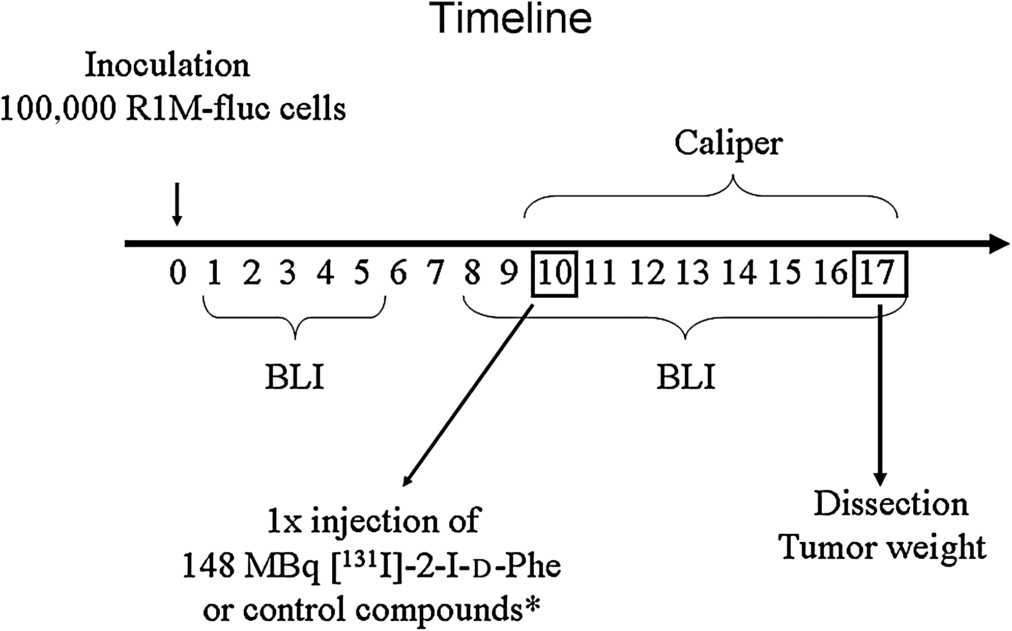
The strategy is represented in Figure 1. Forty (40) NuNu mice (weight ∼30 g, ∼2.1 mL of blood) were inoculated at day 0. The animals were randomly divided into four equal groups and, at day 10 postinoculation, one group was injected i.v. with a bolus of 148 MBq [131I]-2-I-d-Phe in 100 μL kit solution (therapy group), and the other groups were injected with 100 μL kit solution without radioactivity (blank group), with physiological saline (control group), or with 148 MBq [131I]− in physiological saline ([131I]-NaI group), respectively.
Experiment strategy. Control compounds* are 100 μL kit solution without radioactivity, 100 μL physiological saline, or 148 MBq [131I]− in 100 μL physiological saline. BLI, bioluminescent imaging; [131I]-2-I-d-Phe, [131I]-2-iodo-d-phenylalanine.
Mice health and weight were monitored daily throughout the experimental period. BLI measurements were performed from day 2 postinoculation till day 17. Caliper measurements were performed from day 10 to 17. At day 17, the mice were killed and the tumors were dissected and weighed.
Tumor size
The tumor size at day 10 (the time of injection) was about 0.2 cm3 for all groups. This size is a minimum requirement for accurate caliper measurements but allows follow-up on the tumor for a sufficient time until a volume of about 1–2 cm3 is reached, in accordance with the requirements of the ethical committee. Tumor vasculature at day 10 was found to be adequate, and no necrotic tissue were observed at this timepoint.
BLI and caliper measurements
BLI was started at day 2 postinoculation and performed daily until the end of the experiment (day 17). d-Luciferin (Xenogen) was injected i.v. in a tail vein at 30 mg/kg mouse body weight using a stock solution at 6 mg/mL. Left and right tail veins were alternately used for i.v. injections to minimize tail damage. Fifteen (15)–25 seconds after d-luciferin injection, the mice were imaged using a photo imager charge-coupled device camera (Biospace). The photon emission was measured dynamically using a large field-of-view setting and registered using the photon counting technology (Biospace) for 10 minutes. A photographic image in grayscale was obtained at the end of each acquisition, whereupon the bioluminescent color image was superimposed. The color scale of the bioluminescent signal was shown, using red as the most intense signal and blue as the weakest.
Image analysis was performed by drawing a region of interest (ROI) with a constant surface area over the tumors. The images of every acquisition were analyzed using 5-second intervals, allowing a time–activity curve. From this curve, the 95th percentile was used as the maximal photon emission. This was converted to photons per second per steredian using a conversion factor provided by the camera supplier (1 cpm = 28 photons/second/steredian) and referred to as “BLI signal.”
The caliper measurements were performed by 2 independent observers. Tumor volume (cm3) was calculated according to the approximation for ellipsoid tumors:\documentclass{aastex} \usepackage{amsbsy} \usepackage{amsfonts} \usepackage{amssymb} \usepackage{bm} \usepackage{mathrsfs} \usepackage{pifont} \usepackage{stmaryrd} \usepackage{textcomp} \usepackage{portland, xspace} \usepackage{amsmath, amsxtra} \pagestyle{empty} \DeclareMathSizes{10}{9}{7}{6} \begin{document} \begin{align*} V = {{ab}^{2}} / 2, \end{align*} \end{document}
with a as the long axis and b as the short axis of the tumor in cm.
Tumor weight and anatomical pathology
At the end of the therapy experiments, the mice were killed by cervical dislocation. A brief anatomical study of the animals was performed and the tumors were dissected and weighed. No remaining activity in the entire animal was detected at this timepoint.
Statistical analysis
All data are expressed as mean ± standard error. After exclusion of outliers using a Cochran's test, the Kolmogorov–Smirnov test indicated that the results within each group matched a Gaussian distribution. For BLI and caliper measurements, a parametric repeated measures test was used to compare the evolution of each group. A two-tailed paired Student's t-test was used for a day-to-day comparison of mice weight within each group, whereas a two-tailed Student's t-test was used to compare the tumor weight of each group. Differences were considered statistically significant for p < 0.05. The statistical tests were performed using SPSS 16.0.
Results
Radiosensitivity in vitro
At 24 hours after the addition of 370 kBq [131I]-2-I-d-Phe to R1M-fluc cells in growth medium, a 40% loss of viable cells was noted. The radiotoxic effect on R1M cells was therefore rather low, especially when compared with glioblastoma cell lines, where a high radiotoxic effect could be achieved with merely 37 kBq.12
Animal health
The animals showed no severe health problems during the course of the experiment. Food and water intakes were comparable to the control group throughout the experiment, as was general animal behavior. Administration of physiological solution or nonradioactive 2-I-d-Phe in the kit formulation did not cause any weight loss. The injection of [131I]-2-I-d-Phe resulted in a transient weight loss of 0.2 ± 0.3 g (p = 0.07), which was recovered within 1 day. At the end of the experiment, all animals were examined for any pathological problems, but none were found (neither in the control groups nor in the group that received a therapeutic dose).
Pharmacokinetics of [125I]-2-I-d-Phe
The pharmacokinetics of the tracer in the tumor and different organs measured by means of the dissection technique at day 10 postinoculation are represented in Table 1 as the average %IA/g values at appropriate timepoints. The mean tumor weight was 0.13 ± 0.07 g (0.04–0.25). The tumor-to-blood ratio remained constant over time, whereas the transient high activity in the kidneys represented the transfer of activity to the bladder. The high uptake noticed in the pancreas was typical for amino-acid analogs in rodents, but this effect does not occur in humans.
Biodistribution (Measured by Dissection) in R1M Tumor-Bearing Athymic Mice Injected with [131I]-2-Iodo-d-Phenylalanine, Expressed as Percentage Injected Activity Per G
Biodistribution
Organ
0.5 hour
1 hour
2 hours
3 hours
6 hours
24 hours
Blood
0.68 ± 0.04
0.73 ± 0.05
0.72 ± 0.04
0.49 ± 0.03
0.42 ± 0.14
0.008 ± 0.005
Liver
0.83 ± 0.04
0.59 ± 0.04
0.53 ± 0.03
0.51 ± 0.09
0.55 ± 0.06
0.04 ± 0.02
Heart
0.61 ± 0.06
0.58 ± 0.04
0.49 ± 0.04
0.45 ± 0.05
0.53 ± 0.09
0.03 ± 0.01
Kidneys
2.61 ± 0.22
1.59 ± 0.54
1.13 ± 0.25
0.57 ± 0.23
0.79 ± 0.12
0.09 ± 0.02
Brain
0.39 ± 0.07
0.28 ± 0.02
0.25 ± 0.02
0.26 ± 0.08
0.27 ± 0.01
0.008 ± 0.002
Pancreas
2.93 ± 0.15
3.61 ± 0.18
3.87 ± 0.17
2.47 ± 0.60
3.40 ± 0.31
0.08 ± 0.02
Muscle
0.55 ± 0.12
0.77 ± 0.11
0.82 ± 0.23
0.30 ± 0.05
0.33 ± 0.05
0.013 ± 0.002
Tumor
0.96 ± 0.17
0.84 ± 0.04
0.92 ± 0.08
0.80 ± 0.05
0.59 ± 0.03
0.032 ± 0.005
n ≥ 3 for each timepoint.
Assuming the same tumor uptake and kinetics when injecting 148 MBq [131I]-2-I-d-Phe, a tumor dose of 1.26 Gy (8.5 mGy/MBq injected) was calculated.
Influence of nonradioactive kit content on tumor growth
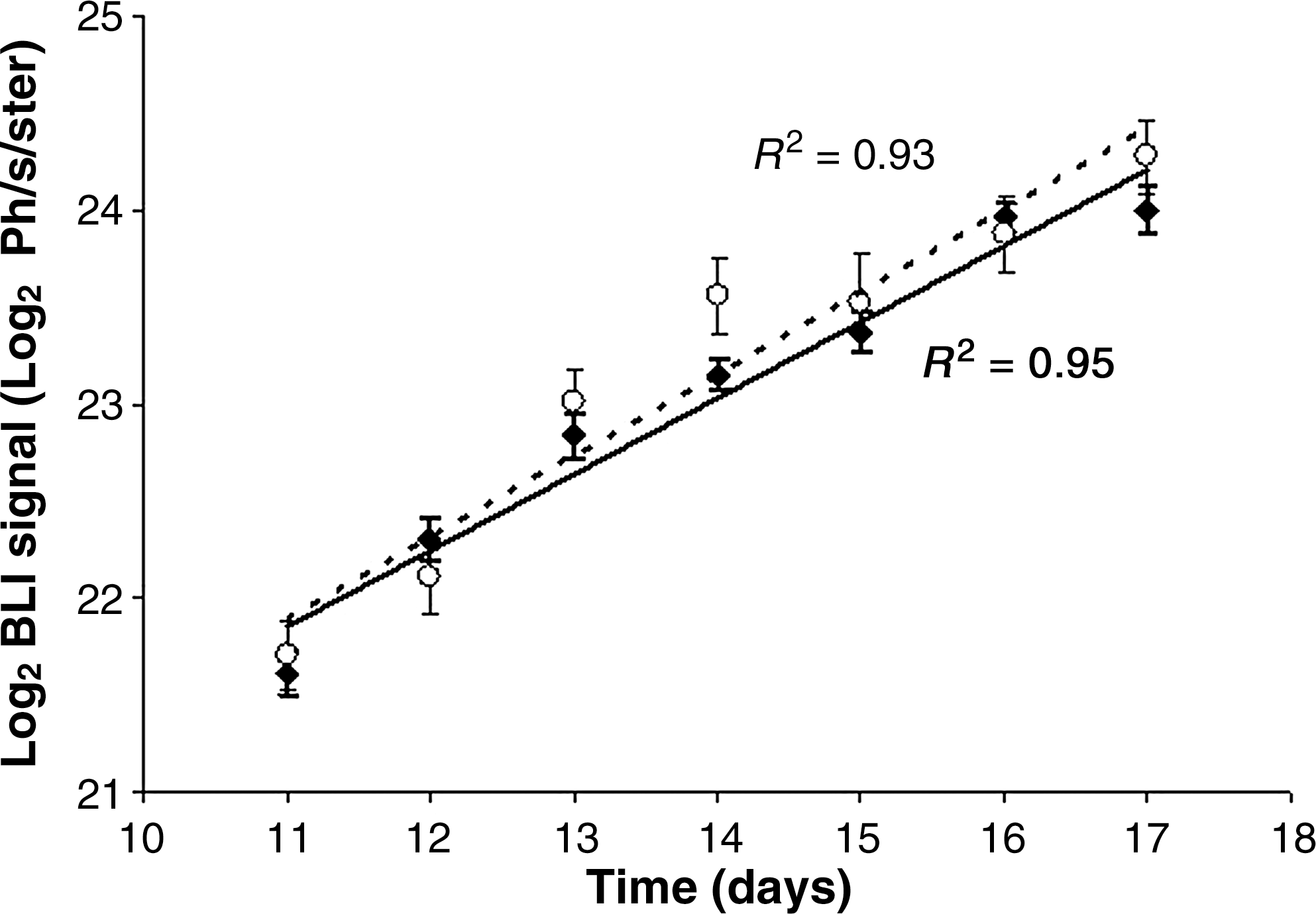
The injection of the “cold-kit” formulation, containing 0.1 mg nonradioactive 2-I-d-Phe and considerable amounts of gentisic acid and citrate (blank group), had no effect on the tumor BLI signal, compared with the injection of isotonic saline. Assuming exponential growth, the doubling time of the blank group and the control group on day 10 (injection of the kit) was calculated to be 61 ± 6 and 56 ± 6 hours, respectively (Fig. 2). Caliper measurements confirmed that the nonradioactive 2-I-d-Phe and the chemicals of the kit formulation had no influence on tumor growth. Also, no influence on the weight and behavior of the mice was detected.
Logarithmic plot of the tumor BLI signal in the blank group (mice injected with kit formula containing no radioactivity; filled diamonds) and the control group (mice injected with physiological saline; open circles) as a function of time. The full line represents a linear fit of the blank group; the dashed line represents a linear fit of the control group. BLI, bioluminescent imaging; Ph/s/ster, photons/second/steredian.
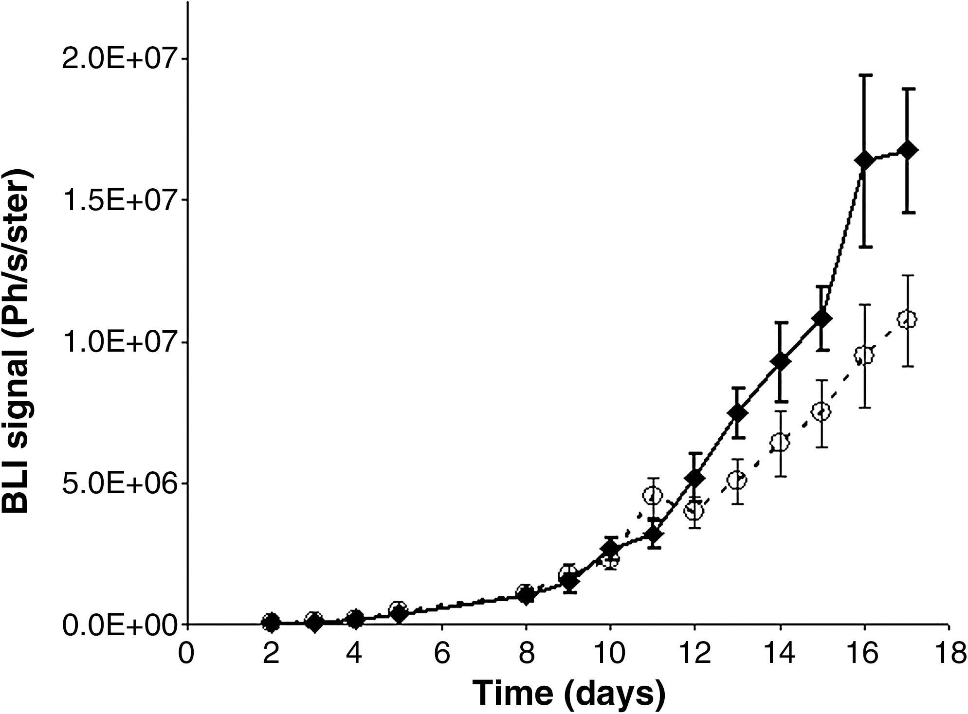
Therapeutic effect of [131I]-2-I-Phe
The injection of the therapeutic dose (148 MBq [131I]-2-I-Phe/kit formulation) produced a significant influence on the tumor growth as observed using both the BLI technique (Fig. 3) and the caliper measurements (Fig. 4). By means of a logarithmic plot of the BLI signal as a function of time after administration, the slope of the group receiving [131I]-2-I-d-Phe was calculated to be significantly smaller than that of the control series, which was treated with only the kit formula (p < 0.05). The tumor volume doubling time after therapy for 85 ± 8 hours was about 30% higher, compared with the control series (61 ± 6 hours). On day 11 (1 day postinjection of the therapeutic dose), a small but reproducible increase of the BLI signal was noticed in the group receiving [131I]-2-I-d-Phe—this data point was omitted from the calculations to avoid an artificial increase of the doubling time.
Tumor BLI signal in the therapy group (mice injected with [131I]-2-I-d-Phe/kit formulation; open circles) and the blank group (mice injected with nonradioactive kit formulation; filled diamonds). A parametric repeated-measures test showed a statistically significant difference between both groups (p < 0.05) (n = 10 for each data point). BLI, bioluminescent imaging; [131I]-2-I-d-Phe, [131I]-2-iodo-d-phenylalanine; Ph/s/ster, photons/second/steredian.
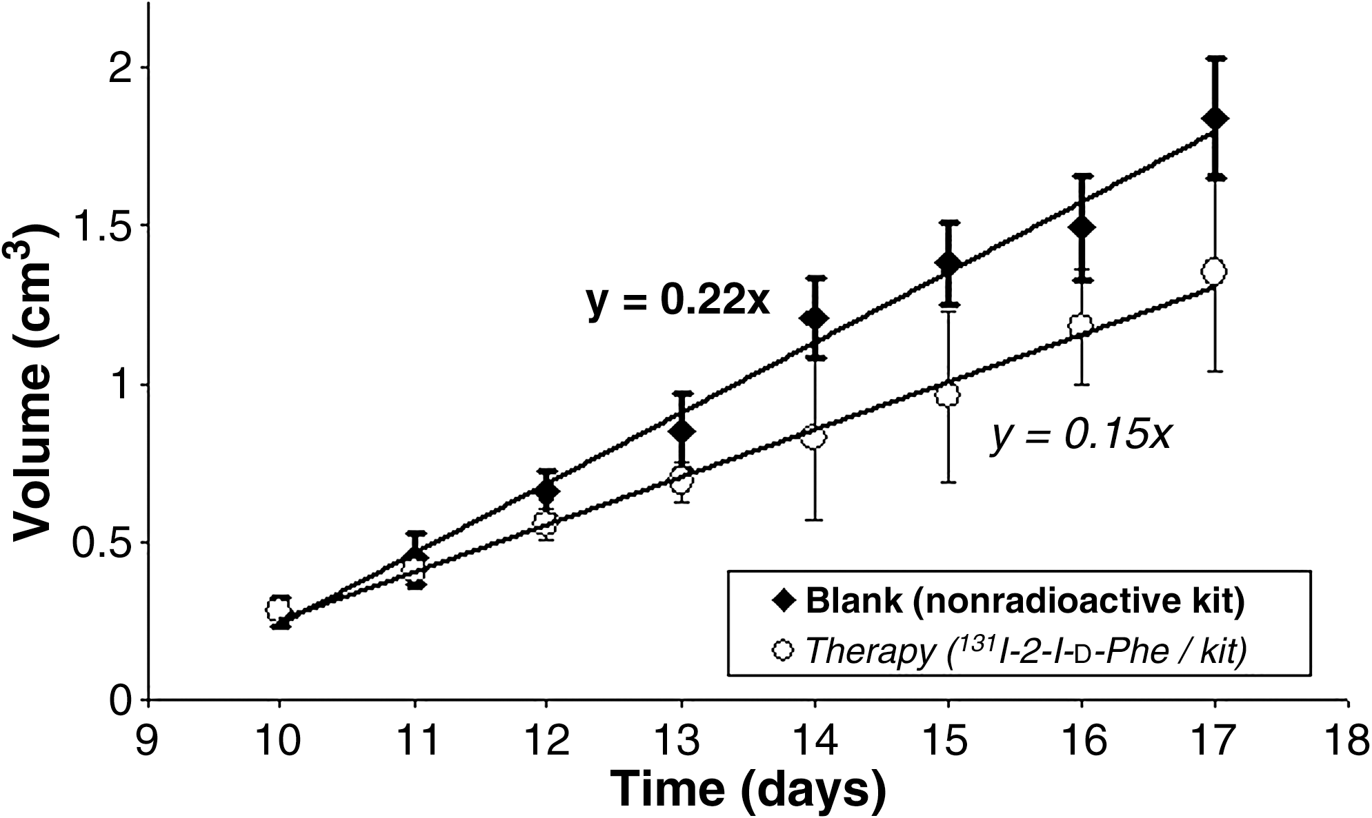
Tumor volume in the therapy group (mice injected with [131I]-2-I-d-Phe/kit formulation; open circles) and the blank group (mice injected with nonradioactive kit formulation; filled diamonds). A parametric repeated measures test showed a statistically significant difference between both groups (p < 0.05). [131I]-2-I-d-Phe, [131I]-2-iodo-d-phenylalanine.
The caliper measurements, represented in Figure 4, showed a pseudo linear growth with a significantly smaller increase in tumor volume with time for the therapy group (average growth of 0.15 cm3/day), compared with the blank group (average growth of 0.22 cm3/day); amounting to a 26% difference in tumor volume at day 17. The mean weight of the dissected tumors at day 17 was 0.9 ± 0.2 g for the treated mice and 1.2 ± 0.2 g for the controls, respectively, meaning a 25% difference (p = 0.29). It has to be taken into account that the weight values and caliper values included necrotic tissue, whereas this was not the case for the BLI measurements.
These results are illustrated in Figure 5, showing an exemplary image series of mice that were untreated (only kit administered) or treated (administration of [131I]-2-I-Phe in kit formulation).
Exemplary image series of a mouse from the blank group (only kit contents administered; upper row) and a mouse from the therapy group (administration of [131I]-2-I-Phe in kit formulation; bottom row). The color scale represents the BLI signal intensity (red = highest; blue = lowest). [131I]-2-I-d-Phe, [131I]-2-iodo-d-phenylalanine.
Discussion
As R1M rhabdomyosarcoma tumor–bearing rodents were shown to be a valuable model for evaluating of [123I]-labeled amino-acid analogs, a luciferase-transduced R1M cell model for the evaluation of [131I]-2-I-d-Phe as an agent for radionuclide therapy in tumor-bearing nude mice became evident. In addition to caliper measurements, the bioluminescence technique was used as a valuable adjuvant technique for follow up on small tumors when assessing the therapeutic effect of a radiopharmaceutical.
The uptake of [131I]-2-I-d-Phe in the R1M tumor after bolus injection at day 10 postinoculation (mean tumor weight: 0.13 g) was about 0.9%IA/g, while the clearance showed a biological half life of 4.6 hours. This clearance follows the blood clearance (t1/2: 4.5 hours), demonstrating that the tumor uptake is a reversible process as expected from the LAT1 characteristics.
The administration of [131I]-2-I-d-Phe significantly reduced tumor growth in the treated mice, compared with the control groups, proving that the therapeutic effect is specifically caused by [131I]-2-I-d-Phe. Both monitoring techniques showed that the mice receiving a therapeutic dose had a tumor growth-rate reduction of at least 33%, and there were no serious adverse events resulting from the therapy, apart from a small and transient weight loss of <1%. These results probably underestimate the potential of the radiopharmaceutical, as they were linked to a small tumor size with only minor vascularization at the time of bolus injection of the radiopharmaceutical. It should also be noted that, although no pathological problems were found at the end of the experiment, late radiation-related effects were not investigated and further experiments are required to confirm long-term animal health.
Our results were compared with those reported for the pharmacokinetics of non–CA [131I]-MIBG in nude mice with SK-N-SH human neuroblastoma xenografts (tumor size: 0.5–2 cm3).21–23 The [131I]-MIBG activity in the tumor remains about constant over the first 24 hours postinjection, whereas in the case of [131I]-2-I-d-Phe this activity becomes almost negligible after 24 hours (3% of initial uptake). The cumulated activity of [131I]-2-I-d-Phe in the tumor was consequently about six times lower compared with that of [131I]-MIBG. However, the cumulated activity in the liver was eight times lower. If the injected amount of [131I]-2-I-d-Phe is increased to achieve an equivalent cumulated activity in the tumors, the liver cumulated activity remains about 35% lower. Moreover, the injection of 25 MBq [131I]-MIBG had a considerable influence on the body weight of mice (up to 10% decrease) up to 5 days postinjection. In contrast, body-weight reduction in the case of 148 MBq [131I]-2-I-d-Phe-injected mice was <1%, which was recovered within 1 day.
Considering that amino acids are of primary use for treating brain tumors because of the easy passage of amino acids via the blood–brain barrier, translation of these preliminary results into patient studies requires an intermediate step, as the biodistribution as well as the dosimetry of [131I]-2-I-d-Phe in glioma-bearing animals needs to be established.
Although the use of [131I]-2-I-d-Phe as a therapeutic agent requires a relatively high amount of radioactivity, considering its negligible effect on mouse health in this study, the amount of radioactivity might not pose a serious difficulty, and [131I]-2-I-d-Phe has a potential as a therapeutic agent, especially in combination with classic radiotherapy or chemotherapy.
Conclusions
[131I]-2-I-d-Phe, injected as a bolus of its radiopharmaceutical kit formulation, is taken up by the R1M tumors in nude mice and significantly reduces tumor growth without noticeable side-effects on the animals' health until at least 1 week post-therapy. The BLI technique, caliper measurements, and tumor weight indicated a reduction in tumor growth rate of at least 33% in the R1M tumors. This is linked to a favorable biodistribution with a fast clearance from the major organs.
Footnotes
Acknowledgments
Financial support for this study was given through grants from GOA/VUB. The research at ICMI was funded by the Interuniversity Attraction Poles Programme—Belgian State—Belgian Science Policy.
Disclosure Statement
No financial conflicts of interest exist.
References
1.
BryanCF, BarriePB. Amino acid transporters ASCT2 and LAT1 in cancer: Partners in crime?Semin Cancer Biol, 2005; 15:254.
2.
VerreyF, ClossEI, WagnerCAet al.CATs and HATs: The SLC7 family of amino acid transporters. Pflugers Arch, 2004; 447:532.
3.
KobayashiK, OhnishiA, PromsukJet al.Enhanced tumour growth elicited by l-type amino acid transporter 1 in human malignant glioma cells. Neurosurgery, 2008; 62:493.
4.
OhnoY, SudaK, MasukoKet al.Production and characterization of highly tumour-specific rat monoclonal antibodies recognizing the extracellular domain of human l-type amino-acid transporter 1. Cancer Sci, 2008; 99:1000.
SLC Tables. www.bioparadigms.org/slc/menu.asp. 2009 September 4.
7.
KanaiY, EndouH. Heterodimeric amino acid transporters: Molecular biology and pathological and pharmacological relevance. Curr Drug Metab, 2001; 2:339.
8.
KanaiY, EndouH. Functional properties of multispecific amino acid transporters and their implications to transporter-mediated toxicity. J Toxicol Sci, 2003; 28:1.
9.
LinJ, RaoofDA, ThomasDGet al.l-type amino acid transporter-1 overexpression and melphalan sensitivity in Barrett's adenocarcinoma. Neoplasia, 2004; 6:74.
10.
Kupczyk-SubotkowskaL, TamuraK, PalDet al.Derivatives of melphalan designed to enhance drug accumulation in cancer cells. J Drug Target, 1997; 4:359.
11.
KillianDM, ChikhalePJA. A bioreversible prodrug approach designed to shift mechanism of brain uptake for amino-acid–containing anticancer agents. J Neurochem, 2001; 76:966.
12.
RomeikeBF, HellwigD, HeimannAet al.Action and efficacy of p-[131I]iodo-l-phenylalanine on primary human glioma cell cultures and rats with C6-gliomas. Anticancer Res, 2004; 24:3971.
13.
KersemansV, CornelissenB, KersemansKet al.123/125I-labelled 2-iodo-l-phenylalanine and 2-iodo-d-phenylalanine: Comparative uptake in various tumour types and biodistribution in mice. Eur J Nucl Med Mol Imaging, 2006; 33:919.
14.
BauwensM, LahoutteT, KersemansKet al.d- and l-[123I]-2-I-phenylalanine show a long tumour retention compared with d- and l-[123I]-2-I-tyrosine in R1M rhabdomyosarcoma tumour-bearing Wag/Rij rats. Contrast Media Mol Imaging, 2007; 2:172.
15.
BauwensM, KeyaertsM, LahoutteTet al.Intra-individual comparison of the human biodistribution and dosimetry of the d and l isomers of 2-[123I]iodo-phenylalanine. Nucl Med Commun, 2007; 28:823.
16.
KeyaertsM, VerschuerenJ, BosTJet al.Dynamic bioluminescence imaging for quantitative tumour burden assessment using IV or IP administration of d-luciferin: Effect on intensity, time kinetics and repeatability of photon emission. Eur J Nucl Med Mol Imaging, 2008; 35:999.
17.
BauwensM, LahoutteT, KersemansKet al.Comparison of the uptake of [123/125I]-2-iodo-d-tyrosine and [123/125I]-2-iodo-l-tyrosine in R1M rhabdomyosarcoma cells in vitro and in R1M tumour-bearing Wag/Rij rats in vivo. Nucl Med Biol, 2006; 33:735.
18.
KersemansV, CornelissenB, BacherKet al.In vivo evaluation and dosimetry of 123I-2-iodo-d-phenylalanine, a new potential tumour-specific tracer for SPECT, in an R1M rhabdomyosarcoma athymic mouse model. J Nucl Med, 2005; 46:2104.
19.
MertensJ, KersemansV, BauwensMet al.Synthesis, radiosynthesis, and in vitro characterization of [125I]-2-iodo-l-phenylalanine in a R1M rhabdomyosarcoma cell model as a new potential tumour tracer for SPECT. Nucl Med Biol, 2004; 31:739.
20.
LoevingerR, BudingerTF, WatsonEE. MIRD Primer for Absorbed Dose Calculations. Reston, VA: The Society of Nuclear Medicine, 1988.
21.
RutgersM, BuitenhuisCK, HoefnagelCAet al.Targeting of meta-iodobenzylguanidine to SK-N-SH human neuroblastoma xenografts: Tissue distribution, metabolism and therapeutic efficacy. Int J Cancer, 2000; 87:412.
22.
VaidyanathanG, FriedmanS, KeirSTet al.Evaluation of meta-[211At]-astatobenzylguanidine in an athymic mouse human neuroblastoma xenograft model. Nucl Med Biol, 1996; 23:851.
23.
MairsRJ, RusselJ, CunninghamSet al.Enhanced tumour uptake and in vitro radiotoxicity of no-carrier-added [131I]-metaiodobenzylguanidine: Implications for the targeted radiotherapy of neuroblastoma. Eur J Cancer, 1995; 31A:576.