
Abstract
This study was designed to establish an interleukin-12 (IL-12)-expressing murine Lewis lung carcinoma (LLC) cell vaccine (LLC/murine IL-12 [mIL-12]) and assess its antitumor efficacy and mechanism in vivo. The recombinant IL-12 plasmid was transfected into LLC cells and screened by G418, and positive clones were obtained. C57BL/6 tumor-bearing mouse model was established and tumor-bearing mice were randomly divided into three groups (n = 20), that is, treated with an intratumoral injection of phosphate-buffered solution, blank plasmid, or LLC/mIL-12 vaccine, respectively, at days 0, 7, and 14. Tumor size was measured before and after treatment. Tumor growth curve was plotted, cytolytic T lymphocyte (CTL) activity assay and natural killer (NK) cell activity assay were performed, CD4+ and CD8+ T lymphocyte were quantitated using flow cytometry, and the expression of interferon-γ (IFN-γ), IL-12, and interferon-inducible protein-10 (IP-10) in serum was detected by ELISA. Microvessel density was determined by immunohistochemistry after all mice were euthanized at day 21. The study revealed suppressed tumor growth, elevated levels of IFN-γ, IP-10, and IL-12, augmented NK and CTL cell activities, and decreased microvessel density of tumor tissues. There were abundant CD4+ and CD8+ T lymphocyte infiltration in the vaccine group. This study demonstrated that the antitumor mechanism of LLC/mIL-12 vaccine was to promote IFN-γ and IL-12 secretion, augment the NK and CTL cell activities, and decrease the microvessel density of tumors.
Introduction
Interleukin-12 (IL-12) is a cytokine mainly produced by antigen-presenting cells such as the macrophage. The local expression of cytokine-induced cellular immune response to tumors has become a focus of biological therapy for cancer. Transfected into tumor cells as a vaccine, the cytokine will generate a remarkable effect in regulating immunological response, including natural killer (NK) cell, T-lymphocyte, and interferon-γ (IFN-γ). 1 –5 Therefore, it has been used to induce antitumor immunity in vivo. As an effective antitumor agent, IL-12 has proven to elicit a significant immunity leading to tumor regression in animal models. 6 –9 Moreover, clinical responses to recombinant human IL-12, administered by subcutaneous, intravenous, or intraperitoneal injection, have been observed in patients with renal cell carcinoma, cutaneous T-cell lymphoma, melanoma, and peritoneal metastasis from ovarian cancer. 10 –16 IL-12 also has significant antiangiogenic properties, which was attributed to secretion of IFN-γ and interferon-inducible protein-10 (IP-10), as well as inhibition of vascular endothelial growth factor. 17 –19
With its combined immunomodulation and strong antiangiogenic effect, IL-12 provides a compelling rationale for further development of this immune-based therapy. Thus, this study investigates the therapeutic effect and the immune response of LLC cell vaccine containing IL-12 DNA in subcutaneous tumor-bearing C57BL/6 mice models.
Materials and Methods
Construction of IL-12 tumor vaccine
The recombinant plasmid pcDNA3.1(+)-murine IL-12 (mIL-12) containing the IL-12 gene was constructed in our laboratory. The plasmid (containing p35 and p40 subunits) and its activity have been described previously. 20,21 The test for endotoxin contamination was performed in the prepared plasmids by using a Limulus amebocyte lysate assay (BioWhittaker). The endotoxin used in this study was <20 EU/mg DNA. Murine Lewis lung carcinoma cells (LLC, a highly metastatic and drug-resistant mouse tumor; Tadao Ohno, RIKEN Cell Bank) were inoculated in Dulbecco's minimum essential medium supplemented with 10% fetal bovine serum and penicillin (100 U/mL). The formulations of plasmid and cationic lipid lipofectin (Invitrogen) were prepared by mixing purified plasmids with lipofectin at a volume ratio of 1:0.5 (approximate weight ratio of 1:1) with 0.01 M phosphate-buffered solution (PBS) as control, and then screened by G418 (600 mg/mL at first, followed by 300 mg/mL 5 days later; Gibco). The positive LLC/mIL-12 clone cells, which steadily express mIL-12 gene, were obtained after 2 weeks. LLC/mIL-12 vaccine was exposed to 50 Gy 60Co before injection to decrease oncogenicity.
Establishment of subcutaneous tumor model
LLC cells in exponential growth phase were collected, harvested with 0.1% trypsin, and washed with and subsequently suspended in 0.01 M PBS. About 2 × 10⁶ tumor cells were directly injected into the right hind leg of female C57BL/6 mice (aged 6–8 weeks and weighing between 18 and 20 g with specified-pathogens free, purchased from animal center of Chongqing Medical University, certificate no. SYK [YU] 20020007) using a sterile technique in a laminar flow hood. The Institutional Review Board of the Chongqing Medical University approved the study design. Each tumor size was measured in three dimensions using a caliper at every 3 days after injection. Intratumor injection of mIL-12 vaccine was performed using the same technique after growth of the tumor.
Therapeutic regimen
The C57BL/6 mice bearing LLC were divided into three groups (n = 20) randomly when the tumor reached 0.5–1.0 cm in diameter, namely the experimental group, the blank carrier group, and the control group, treated with an intratumoral injection of 1 × 10⁶ mIL-12/LLC cell vaccine, 0.2 mL pcDNA3.1(+), or 0.2 mL of 0.1 M PBS, respectively, at days 0, 7, and 14. Animals were monitored for survival and sacrificed after 21 days. Blood, spleen, and tumor tissue were harvested for corresponding detection and analysis. Tumor volume was calculated by the following formula: V = 1/2ab 2, where a and b represent two greatest perpendicular axial dimensions of a tumor.
IP-10, IL-12, and IFN-γ were quantitated by ELISA
The quantity of cytokines was determined using mouse IP-10, IL-12, and IFN-γ ELISA kit (Endogen Com). One hundred microliters (100 μL) of blood was obtained through an orbital puncture and added into a 96-well plate. Diluted samples were used to quantify IP-10, mIL-12, and IFN-γ proteins according to an instruction manual. The absorbance (A) values of variant concentration standard preparation were detected within 30 minutes. IP-10, IL-12, and IFN-γ concentrations in the sample were counted according to a standard curve.
The activity of cytolytic T cell and NK cell
Spleen was taken from 10 mice in each group at the end of treatment to serve as effectors. Spleen cells were isolated and washed with culture medium and then suspended in culture medium at 4 × 10⁶ cells per well. LLC target cells were prepared by incubation for 1 hour in sodium chromate and then washing with culture medium three times. Cr-loaded LLC cells were seeded into a 96-well plate at 3 × 10³ cells per well containing 100 μL of medium, and then effector lymphocytes were added according to the effector-to-target cell ratios of 25:1 and 50:1 in a final volume of 200 μL per well. The assay was done three times. The plates were incubated for 4 hours and then centrifuged. The culture supernatant was used to determine 51Cr release using a γ-counter. Specific lysis percentage was calculated according to the following formula: (sample release − spontaneous release)/(maximum release −spontaneous release) × 100%.
The method to determine NK cell activity in spleen cell was the same as cytolytic T lymphocyte (CTL) except that the target cell used was the Yac-1 cell (sensitive to the cytotoxic activity of natural killer cells in mice).
NK cell activity = (sample release − spontaneous release)/(maximum release − spontaneous release) × 100%.
CD4+ and CD8+ T lymphocyte quantitation
Animals were euthanized at the end of treatment and tumor tissue was separated by surgical scissors. Tumor cells were isolated and washed with culture medium and then suspended in culture medium. Immunofluorescence analysis was performed by following the method recommendated by the National Committee for Clinical Laboratory Standards. 22
Detection of microvessel density
To evaluate the antiangiogenic effect of IL-12 expression driven by mIL-12 vaccine, immunohistochemical staining for microvessel density was performed on slices of tumor from animals euthanized at 21 days after treatment. Anti-CD34 antibodies were employed to stain the microvessel endothelial cell of the tumor.
Statistical analysis
All data were analyzed by using SPSS 13.0 software. Data were expressed as mean ± standard deviation. Statistical significance was established at p < 0.05.
Results
Generation of hypsi-bioactivity mIL-12 vaccine
An in vitro assay was used to confirm the functional properties of the IL-12 vaccine. In this study, hypsi-concentration and hypsi-bioactivity mIL-12 vaccine was generated.
Therapeutic effect of tumor vaccine
We studied the effect of mIL-12 vaccine on the tumor inhibition rate in mice bearing LLC. Treatment with 1 × 10⁶ mIL-12 vaccine led to significant decrease of tumor weight and volume compared with mice treated with blank carrier or PBS (Table 1). The weight of tumor in the vaccine group was the smallest and there were significant differences between the vaccine and the control groups (p < 0.05).
PBS, phosphate-buffered solution; mIL-12, murine interleukin-12.
IP-10, IL-12, and IFN-γ expression in mice after tumor vaccine injection
Of note, IP-10, IL-12, and IFN-γ protein expression in mice blood serum was elevated with intratumoral injection of mIL-12/LLC vaccine at the end of treatment, but there was less IP-10, IL-12, and IFN-γ protein expression in the pcDNA3.1(+) and the PBS groups (p < 0.05; Table 2).
IL-10, interferon-inducible protein-10; IFN-γ, interferon-γ; PBS, phosphate-buffered solution; mIL-12, murine interleukin-12; LLC, Lewis lung carcinoma.
CTL and NK cell activity
MIL-12/LLC vaccine can induce mice spleen cell specificity: CTL activity (69% ± 5.9% and 52% ± 4.5% at E/T = 50/1 and E/T = 25/1, respectively) (Fig. 1A) and NK cell activity (52% ± 6.7% and 35% ± 3.1% at E/T = 50/1, respectively) (Fig. 1B).
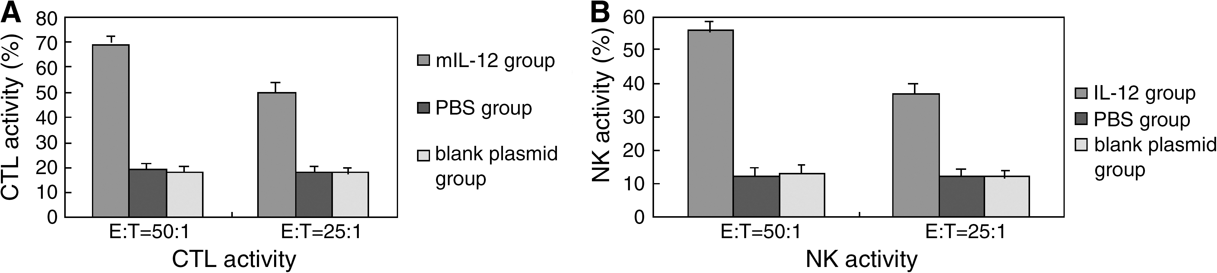
CTL and NK cell activities in mice spleen cell of different groups after treatment with mIL-12/LLC vaccine and PBS or blank carrier pcDNA3.1(+). mIL-12/LLC vaccine can induce mice spleen cell-specific CTL activity (69% ± 5.9% and 52% ± 4.5% at E/T = 50/1, respectively) (
CD4+ and CD8+ T cells infiltrate in tumor tissue
FCM analysis of CD4+ and CD8+ showed positive infiltration of both cell populations in tumors treated with mIL-12 vaccine, in contrast to the blank carrier pcDNA3.1(+) group and the PBS control group (p < 0.05).
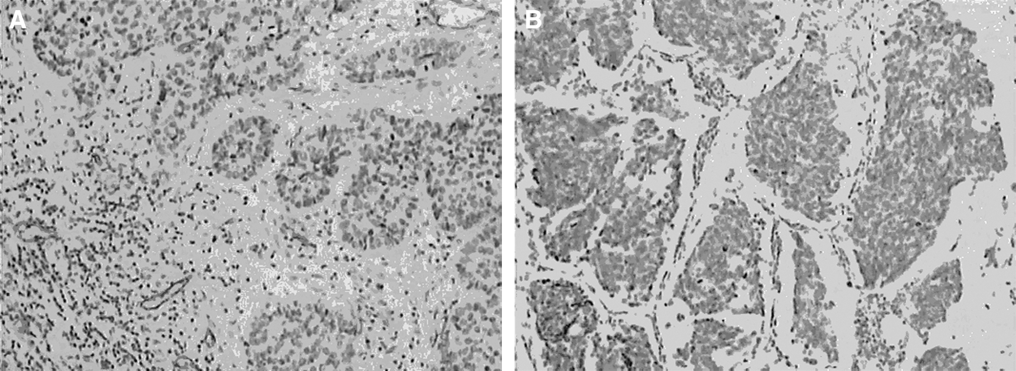
Microvessel density of tumor
The tumor of animals treated with PBS or blank carrier pcDNA3.1(+) did not show any decrease in microvessel density (45.58 ± 15.60/10 HPF; Fig. 2A). In contrast, the tumor of mice intratumorally injected with mIL-12/LLC showed significant decrease in microvessel density of 12.64 ± 3.22/10 HPF (Fig. 2B).
(
Discussion
In this study, we examined the feasibility of delivering a tumor cell vaccine into experimental animals by intratumoral injection. The concentrated recombinant mIL-12 plasmid proved to be compatible with transgenic expression in murine tumor. mIL-12/LLC vaccine seems to be well tolerated by this route. In this study, we hypothesized that local intratumoral delivery of a highly toxic therapeutic agent may be better tolerated and show increased efficacy versus systemic delivery. Further, local delivery of mIL-12 vaccine bypasses some of the systemic side-effects of vaccine. 23
It has been shown that IL-12 can be produced in sufficient quantity to guarantee a significant therapeutic effect after intratumoral injection. Treatment with mIL-12/LLC led to a significant decrease in tumor volume and weight compared with the control group. Tumor inhibition rate was 51%. In this study, irradiated LLC cells that produced IL-12 were shown to be effective in the treatment of tumor-bearing mice. It might be related to the durative secretion of IL-12 after intratumoral injection of vaccine. 23 For an optimal antitumoral effect, cytokines must be secreted over an extended period of time. 24,25 In a previous study, IL-12 was shown to inhibit tumor establishment at a distant site in a murine renal carcinoma model. Systemic administration of recombinant IL-12 had a relatively weaker effect than local injection of IL-12, because there was no persistent and local expression of IL-12 protein after systemic administration; therefore, in this study, IL-12 was administered by intratumoral injection.
This study also found that IL-12 expression as a result of a single local injection of mIL-12/LLC vaccine lasted for at least 21 days after injection. But systemic IL-12 administration led to a half-life of <24 hours. Gene therapy with mIL-12/LLC might thus offer a significant advantage over conventional recombinant protein therapy by systemic administration, in that it can provide more sustainable high concentrations of IL-12 at the local tumor and can generate a stronger therapeutic effect. 26 –28 In the other hand, the observation that the cytokine kept bioactivity until the mice were euthanized suggests that the local expression of IL-12 in tumor vaccine does not cause lethal toxicity in mice—thus, a potentially safer and more effective treatment for solid tumors.
IL-12 with various gene delivery systems has been used for cancer gene therapy. Previous studies demonstrated that IL-12 stimulates the proliferation and activation of several types of leukocytes with antitumor activity, including NK cells, lymphokine-activated killer cells, antigen-specific T-helper cells, cytotoxic lymphocytes, macrophages, and B cells. 29,30 Studies in other murine tumor models have shown that both NK cells and cytotoxic T lymphocytes play vital roles in the antitumor activity of IL-12. 1,2,31,32 The mechanism of IL-12 that leads to tumor extinction in mice has been previously described in the setting of cancers. Recent studies have suggested that the mechanism may involve increase in blood cytotoxic lymphocytes and NK cell numbers and IFN-γ expression. 2,8,23 The present study has documented CD4+ and CD8+ cell infiltration in mice. The results revealed that total IL-12 and IFN-γ levels were greater in tumors treated with IL-12 vaccine than in tumors treated with PBS, presumably because the IL-12 vaccine induces IL-12 and IFN-γ secretion. Compared with the control group, the expression of the IL-12 gene played a major role in enhancing antitumor effect. IL-12 gene has enhanced cell-mediated immune responses in spleen cells, which eventually improve the activity of NK and CTL cells.
On the other hand, the antiangiogenic feature of IL-12 remains a possibility. 33,34 It can enhance the expression of the antiangiogenic chemokine IP-10, inhibit vascular endothelial growth factor levels, and inhibit tumor angiogenesis consequently.
The combination of IL-12 and tumor vaccine shows promise in the setting of established tumors. 34 –36 A combined treatment of IL-12 and tumor vaccine clearly works in synergy to extend the tumor inhibition rate in experimental animals. These results provide a basis for human clinical trials on the specific IL-12 vaccine formulation. This novel approach utilizing nonviral gene immunotherapy can be used in the treatment of lung cancer, alone or in combination with standard therapy.
Footnotes
Acknowledgments
The authors thank Dr. Eddy S. Yang in the Department of Radiation Oncology, Vanderbilt University Medical Center, for his kind help in editing the manuscript. This study was supported by the Nation Natural Science Foundation of China (no. 30370422) and the Science and Technical Committee of Dadukou District, Chongqing.
Disclosure Statement
No competing financial interests exist.