
Abstract
This study was conducted to determine the possible radiopharmaceutical potential of morphin labeled with 131I. Morphine was extracted from dry capsules of the opium poppy (Papaver somniferum L.), purified by high-performance liquid chromatography, and characterized with nuclear magnetic resonance and infrared spectroscopy. The purified compound was labeled with 131I. Male Albino Wistar rats (18) were used for receptor blockage and unblockage biodistribution studies. Tissue distribution studies showed that radiolabeled morphine had higher uptake in lung, liver, small intestines, large intestines, and stomach than the other tissues. The highest uptake of radiolabeled compounds in rats' brain was found to be in the midbrain and hypothalamus. After receptor blockage with morphine, uptake of 131I-morphine decreased in the lungs, liver, kidney, testis, prostate, spinal cord, cerebellum, hippocampus, striatum, and temporal cortex with respect to receptor unblockage studies of rats. This study concludes that the labeling yield of 131I-morphine was high, high amount of 131I-morphine was found in the hypothalamus, and 131I-morphine has enough stability for diagnostic scanning.
Introduction
Morphine has been extracted from opium poppy, which is classified botanically as Papaver somniferum. It comes from a great botanical family of 28 genera and over 250 individual species, most of which are cultivated in the mild and subtropical regions of the Northern Hemisphere. 1
In 1803, the Parisian Derosne illustrated the separation of the first crystalline compound “salt of opium” from opium. 2 Separation of morphine from opium poppy had been made by Serturner at the beginning of the 19th century and he used first the name “morphine” (Morpheus—Greek god of dreams) for the new substance. 2 In 1818, another German pharmacist, Karl Friedrich Wilhelm Meissner, used the name “alkaloid” (“alkali-like”) for these new salifyable bases, a name that continues from history to this day. 3 –6 The alkaloid content of opium is roughly 10%–20%. Only five of these alkaloids, such as morphine, codeine, thebaine, papaverine, and noscapine, are important for nearly all of the quantitative alkaloid content in opium. 2,7,8
Morphine is a classical exogenous opioid and produces a well-characterized analgesia, as well as certain other pharmacological actions, as a result of its affinity to bind to receptors normally acted upon by endogenous opioids. Receptors comprise of long-chain protein molecules. The body contains a lot of receptor proteins for their normal processes. Numerous studies have demonstrated that there are three well-defined types of opioid receptors, namely the mu (μ), kappa (κ), and delta (δ), and the genes encoding for these receptors have been cloned. 9 –13
Opioid receptors are found in nerve cells located in the various regions of the brain and the spinal medulla, as well as in intramural nerve plexuses that are involved in the regulation of gastrointestinal and urogenital motility. There are selective agonists and antagonists to these types, as well as various subtypes. Radioligand-binding assays of brain and other tissues have been particularly useful in the development of opioid receptor selective agonists and antagonists. 5,14,15
Morphine and narcotic agonists have agonistic actions at the μ, κ, and δ receptors. Nociceptive effects result from actions at the level of both the brain and the spinal cord. The morphine is responsible for the attenuation of impulse spread and the inhibition of pain perception, whereas the narcotic agonists are responsible for inhibition of transmission of nociceptive impulses. 14 Therefore, the morphine has been adopted extensively throughout the world for many years to relieve moderate to severe pain. According to the program of the WHO's Cancer Pain Relief Program, 1 morphine is the main drug utilized in treating cancer pain.
Different groups had studied with morphine for different purposes. According to the study of Motamedi et al., chronic morphine administration decreases spatial learning but has no effect on spatial memory and motor activity. 16 Slamberová's group 17 studied the effects of prenatal morphine exposure on learning and memory in male and female rats, and their results showed that prenatal morphine exposure altered differential performance of adult male and female rats on tasks requiring learning and spatial memory. Taking opiates during critical periods of prenatal development leads to long-lasting alterations in the neuroendocrine control systems of the animal. These alterations may have significant consequences on the physiological maturation and adult behaviors of the animal. 18 Another study was conducted to determine the effect of chronic morphine on plasma and brain beta endorphin and methionine enkephalin in pregnant rats and their fetuses or newborn. 19 The effects of acute coronary artery ligation on cardiac rhythm and hemodynamic were studied in rats receiving either acute or chronic morphine treatment. Chronic morphine treatment decreased the occurrence of early ventricular arrhythmias caused by acute myocardial ischemia in rats. 20
The aim of this study was to label morphine extracted from dry capsules of the opium poppy with 131I and to investigate its radiopharmaceutical potential with biodistribution studies in rats.
Materials and Methods
All reagents were commercially available and of analytical grade. 131I was obtained from the Department of Nuclear Medicine of Ege University.
Extraction of morphine
Plant materials were harvested near localities of Afyonkarahisar, Turkey. Five (5) grams of opium were cut into small pieces and extracted by reflux with 0.4 g of sodium hydroxide in 25 mL of methanol for 2 hours. The methanol extract was then filtered, and methanol was removed under reduced pressure. The residue was then mixed with 5.5 mL of 1.0 N sodium hydroxide solution at 35°C for 10 minutes. pH was adjusted to about 11.7 with 50% acetic acid in water. This aqueous extract was filtered and the precipitate was washed with 5.5 mL of 0.01 N sodium hydroxide solutions. The combined aqueous filtrate was stirred with 0.5 g of sodium acetate for 15 minutes and filtered again. The filtrate was then extracted twice with 5 mL of toluene, and the pH of the aqueous filtrate was then adjusted to pH 9.2 with 50% acetic acid in water. The mixture was allowed to remain for a period of 7 hours at room temperature for complete precipitation and was then filtered. This precipitate was washed with water and dried at room temperature. The dried substance was dissolved with toluene and the precipitate was filtered. The morphine was purified by high-performance liquid chromatography (HPLC) (morphine yield: 0.52%).
Chromatography
A low-pressure-gradient HPLC system (LC-10ATvp quaternary pump and SPD 10A/V UV detector), a syringe injector equipped with a 1-mL loop, and 7-μm RP-C-18 column (250 × 4.6 mm inner diameter; Macharey-Nagel) were used for preparative procedures. The elution was collected with an FRC-10A fraction collector (Shimadzu). The flow rate was set at 5.0 mL/min. The column was eluted with 100% of 10 mM tetraethylammonium hydroxide. UV detection was achieved at 254 and 280 nm.
Labeling procedure
Morphine was radioiodinated with 131I using the iodogen method under the same conditions as described earlier by Ünak and Ünak. 21 To label with 131I, morphine (1 mg morphine in 1 mL of 0.5% acetic acid solution) was added into the iodogen (1 mg)-coated tube and then 500 μCi of Na131I was added. This reaction mixture was kept at room temperature without stirring for 25 minutes. At the end of this time, quality control of radioiodinated morphine was performed.
Quality controls
The following quality control studies were done with thin layer radiochromatography (TLRC) and paper electrophoresis.
TLRC procedures
Each TLRC cellulose-coated sheet was covered by a celloband after its development and cut into sections of 0.5 cm width. Then these samples were counted using the Cd(Te) detector equipped with a RAD 501 single-channel analyzer. TLRC chromatograms were obtained from these figures by plotting counts versus distance. The retention factor (R f ) values and labeling efficiencies were derived from these chromatograms. Isopropyl alcohol, n-butanol, and 0.2 M ammonium hydroxide (2/1/1, v/v/v) were used as a solvent.
Electrophoresis procedures
Electrophoresis was conducted with a Gelman electrophoresis chamber. The cathode and anode poles and application points were indicated on the cellulose acetate strips, which were moistened by buffer solution (n-butanol/water/acetic acid: 4/2/1, v/v/v). After each compound was set on the strips, they were placed in the electrophoresis chamber. The standing time and applied voltage were 2.5 hours and 300 V, respectively. The developed strips were dried and cut into 1-cm pieces and each one was counted in the Cd(Te) detector.
Lipophilicity
A 100 μL aliquot of radiolabeled morphine was added to a test tube containing 3 mL of each of n-octanol and water. The tube was vortexed for 1 hour at room temperature and then centrifuged at 3000 rpm for 5 minutes. Aliquots (0.5 mL) of each phase were taken for counting. The partition coefficient was determined by the following function: partition coefficient = log10 (counts in n-octanol layer/counts in aqueous layer).
Serum stability of 131I-morphine
131I-morphine solution (300 μL) was incubated with fresh human serum (600 μL) in duplicate at 37°C through 24 hours. During incubation, the sample was analyzed at 15 minutes, 1, 2, 3, 4, and 24 hours by TLRC method.
Biodistribution studies in rats
Biodistribution studies in rats were approved by the Institutional Animal Review Committee of Ege University.
The biodistribution data are expressed as percentage of injected radioactivity per gram of tissue for some selected organs as the mean value of 3 rats. Male Albino Wistar rats weighing ∼150–200 g were used for the study. For blocking of iodine uptake in the thyroid gland, 10 mg of potassium iodide was added to l L drinking water of rats. Morphine was labeled with 131I at pH 2 using two consecutive vials that contained 5 mg iodogen. Then, radiolabeled morphine was adjusted to pH 7, using 0.1 N ammonium hydroxide, sterilized by using a Millipore filter (0.22-μm pore), and then injected into the tail vein of the animals (131I-morphine, 21.11 GBq/μL; 1 μg morphine/rat).
In vivo blocking experiments
Ten micrograms of nonlabeled ligand (morphine) was injected into the tail vein of each animal at 30 minutes before injecting 131I-morphine to determine whether uptake in receptor-expressing target tissue was specific. The same procedure was repeated as indicated above.
Then, the rats were sacrificed at 15, 60, and 180 minutes under ether anesthesia and the tissues of interest were removed. All tissues were weighed and counted for radioactivity with a Cd(Te) detector. The percent of radioactivity per gram of tissue weight (i.e., % injected activity/g tissue) was determined.
Statistical analyses
Data were analyzed statistically by using SPSS statistical software (SPSS for Windows; Release 10.0.1 Standard Version). Comparisons between different groups were performed by Pearson correlation and one-way analysis of variance (ANOVA); if ANOVA revealed significant differences, post hoc comparisons were performed by Duncan multiple range tests. A p-value of <0.05 was considered significant.
Results
Structural parameters
The structural parameters of morphine were obtained by nuclear magnetic resonance (NMR), infrared (IR) spectroscopy, and HPLC (Fig. 1).

High-performance liquid chromatography result of morphine.
The characteristics of morphine are IR (KBr, cm−1) 3400 (hydroxyl, OH), 2950 (alkyl, CH), 1633 (alkenyl, C=C), 1260 (N–C) and 1H-NMR (DMSO) δ(ppm) values: 6.42 (d, 1H, 11-H), 6.32 (d, 1H, 10-H), 5.52 (t, 1H, 4-H), 5.21 (t, 1H, 7-H), 4.65 (t, 1H, 8-H), 4.08 (s, 1H, 1-H), 3.37 (s, 1H, 14-H), 3.21 (d, 1H, 2-H), 2.84 (d, 2H, 5-H), 2.70 (t, 2H, 9-H), 2.48 (m, 1H, 3-H), 2.25 (s, 3H, NCH3), 1.95 (d, 1H, 13-H), 1.61 (t, 2H, 6-H), 13C-NMR (DMSO): δH 20.8, 36.2, 41.1, 43.4, 46.08, 55.2, 59.3, 67.3, 92.1, 117.1, 119.5, 126.3, 129.1, 131.7, 134.1, 139.2, 146.3.
R f values were obtained from the TLRC chromatograms (Table 1). The experimental results indicated that the labeling yield of 131I-morphine was 95% ± 2.0% under optimal conditions (pH 2, n = 10, reaction time: 25 minutes, reaction temperature: room temperature).
The retention factor (R f ) is defined as the distance traveled by the compound divided by the distance traveled by the solvent. Bath solvent of TLRC is isopropyl alcohol/n-butanol/0.2 M ammonium hydroxide (2/1/1, v/v/v). Buffer solution of electrophoresis is n-butanol/water/acetic acid (4/2/1, v/v/v).
TLRC, thin layer radiochromatography.
Data obtained from electrophoresis study are shown in Table 1. Iodine (131I−) moved 2 cm toward the anode, whereas 131I-morphine and oxidized 131I remained at the point of application.
Effect of pH
Effect of pH on the radiolabeling yield was examined for pH 2–7. Amount of morphine (1 mg) and iodogen (1 mg) were fixed during the labeling. The highest labeling yield (95% ± 2.0%) was obtained at pH 2. The obtained data showed that radiolabeling yield decreased with increasing pH (pH = 4 [59.7% ± 2%] and pH = 7 [51.7% ± 2%]).
Effect of iodogen amount
The effect of iodogen on radiolabeling was tested between 0.1 and 1.0 mg. Radiolabeling yield increased depending on the amount of iodogen (0.1, 0.5, and 1 mg iodogen, radiolabeling yield: 66% ± 6%, 74% ± 6%, and 95% ± 5%, respectively).
Stability of 131I-morphine in human serum
The results of 131I-morphine in human serum showed that the labeling yield of radioiodinated morphine decreased to 70% ± 8% and 26% ± 5% at the 3rd and 24th hour, respectively, at room temperature (Fig. 2). Hence, the period of stability of 131I-morphine is sufficient for imaging procedures.
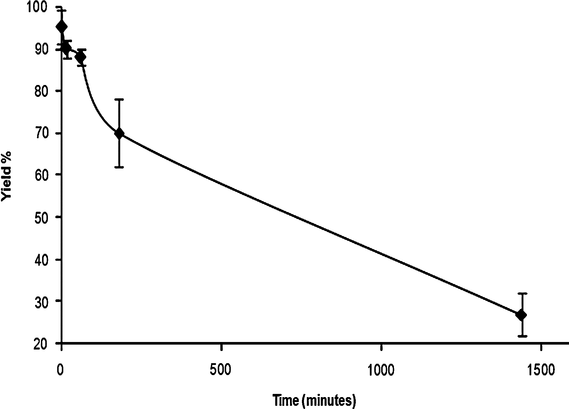
Stability of 131I-morphine in serum with respect to time.
Biodistribution results
Biodistribution studies were performed with high radiolabeling yield (95% ± 5%) and 21.11 GBq/μL specific activity of 131I-morphine.
The normal biodistribution data of 131I-morphine are shown in Figure 3. The highest %ID/g value of radiolabeled morphine was obtained for large intestines (L.intestine) at 180th minute and the value reached 17.6%. Its concentration significantly increased in L.intestine (p < 0.000) through time, whereas the values significantly decreased for lungs (p < 0.001), liver (p < 0.003), muscle (p < 0.03), and spinal cord (p < 0.004) through time.
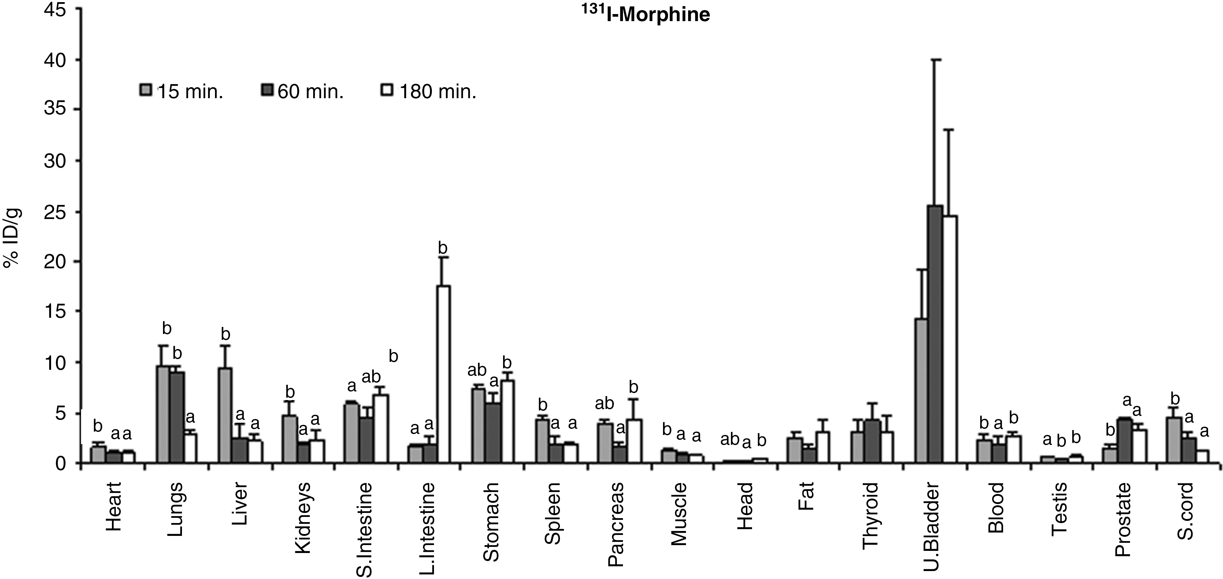
Biodistribution of 131I-morphine in rats as a function of time. Results are expressed as means ± standard deviation. Different superscript letters indicate significant differences with respect to groups and time. %ID/g, % injected dose per g.
A significantly positive correlation of 131I-morphine uptake was observed for heart–liver (r = 0.90, p < 0.002), heart–kidneys (r = 0.85, p < 0.004), stomach–testis (r = 0.91, p < 0.001), prostate–L.intestine (r = 0.95, p < 0.001), and medulla pons–temporal cortex (r = 0.81, p < 0.008), whereas a significantly negative correlation of 131I-morphine uptake was observed for lung–L.intestine (r = −0.93, p < 0.0003), medulla pons–frontal cortex (r = −0.82, p < 0.007), and lung–prostate (r = −0.85, p < 0.003) in rats.
There are three well-defined opioid receptors, namely the mu (μ), kappa (κ), and delta (δ). Morphine is a classical exogenous opioid that produces a well-characterized analgesia, as well as certain other pharmacological actions, as a result of its affinity to bind to receptors normally acted upon by endogenous opioids.
The results of receptor blockage experiment showed that the values of 131I-morphine uptake in the small intestine (s.intestine), lung, liver, and spinal cord were 11.40, 6.98, 4.74, and 4.33 (%ID/g), respectively, at 15 minutes (Fig. 4). After receptor blockage, 131I-morpine uptake (Rec-131I-mor) significantly decreased in heart (p < 0.000), lungs (p < 0.02), and kidney (p < 0.001), whereas it increased in the spinal cord (p < 0.009) through time (Fig. 4).
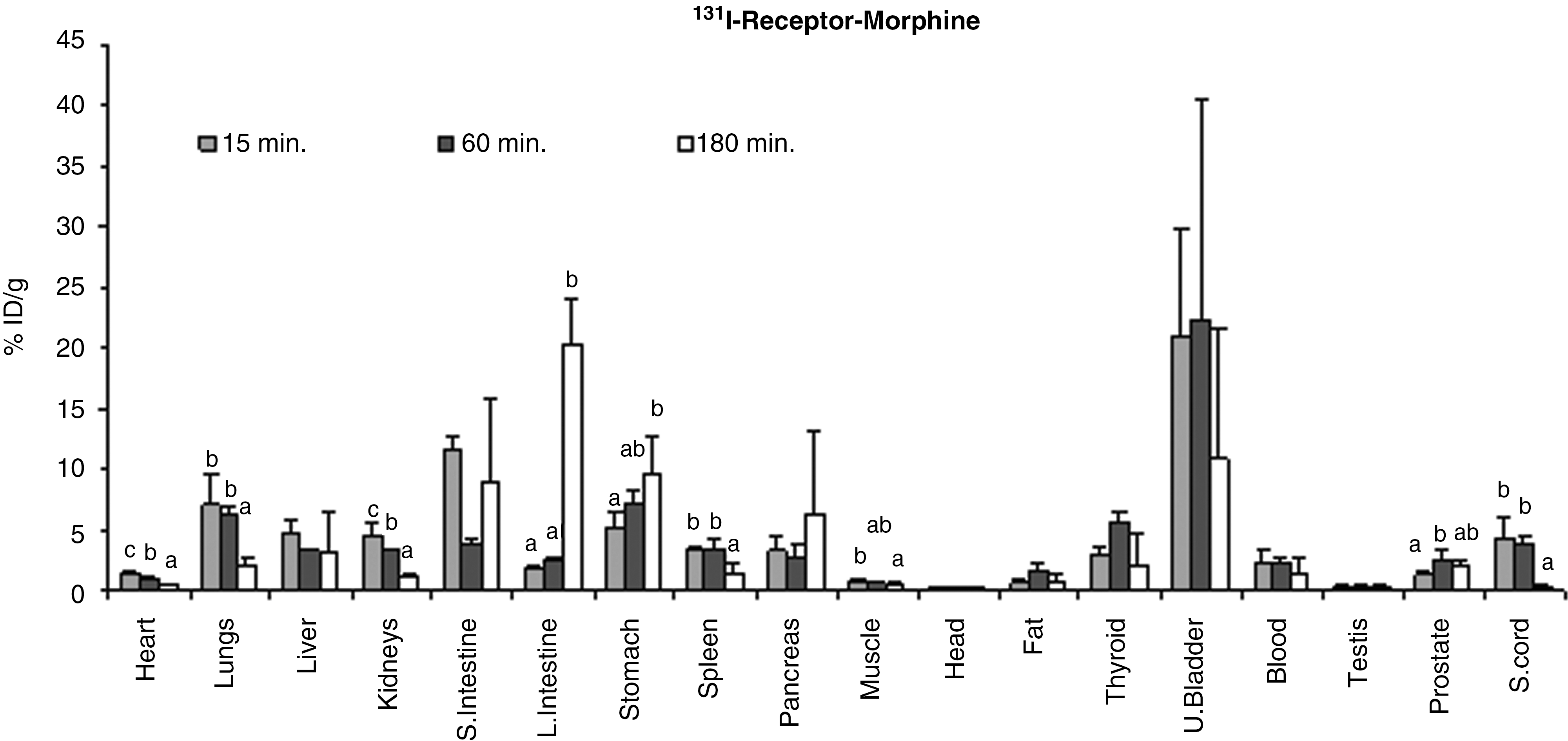
Biodistribution of 131I-morphine in receptor binding studies in rats as a function of time. Results are expressed as means ± standard deviation. Different superscript letters indicate significant differences with respect to groups and time. %ID/g, % injected dose per g.
The results of receptor blockage and unblockage of 131I-morphine showed a positive correlation in the L.intestine–receptor (L.intestine–rec.L.intestine) (r = 0.99, p < 0.000) and prostate–rec.L.intestine (r = 0.95, p < 0.000), but a significantly negative correlation was observed for the prostate–rec.L.intestine (r = −0.94, p < 0.000) and rec.L.intestine–rec.spinal cord (r = −0.86, p < 0.002).
Receptor blockage study in rats showed a significantly negative correlation of 131I-morphine for rec.kidney–rec.L.intestine (r = −0.86, p < 0.003) and rec.spinal cord–rec.L.intestine (r = −0.87, p < 0.002), whereas a positive correlation was observed for rec.kidney–rec.heart (r = 0.95, p < 0.0001) and rec.lung–rec.kidney (r = 0.95, p < 0.000). According to Enginar et al., the lung, liver, stomach, prostate, and spinal cord showed sensitivity to receptor blockage with codeine. In the present study, uptake of 131I-morphine decreased in some tissues (lungs, liver, kidneys, testis, prostate, spinal cord, cerebellum, hippocampus, striatum, and temporal-cortex) in the receptor blockage with respect to receptor unblockage studies of rats. The value of 131I-morphine concentration in these organs decreased after receptor blockage studies.
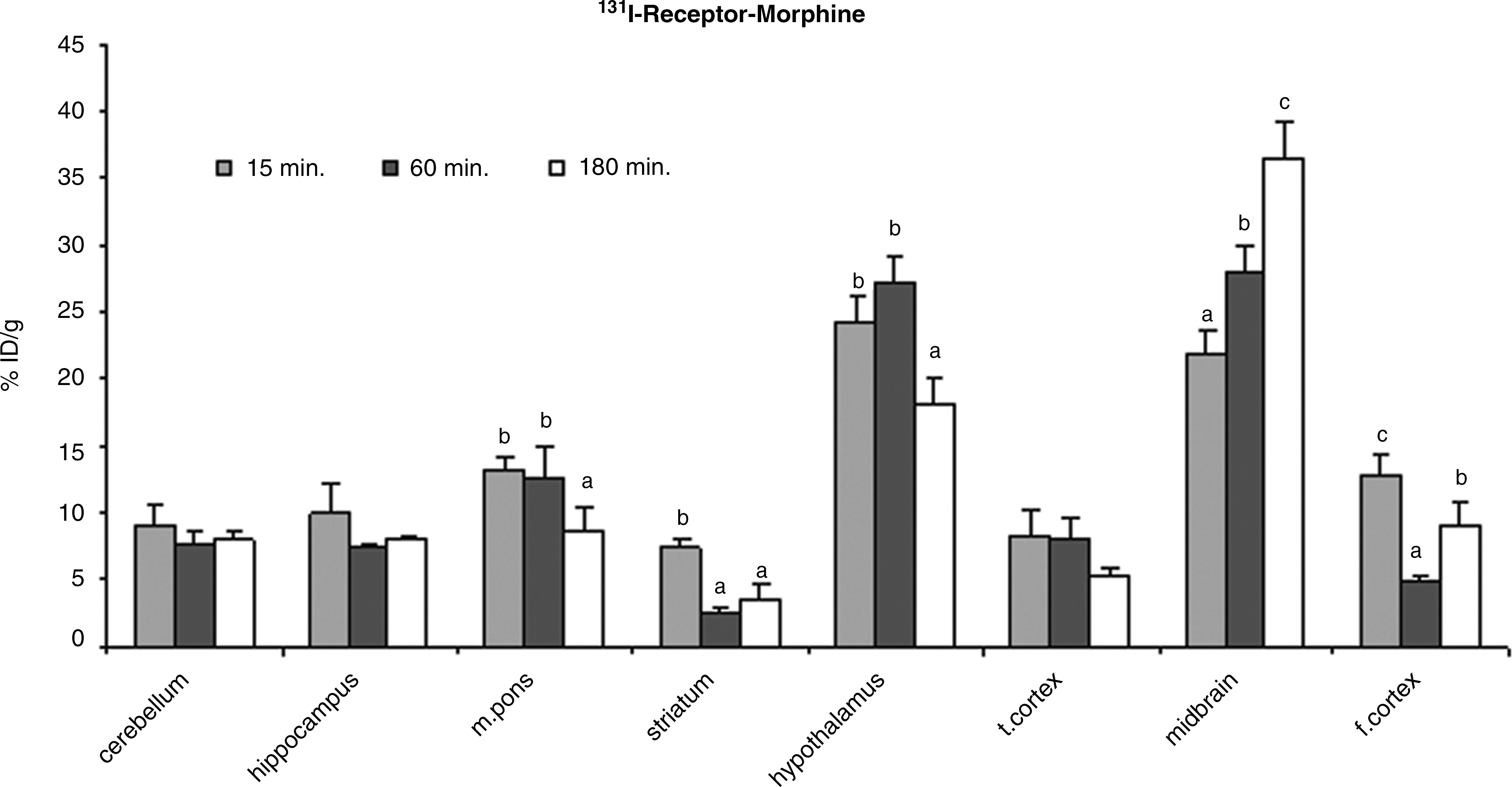
The results indicate that 131I-morphine accumulated in some brain regions (Fig. 5). The data showed that the highest uptake of labeled morphine in the midbrain (%ID/g = 36.99, %ID/g = 30.27, and %ID/g = 37.46) were seen at 15, 60, and 180 minutes, respectively. The uptake of 131I-morphine increased in the striatum and midbrain, but it significantly decreased in the hippocampus (p < 0.003) during the study period. A significantly negative correlation was observed for the hypothalamus–f.cortex (r = −0.81, p < 0.008) and f.cortex–t.cortex (r = −0.85, p < 0.003) in rat brain.

Biodistribution of 131I-morphine in rat brain as a function of time. Results are expressed as means ± standard deviation. Different superscript letters indicate significant differences with respect to groups and time. %ID/g, % injected dose per g.
The results of receptor blockage and unblockage studies showed that there are statistically significant relations between the cerebellum and m.pons of rat brain (Figs. 5 and 6). Midbrain uptake significantly increased through time (p < 0.000), but uptake in m.pons (p < 0.003) and t.cortex (p < 0.05) significantly decreased.
When receptors were blocked, radioiodinated morphine showed the maximum uptake in the hypothalamus, midbrain, and midbrain: 27.25 (%ID/g) at 15 minutes, 27.91 (%ID/g) at 60 minutes, and 36.55 (%ID/g) at 180 minutes, respectively (Fig. 6). A positive correlation was observed for the hippocampus–rec.midbrain (r = 0.72, p < 0.03), hippocampus–rec.striatum (r = 0.88, p < 0.002), t.cortex–rec.m.pons (r = 0.87, p < 0.002), and m.pons–rec.hypothalamus (r = 0.88, p < 0.002), but a negative correlation was observed for m.pons–rec.midbrain (r = −0.74, p < 0.02), hippocampus–rec.midbrain (r = −0.73, p < 0.02), and f.cortex–rec.hippocampus (r = −0.88, p < 0.03). A significantly positive correlation was observed between cerebellum and hippocampus (r = 0.84, p < 0.005) when receptors were blocked.
Biodistribution of 131I-morphine in receptor binding studies in rat brain as a function of time. Results are expressed as means ± standard deviation. Different superscript letters indicate significant differences with respect to groups and to time. %ID/g, % injected dose per g.
Discussion
According to the electrophoresis results, 131I-morphine has neutral charge because it did not move toward any pole from the application point. The n-octanol/water partition coefficient (lipophilicity) of 131I-morphine was determined and the lipophilicity was found to be 0.83 ± 0.6 (n = 3) at pH 7.0. The lipophilicity for morphine and iodomorphine was found to be 0.43 and 1.38, respectively, by the ACD programmer. The results showed a good agreement for lipophilicity between the calculated and experimental values and that iodine attachment increased the lipophilicity of morphine. If any of the unpolar substances attach to organic molecules, solubility of the product increases in organic solvent. Iodine attachment to molecules increases the lipophilicity, and highly lipophilic substances enter the brain by directly crossing the membrane. 12,22 Therefore, the blood–brain barrier permits selective entry of substances into the brain. Abbruscato et al. 23 showed that the penetration of [3H]morphine by crossing the blood–brain barrier increased in the central nervous system with increasing lipophilicity of morphine. Therefore, the present study suggests that idomorphine can penetrate the blood–brain barrier more than morphine.
Opioids are primarily eliminated via the kidneys after hepatic metabolism. Renal elimination generally accounts for ∼90% of an opioid and its metabolites. 24 Morphine is metabolized chiefly through glucuronidation by uridine diphosphate glucuronosyl transferase (UGT) enzymes. Morphine has two main metabolites, namely morphine-3-glucuronide (M3G) and morphine-6-glucuronide (M6G). 25 UGT 2B7, which primarily produces the 6-conjugate, and UGT 1A3, which produces the 3-conjugate, are the major enzymes involved in the glucuronidation of morphine. 26 Morphine is glucuronidated to M3G in the gastric, intestinal, colonic, liver, and kidney cells and to M6G in the all except gastric cells. 27,28
Donnerer et al. 29 observed that the uptake of morphine was high in intestine, liver, kidney blood, and brain at 1 hour after intravenous injection. Enginar et al. 30 showed that the concentration of 131I-codeine reached the highest value in lung, liver, kidney, small intestine, stomach, and prostate after 15 minutes. In the present study, 131I-morphine activity was considerably high in the lung, liver, small intestine, and stomach at 15 minutes after the injection.
According to Lee et al., 31 the opiate content of the cerebellum showed a tendency for a continuous increase during the 4 days. Enginar et al. 30 observed that the uptake of codeine was high in m.pons, hypothalamus, and midbrain at 180 minutes after the injection. In our study, active morphine concentration in rat brain was found in the hypothalamus and midbrain at 180 minutes.
Opioid receptors are found in nerve cells located in the various regions of the brain and the spinal medulla. Radioligand-binding assays of brain and other tissues have been particularly useful in the development of opioid receptor-selective agonists and antagonists. 5,14,15 After receptor blockage with morphine, uptake of 131I-morphine decreased in the lungs, liver, kidneys, testis, prostate, spinal cord, cerebellum, hippocampus, striatum, and temporal-cortex with respect to receptor unblockage studies of rats. The uptake of 131I-morphine concentration in the unblockage receptor studies is higher than that in receptor blockage studies for cerebellum, hippocampus, striatum, and t.cortex in rat brain. The results of the present study agree with those of the study of receptor blockage with codeine. 30
Donnerer et al. observed that morphine accumulated in the cortex, midbrain, pons medulla, and cerebellum in the biodistribution study of rats. 29 In the present study, according to the biodistribution of 131I-morphine in the brain, uptake of 131I-morphine was high in the mid-brain and hypothalamus. These results supported the results of Donnerer et al.
Conclusions
In conclusion, the original feature of the present study is the biodistribution of 131I-morphine in rats. Additionally, it was also shown that the labeling yield of 131I-morphine was high. Moreover, 131I-morphine accumulated highly in the brain. Thus, the present study concludes that 131I-morphine has enough stability for diagnostic scanning. Morphine radiolabels with 123I instead of 131I may be used as a radiopharmaceutical for brain imaging in SPECT.
Footnotes
Acknowledgments
This work was supported by the Scientific and Technical Research Council of Turkey (TUBITAK, Project Number 2004-104T187) and T.R. Prime Ministry State Planning Organization (DPT, Project Number 2006 DPT 06).
Disclosure Statement
There are no conflicts of interest among the authors in this study.