
Abstract
Melanoma is one of the most aggressive forms of tumor, being responsible for about 80% of skin cancer deaths. Much effort is being directed at obtaining less-toxic anticancer therapies, and the combination between low cytotoxic doses of chemotherapeutic drugs and natural differentiative compounds seems to be of particular importance. The present study was undertaken to examine the possible role of a combination therapy using paclitaxel (PTX) as chemotherapeutic molecule and theophylline (TH) as differentiative agent in the prevention of metastasis in B16-F10 melanoma-bearing C57BL6/N mice. In vitro proliferation studies demonstrated that TH enhanced the antiproliferative effect of PTX. In the in vivo experiments, a highly sensitive computerized image analysis method, performed on histological lung sections of mice injected with melanoma cells, was used to quantify the efficacy of the treatments. This study demonstrated that the simultaneous treatment of mice with TH and a low dose of PTX produced a similar anti-invasive effect than that caused by highly toxic PTX concentration.
Introduction
Melanoma is the most aggressive, metastasizing, therapy-resistant, and deadly form of skin cancer, and its incidence is increasing at a rate greater than any other form of cancer. 1 Melanoma patients generally undergo surgical therapy, chemotherapy, radiotherapy, or a combination of these treatments. Although the effects of these treatments are significant, it is a fact that most patients suffer from side-effects. In some countries, herbal medicine is used as a supplemental therapy for many kinds of diseases. Recent reports clearly showed that when herbal medicines are used for cancer treatment, many patients experience fewer or diminished side-effects induced by other toxic therapies such as chemotherapy and radiotherapy, and the survival period is longer. 2,3 Some of the herbal medicines of interest are contained in common beverages such as tea, one of the most popular beverages in the world, obtained by an infusion from Camellia sinensis (L.) Kuntze leaves. Although tea has been consumed for centuries, it has only recently been studied extensively as a health-promoting beverage, which may act to prevent a number of diseases. Additionally, the cancer-preventive effects of green tea have been widely supported by results from epidemiological, cell culture, animal, and clinical studies. 4 Tea leaves produce organic compounds that may be involved in the defense of the plants against invading pathogens including insects, bacteria, fungi, and viruses. These “secondary metabolites” include polyphenolic compounds (catechins) and methylxanthine alkaloids, such as theophylline (1,3-dimethylxanthine, TH), theobromine (3,7-dimethylxanthine), and caffeine (1,3,7-trimethylxanthine). These molecules are of interest because of the widespread ingestion of common beverages (coffee, tea, etc.), whereas others have been used for therapeutic advantage. 5,6 TH has been used over the last 70 years for treating patients with asthma. It is well known as a bronchodilator and the current asthma guidelines recommend it as an add-on therapy in noncontrolled asthmatics. 7 TH acts as cAMP-phosphodiesterase inhibitor, 8 involving alterations in cAMP system tumor cell, but also as an adenosine antagonist. 9 A need exists to develop a better understanding of the involvement of dietary methylxanthines in cancer prevention. A number of publications have demonstrated the role of TH in negatively affecting cancer cell growth and invasiveness in vitro and in vivo 10 –15 and in the induction of tumor cell differentiation.
The development of new chemotherapeutic and molecular targeting agents has opened the door to various clinical trials in search for novel therapeutic strategies to improve the outcome of cancer patients. Among chemotherapeutic agents, paclitaxel (taxol, PTX), an alkaloid ester, emerged as one of the most powerful compounds. 16 It was found that a crude extract from the bark of the Pacific yew, Taxus brevifolia Nutt., proved to have cytotoxic activity against many cancer cells. 17 PTX promotes the polymerization of tubulin, and microtubules formed are firmly stable and dysfunctional, thereby disrupting the normal microtubule dynamics required for cell division and interphase processes. 18,19 As a consequence, cells exit from mitosis and DNA fragmentation, indicative of apoptosis, occurs. 20,21
Despite increasing knowledge of cancer biological properties, its eradication remains a puzzling clinical problem. Clinical behavior and therapeutic response are difficult to predict because of tumor heterogeneity. 22 Chemotherapy is a common type of cancer treatment that uses drugs or medications by targeting and eliminating rapidly dividing cancer cells. Nevertheless, this aggressive and cytotoxic treatment causes many side-effects in cancer patients. In contrast, differentiation therapy is based on the concept that drugs and natural substances can inhibit carcinogenesis and development of tumors through the induction of cellular terminal differentiation 23 –25 and do not possess the same cytotoxicity typical of chemotherapeutic drugs. 26 The potential use of combined therapy is under intensive study, including the association between classical cytotoxic agents and differentiative inducers, which might be able to enhance the antitumor activity. The main purpose of the present study was to evaluate the possible synergistic antineoplastic effect of PTX in combination with TH. First, the in vitro antiproliferative and cytotoxic properties of these compounds on B16-F10 murine melanoma cells were investigated. Then, the effect of the in vivo treatment of melanoma-bearing C57BL6/N mice with high PTX dose was compared with that obtained using TH in combination with a lower PTX concentration.
Materials and Methods
Materials
Dulbecco's modified Eagle's medium (DMEM), fetal calf serum (FCS), glutamine, penicillin/streptomycin (10,000 IU/mL), trypsin/ethylenediaminetetraacetic acid, and phosphate-buffered saline (PBS) were obtained from Gibco Laboratories. PTX, TH, and Hoechst 33258 were from Sigma Chemicals. All other chemicals were provided by Merck.
Cell line
Highly metastatic murine B16-F10 melanoma cell line was purchased from the Division of Cancer Treatment at Tumor Repository NIH, cultured in DMEM with 10% FCS, supplemented with 200 mM glutamine, 100 U/mL penicillin, and 100 μg/mL streptomycin, and maintained in a humidified atmosphere of 5% CO2 at 37°C. The absence of Mycoplasma spp. was confirmed by direct fluorescence with a specific DNA dye (Hoechst).
Proliferation and cytotoxicity studies
For proliferation studies, cells were plated and grown in 35-mm dishes and treated with PTX (0.05, 0.25, and 0.5 μM) or TH (50 μM, 500 μM, and 1 mM) for 24, 48, and 72 hours. Cells were detached and counted using a Neubauer modified chamber. Cytotoxicity was assessed after trypan blue staining. Briefly, 5 × 10⁴ cells in complete medium were plated in six-well plates and allowed to attach overnight. The medium was replaced and cells were treated as described, and the plates were incubated for 24, 48, or 72 hours at 37°C. Both floating and adherent cells were collected, suspended in 250 μL of PBS, and mixed with 50 μL of 0.4% trypan blue solution. Cells were counted under a light microscope. Cell counts were done by 2 researchers in a blinded fashion. The slides were projected onto a grid comprising 12 fields, and 6 randomly chosen fields were used. Only cells that could be clearly identified as individual cells were counted as viable cells. Trypan blue-stained cells (necrotic + apoptotic cells) did not fulfill this criterion and were considered as background staining.
Animals
Male C57BL6/N 6–8-week-old mice, with a mean body weight of 24.6 g at the outset of the experiments, were purchased from Iffa-Credo. Mice were maintained in a pathogen-free environment at 25°C under controlled lighting (12 hours light/12 hours dark) and allowed free access to water and food pellets. About 10–20 animals for each group were used. All experimental protocols have been carried out following the Guidelines for the Welfare of Animals in Experimental Neoplasia 27 and the EEC Council Directive 86/609.
Agents
For animal studies, TH was dissolved in tap water at 20 mM concentration and administered ad libitum. Plasma levels of TH, measured using high-performance liquid chromatography as described elsewhere, 28 were calculated to be steadily about 100 μM. 12 PTX was dissolved in dimethylsulfoxide, subsequently diluted in PBS, and administered intraperitoneally.
Experimental lung metastasis
Lung metastases were produced by intravenous injection of 2 × 10⁵ viable B16-F10 cells, resuspended in 0.2 mL DMEM, through the lateral tail vein of mice. Oral administration of TH was started at 3 days before tumor cell injection and maintained until animal sacrifice. PTX was administered (15 or 150 mg/kg body weight) by intraperitoneal injection on days 1 and 8. Tumor-injected mice were divided into 5 groups: (1) untreated; (2) orally administered with TH; (3) injected with 15 mg/kg PTX; (4) injected with 150 mg/kg PTX; (5) orally treated with TH + injected with 15 mg/kg PTX. The animals were carefully monitored and sacrificed by cervical dislocation after 3 weeks from tumor cell injection. Lungs were rapidly excised, rinsed in PBS, and further processed for macroscopical analysis and histology.
Macroscopic analysis
The metastatic nodules on the pleural surface of the lung lobules were counted by using a Wild Heerbrugg M3 stereomicroscope (homogeneous brown structures on the lung surface, of variable size and sufficiently separated from other structures to be counted individually). The evaluation was performed by 2 independent operators. A total of 500 lung foci were considered as the maximum value (100%).
Histology and morphometric analysis of the tumors
Samples were fixed in 10% formalin for 48 hours, dehydrated in ethanol, and embedded in paraffin. Ten (10)-micrometer serial sections from each lung lobule, performed every 100 μm, were obtained with an ultramicrotome Leitz 1512 and stained with hematoxylin and eosin. Morphometric calculations on B16 melanoma lung metastases were performed on ∼100 random lung sections in each lung. Quantification of the portion of lung section occupied by metastatic tissue (% implantation), the growth index (GI), and the invasion index (II) were obtained using an integrated image analysis system (Quantimet 970; Cambridge Instruments), as previously described. 29 As the area of a metastatic focus is related to its growth, GI was considered to be the ratio between the average area of metastatic foci over the total area of the histological sections. Further, the number of lung foci, related to the invasion of tumor cells, is expressed by the II, calculated as the ratio between the total metastatic area and the average area of the metastatic foci.
Statistical analysis
For the in vitro proliferation studies, data were analyzed by Student's t-test and differences were considered significant when p < 0.01. Values for untreated tumor-bearing mice were compared with those for TH- or PTX-treated animals. The results were expressed as the mean ± standard error of the values obtained for each lung. All in vivo experimental data were analyzed by nonparametric Mann–Whitney test (p-values lower than 0.01 were considered as significant).
Results
Cell proliferation
The treatment with PTX affected B16-F10 melanoma cell growth in a time- and concentration-dependent manner. In particular, proliferation was progressively reduced, with respect to the control, by about 20%, 28%, and 38% after 72 hours treatment with 0.05, 0.25, and 0.5 μM PTX, respectively (Fig. 1A). TH treatment (50 and 500 μM) caused a slight decrease in tumor cell growth, with respect to the control, whereas a significant reduction of cell number by 30% after 72 hours was detected in the presence of 1 mM TH (Fig. 1B). Figure 2 shows the comparison between the antiproliferative effects of 1 mM TH, 0.05 μM PTX, and 0.5 μM PTX and the simultaneous treatment of cells with 1 mM TH and 0.05 μM PTX. Interestingly, the antiproliferative role of the combined treatment was remarkably higher than the single compounds used alone (55% reduction vs. control after 72 hours of exposure).
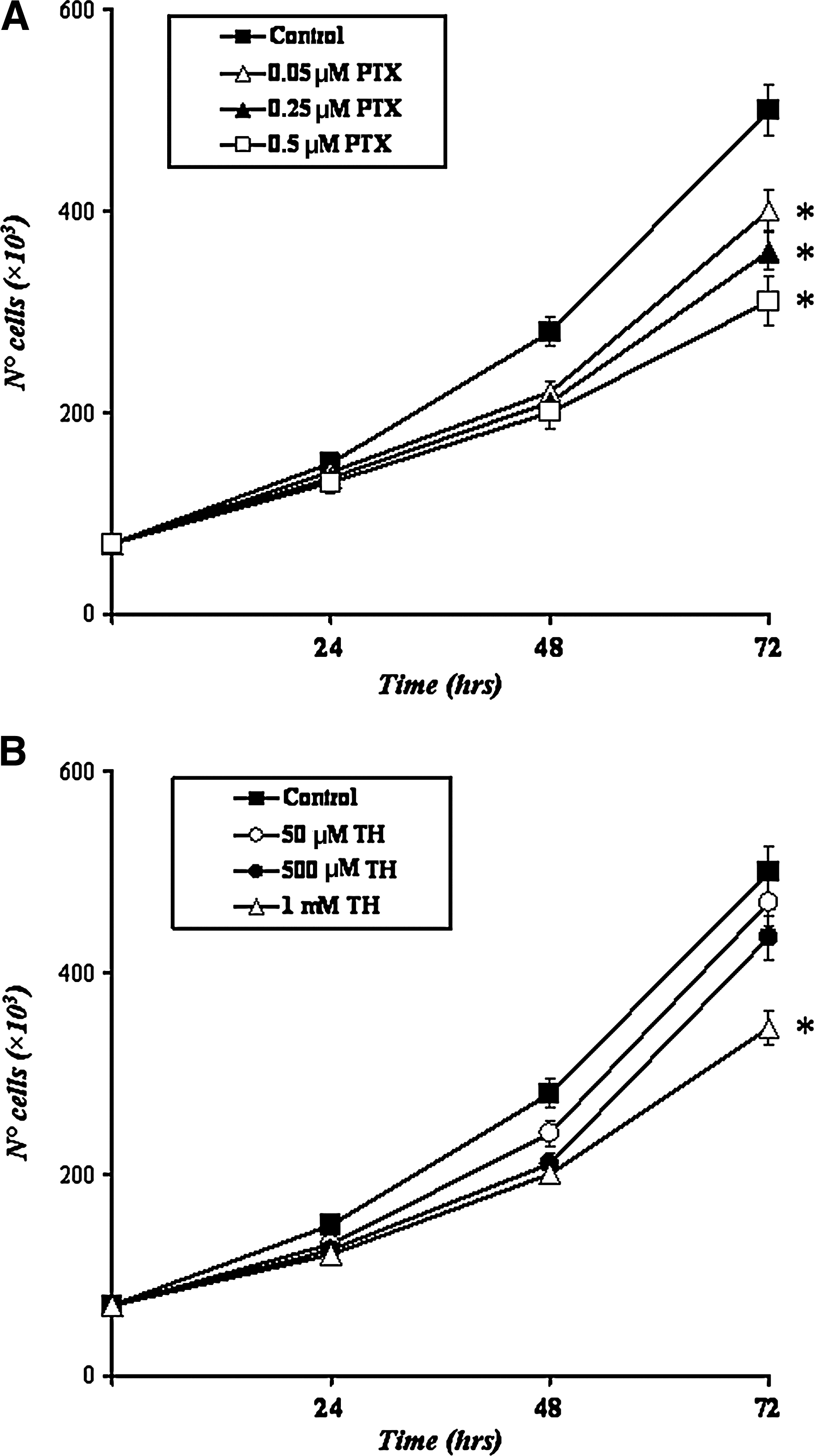
Time course curve of murine B16-F10 melanoma proliferation induced by PTX and TH. After drug treatment, the cells were incubated for various times before they were counted using a hemocytometer. (
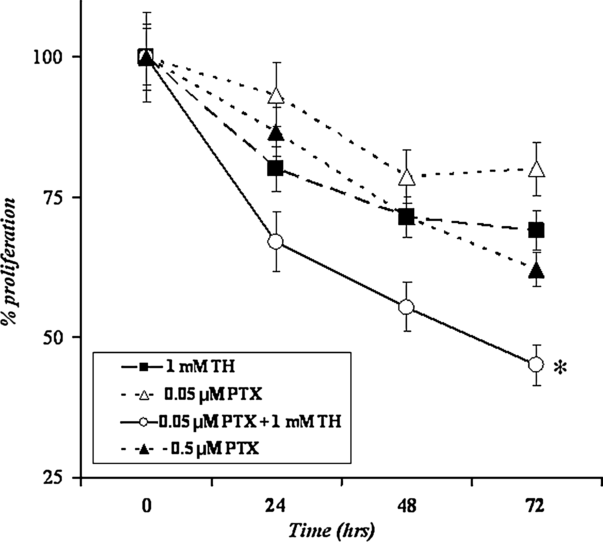
Proliferation curve of murine B16-F10 melanoma cells treated with 1 mM TH (filled square), 0.05 μM PTX (open triangle), 0.5 μM PTX (filled triangle), and 1 mM TH + 0.05 μM PTX (open circle) for 24, 48, and 72 hours. Data are expressed as % proliferation with respect to the control (100%). Results represent the mean of three different determinations ± standard deviation ( * p < 0.01). TH, theophylline; PTX, paclitaxel.
Cell viability
The effects of different treatments on the viability of B16-F10 melanoma cells were analyzed by using the trypan blue dye exclusion test. As shown in Figure 3, cytotoxic effects were observed after 48 and 72 hours of incubation with the drugs. In particular, TH treatment did not have remarkable effects on cell viability at all concentrations used. In contrast, cytotoxicity was strongly enhanced after exposure to a high concentration of PTX (0.5 μM). The simultaneous treatment of tumor cells with 1 mM TH and the lowest concentration of PTX (0.05 μM) increased cytotoxicity, with respect to the single treatments, but it was lower than the toxicity caused by 0.5 μM PTX.

Cytotoxic effect of TH and PTX. B16-F10 cells were grown to 50% confluence and treated with increasing concentrations of TH or PTX for different incubation times. Cell mortality was evaluated by trypan blue dye exclusion assay. In the control cells, mortality was always lower than 5%. Combined treatment (TH + PTX, black bars) was performed by treating cells with both 1 mM TH and 0.05 μM PTX. All values, expressed as the ratio over control, represent the mean ± standard deviation of at least three different experiments in triplicates (p < 0.01). TH, theophylline; PTX, paclitaxel.
Quantitative evaluation of B16-F10 lung metastases
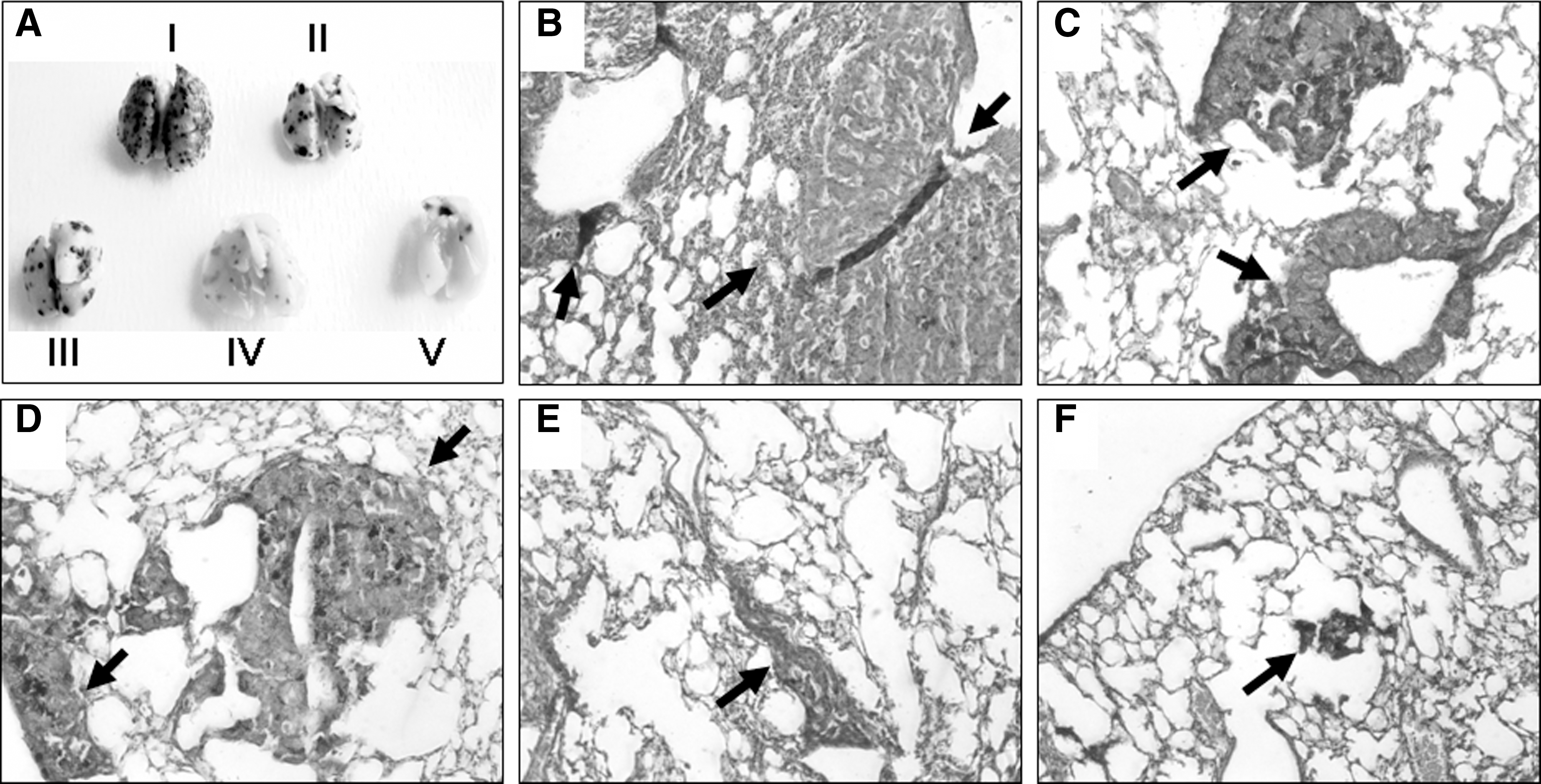
Figure 4A shows the appearance of representative mouse lungs at 3 weeks after the inoculation of B16-F10 melanoma cells. The histological sections of lungs invaded by treated and untreated mice showed that the melanoma colonies were usually located either inside or superficially near pulmonary blood vessels and bronchioles (Fig. 4B–F). The quantitative evaluation of the antineoplastic activity of the compounds under examination has been performed by counting the number of surface metastases (Fig. 5), which was found to be reduced, with respect to the control, by 75% in TH-treated mice and by 50% and 90% in 15 mg/kg PTX and 150 mg/kg PTX treatments, respectively. The combined treatment with TH and 15 mg/kg PTX reduced the frequency of lung superficial foci by about 92% versus control. The histological sections of lung show that in 150 mg/kg PTX and 15 mg/kg PTX + TH groups, the sizes of melanoma foci appear lower than that found in all other experimental groups. The area of lung tissue occupied by metastases was calculated on each histological section by means of a computerized procedure. In Table 1, the percentage of implantation of melanoma cells into the target organ is shown. Tumor implantation was inhibited, with respect to the control, by 77% after TH treatment of mice, by 66% after 15 mg/kg PTX treatment, by 85% after 150 mg/kg PTX administration, and by about 89% after the simultaneous exposure to 15 mg/kg PTX and TH. To give a measure of the rate of tumor growth in the target organ, expressed as the GI, and of the frequency of foci in the lung, expressed as II, all the histological sections obtained from B16-F10-invaded lungs were further analyzed by a computer-assisted image analysis system (Quantimet), and the data are given in Table 1. As shown, the only treatments able to negatively affect metastases growth were the high concentration of PTX (150 mg/kg) and the PTX + TH treatments, which reduced the average metastatic area, compared with the control, by 39% and 57%, respectively. On the contrary, the invasive potential (II) of B16-F10 melanoma cells was markedly reduced (about 80% reduction) in mice after all treatments.
(
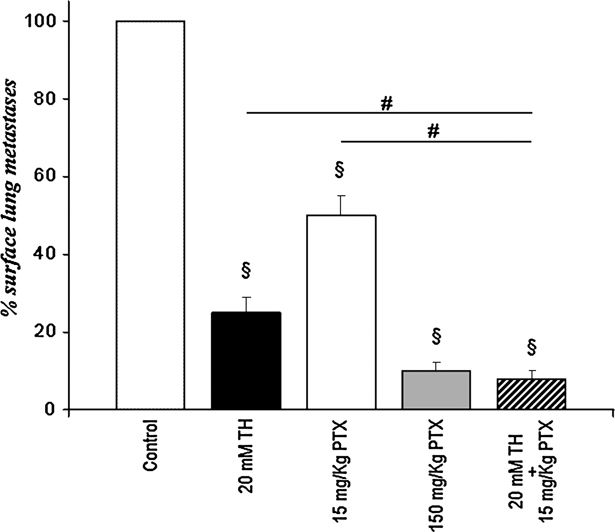
Quantitative evaluation of the frequency of subpleural lung melanoma metastases from B16-F10 cell-injected mice untreated or treated with TH or PTX. Data are expressed as % surface lung metastases with respect to the control (100%). §Statistical significance versus control (p < 0.01).
Each value represents the mean ± standard deviation of three different determinations.
Highly significant versus control (p < 0.005).
Significant versus control (p < 0.01).
Significant versus TH or 15 mg/kg PTX (p < 0.01).
TH, theophylline; PTX, paclitaxel.
Discussion
Throughout history, natural products have afforded a rich source of compounds that have found many applications in medicine, pharmacy, and biology. Within the sphere of cancer, a number of important new molecules, obtained from natural sources, displays marked differentiative properties on tumor cells. 30,31 The application of differentiation therapy seeks to reverse the loss of the differentiated state and forces cancer cells to resume a more mature phenotype, allowing them to regain the morphology and function of mature cells, typical of the organ where they were originated. 26 Although this would not eradicate cancer, the present study suggests that this kind of approach may be combined with the more conventional cytotoxic chemotherapy to interfere with cancer progression. For instance, many studies have indicated that combining antiangiogenic agents with conventional chemotherapeutics enhanced the inhibition of tumor growth and metastasis. 32 –34 Then, cisplatin and its analogs have been widely used in combination with natural compounds, such as vindesin in non-small-cell lung cancer, 35 curcumin in human ovarian carcinoma cells, 36 or TH as nephroprotective agent. 37 Others associated PTX to plant-derived molecules, such as genistein 38 or flavopiridol. 39 In the present study, PTX in combination with TH, a natural compound known to have differentiative properties in melanoma, 40 was used to test the possible improvement of the antiproliferative and anti-invasive effect on B16-F10 melanoma cells. The experimental metastasis model associated to the computerized image analysis procedure for the analysis of foci in histological lung tissue sections, more accurate than the macroscopical surface counting, appears extremely useful for the in vivo discrimination between inhibition of tumor growth and the anti-invasive capability of the treatment under investigation.
The in vitro data showed that the use of a low concentration of PTX, combined with TH, exerts an antiproliferative action on B16-F10 melanoma cells higher than the single treatments, showing a cytotoxicity lower than PTX used alone at high concentration. On the basis of these results, the in vivo effect of TH and PTX on experimental lung metastasis was further investigated. Although the metastatic potential (II) is largely affected by all treatments, the simultaneous administration of low-dose PTX and TH is able to decrease the GI more than the single treatment with high-dose PTX, as also highlighted by the macroscopical analysis. The present study demonstrates that the combination of a differentiative molecule with a low-toxic dose of a chemotherapeutic exhibits similar antineoplastic properties to those exerted by the same cytotoxic drug at high concentration. This evidence may assume particular importance in the light of the sensible reduction of the side-effects due to the cytotoxicity of chemotherapeutical agents.
Conclusions
Taken together, the combination of TH and PTX markedly decreases tumor cell proliferation and dramatically reduces tumor burden and pulmonary metastasis. The data of the present study suggest that the combination of TH and PTX might have novel clinical application in reducing tumor growth and inhibiting metastasis over a prolonged period in patients with advanced melanoma, supporting the hypothesis that the induction of tumor cell differentiation may represent an advantageous therapeutic strategy to be coupled to the conventional chemotherapy.
Footnotes
Disclosure Statement
No competing financial interests exist.
*
EEC Council Directive 86/609, Official Journal of the European Communities L358, 1.12.1987.