
Abstract
Hepatocellular carcinoma (HCC) is one of the most prevalent forms of cancer with high morbidity. 131I-lipiodol is used clinically and has been found to be effective for the treatment of HCC. However, this preparation has its limitations, including compromised yield and stability of exchange labeling and unnecessary dose burden from gamma emissions. In the present study, 177Lu-oxine in lipiodol was considered as a possible alternative for radioiodinated lipiodol. Oxine or 8-hydroxyquinoline was labeled with 177Lu obtained by neutron irradiation of natural lutetium. Under optimized conditions, the radiolabeled complex was obtained with yields >98% and adequate in vitro stability. 177Lu-oxine dispersed in lipiodol showed appreciable uptake into rat liver cells (normal and HCC-induced) in vitro. 177Lu-oxine-lipiodol showed initial localization in the liver, but subsequent leakage of radioactivity with deposition in the skeletal tissue was seen. The studies suggest that 177Lu-oxine dispersed in lipiodol might not be suitable for treatment of HCC.
Introduction
Hepatocellular carcinoma (HCC) is one of the most clinically significant forms of cancer in humans, especially in the Asian region. It has a high associated mortality rate (≥5 years survival in <5% of cases). 1,2 Therapy for HCC falls into three major categories: surgery, chemotherapy, and radiotherapy. 3 In the recent years, an increasing amount of clinical research toward treatment of HCC is being focused on the application of radiolabeled compounds containing therapeutically relevant beta-emitting radioisotopes directly injected into the hepatic artery, which are subsequently localized in the liver. 4
There are two approaches to the above-referred radioembolization. In one approach the therapeutic isotope is incorporated into nonbiodegradable or very slowly degrading particulates of several microns in diameter, which get localized in the capillaries, for example, 90Y-labeled glass microspheres. 5 Alternatively, the radioactivity is incorporated into a strongly lipophilic preparation, which permeates through the hepatocellular tissue and is retained there to deliver the therapeutic dose. A well-known example of this is 131I-labeled lipiodol. Lipiodol, an iodinated and esterified lipid of poppyseed oil, is known to accumulate in the liver tissue when injected via the hepatic artery and has been used as a magnetic resonance imaging contrast agent. 6 Lipiodol labeled with β-emitting radionuclides such as 131I, 188Re, and 166Ho has been evaluated for targeting liver cancer in both animal models and human patients. 7 –15 131I-labeled lipiodol has been proven to be clinically effective and is a commercially available product. 9,14 However, a drawback in the use of 131I is its high-energy gamma photon emission with high abundance (364 keV, 81%). 10,16 Moreover, the radiolabeling yield of 131I-labeled lipiodol is also poor because of the exchange labeling method used.
Lutetium-177 has recently emerged as a promising radionuclide for in situ therapeutic applications because of its suitable decay properties (T 1/2 = 6.73 days, E β(max) =0.49 MeV, E γ = 208 keV [11%]) and the feasibility of its large-scale production in moderate flux reactors with adequate specific activity and radionuclidic purity. 17 The half-life of 177Lu is comparable to that of 131I and facilitates its wider global distribution without significant decay loss. The presence of low-energy gamma photons with relatively low abundance is useful for simultaneous scintigraphy and dosimetry studies without significant additional dose burden to the patients, making for a potential advantage over 131I. 17,18 Thus, a 177Lu complex in lipiodol could be envisaged as a viable alternative to 131I-lipiodol for HCC therapy.
In the present study, the authors have attempted to prepare 177Lu-labeled lipiodol for potential application in HCC therapy and study its biological behavior in an animal model. As direct incorporation of 177Lu into lipiodol is not feasible, radiolabeling of lipiodol was achieved via the incorporation of a lipophilic complex of lutetium. Oxine (8-hydroxyquinoline) was selected as the ligand for complex formation, as it is known to form stable lipophilic complexes with lanthanides, and has been used for extraction of Ln+3. 13,16,19
Experiment
Materials and methods
177Lu used in the study was produced by irradiating natural Lu2O3 target following the procedure reported in the literature. 17,18 8-Hydroxyquinoline (oxine) was purchased from Aldrich Chemical Company. Lipiodol was obtained from Guerbet. Dulbecco's modified Eagle's medium (DMEM) used for in vitro cell experiments and the cell dissociation kit for preparing single-cell suspension of liver cells were obtained from Sigma. Horse serum used as an in vitro growth supplement for liver cells was obtained from Hi-Media. All other chemicals used were of AR grade and supplied by reputed chemical manufacturers.
Scintigraphic images were recorded on the Millennium MPS Image Acquisition System (Wipro–GE Medical). The gamma camera was calibrated for 208 keV gamma photon of 177Lu with 20% window prior to the acquisition of the images.
Preparation of 177Lu-oxine-lipiodol
177Lu-oxine-lipiodol was prepared by a two-step process. In the first step, a highly lipophilic 177Lu-oxine complex was prepared by radiolabeling oxine with 177Lu and subsequently this lipophilic complex was dispersed in lipiodol in the second step.
Preparation of 177Lu-oxine complex
For the preparation of 177Lu-oxine complex, 100 μL 177LuCl3 (∼370 MBq of 177Lu, 4 μg Lu) was added to a 375 μL solution of oxine (20 mg/mL concentration) in ethanol after the addition of 525 μL of 0.1 M ammonium acetate buffer of pH ∼5. The pH of the reaction mixture was adjusted to ∼7 by using dilute NaOH solution and it was incubated at 50°C for 1 hour for the completion of reaction. Several parameters such as concentration of oxine, pH of the reaction mixture, incubation time, and temperature were varied extensively to arrive at the optimized protocol.
Dispersion of 177Lu-oxine complex in lipiodol
For the preparation of 177Lu-lipiodol, 177Lu-oxine complex prepared under optimized reaction conditions was extracted in dichloromethane. The dichloromethane layer was carefully separated from the aqueous layer and subsequently evaporated by controlled heating in a water bath. Lipiodol (500 μL) was added to the residue after cooling and the mixture was mechanically stirred at 50°C for 1 hour to ensure homogeneous dispersion of the 177Lu-oxine complex in lipiodol. The final preparation was washed twice with normal saline.
Determination of complexation yield of 177Lu-oxine
The complexation yield of 177Lu-oxine complex prepared was determined by solvent extraction technique. A 100 μL aliquot of the reaction mixture was withdrawn and diluted with 400 μL distilled water. The resulting solution was extracted using 500 μL dichloromethane. Finally, aliquots of 20 μL of each phase were withdrawn and radioactivity associated with each layer was counted. These data were used to determine the complexation yield.
Determination of percentage dispersion of 177Lu in 177Lu-oxine-lipiodol
The percentage of 177Lu activity dispersed in lipiodol was determined by following a simple procedure. Aliquots of 20 μL were withdrawn from the 177Lu-oxine complex (prior to mixing with lipiodol) and 177Lu-oxine-lipiodol after the dispersion was complete. The radioactivity associated with each layer was counted, and from these data, total activity of 177Lu-oxine prior to mixing and 177Lu-oxine-lipiodol were determined. The ratio of the counts observed in 177Lu-oxine-lipiodol to 177Lu-oxine provided the percentage dispersion of 177Lu in 177Lu-lipiodol.
In vitro stability of 177Lu-oxine-lipiodol
To ascertain the in vitro stability of 177Lu-oxine in lipiodol, 500 μL of normal saline was added to 500 μL of the preparation and mixed vigorously. The mixture was allowed to settle at room temperature. Twenty (20) microliters of aliquots were withdrawn from both the layers at different time intervals post-preparation and the associated activity was counted. The stability of the radiolabeled preparation at various time points was determined by calculating the percentage of activity associated with the lipid phase from these data.
Biological studies
Biological studies on 177Lu-oxine-lipiodol were performed using Wistar rat as the animal model. HCC was induced in Wistar rats through the use of the known HCC-inducing agent diethylnitrosamine. Animals weighed ∼200 g at the start of the carcinogenesis regimen. Separate sets of animals labeled “HCC-induced” and “Control” were maintained. They were provided a normal diet and housed under identical conditions except that the HCC-induced set was provided water containing 0.01% diethylnitrosamine. The water was replaced every week, or before if required. They were thus maintained for up to 60 days, during which animals in the HCC-induced set developed multiple foci of tumor in the liver. This was confirmed by histological studies. At the end of the carcinogenesis period the animals were immediately taken for further experiments. All experimental protocols involving animals were performed in accordance with the national laws governing conduct of animal experiments.
In vitro cell uptake
For the in vitro cell uptake studies, liver cells were harvested from Wistar rats in the control and HCC-induced sets. Briefly, the liver tissue was excised and immersed in plain DMEM. In the HCC-induced set, tissue was taken from the tumor lesions in the liver. Using a cell dissociation kit, the liver tissue was processed and single-cell suspension was obtained. The cells were centrifuged and suspended in DMEM supplemented with 25% horse serum for use in the cell uptake studies.
The protocol for the in vitro cell uptake studies was as follows: 106 liver cells (of normal or HCC origin in respective sets) were taken per reaction tube in 200 μL of suspension medium. 177Lu-oxine in lipiodol was added (∼3.8 μCi in 25 μL per reaction), which settled at the bottom of the reaction tube. The cells were spun down at 1000 g to form a layer at the interface of lipiodol and aqueous medium. The cells were kept incubated at 37°C for various time points (1, 3, and 24 hours). For up to 3 hours, the reaction tubes were gently agitated every hour to redisperse the cells and spun down again. At the end of the respective incubation periods, the cell suspensions were gently agitated and removed without disturbing the lipiodol layer. The cells were spun down and incubated in fresh DMEM (1 mL) with 25% horse serum at 37°C for 60 minutes. Then the cells were washed twice with ice-cold Hank's balanced salt solution and taken for estimation of bound activity.
In vivo distribution
Normal and HCC-induced Wistar rats were employed for the in vivo studies. The animals were administered with the 177Lu-labeled oxine as per the following protocol. The animals were anesthetized with xylazine–ketamine (1:10, v/v). A surgical incision was made to expose the hepatic region and 50 μL of the labeled preparation was injected directly through the hepatic vein. The incision was sutured and the animals were allowed to recover from the effects of the operation. Scintigraphic imaging was performed on the Millennium MPS medical imaging system (Wipro–GE Medical). Images were acquired using the Genie Acq software (Release 3.0); acquisition settings were as follows: 256 × 256 matrix, 1.33 zoom, 15 minutes acquisition time. Image processing was done on the Xeleris software (Version 1.0272).
Results
Production of 177Lu
Lutetium-177 used for the present study was obtained with a specific activity of ∼18.5 GBq/mg (∼500 mCi/mg) at 24 hours after end-of-bombardment by irradiation of natural Lu2O3 target at a thermal neutron flux of ∼6 × 10¹³ n/(cm2 s) for 21 days. The radionuclidic purity of 177Lu produced was determined to be 99.985% by analyzing the γ ray spectrum of the irradiated target after radiochemical processing.
Optimization of 177Lu-labeling of oxine and dispersal in lipiodol
To obtain maximum complexation yield of 177Lu-oxine, reaction parameters such as ligand concentration, pH, reaction time, and temperature were varied and their effects were studied. Concentration of oxine in the reaction mixture was varied from 500 μg/mL to 10 mg/mL to determine optimum ligand concentration. It was observed that the complexation yield, which was only 16.4% when using 500 μg of the ligand, increased to 98.8% when the ligand concentration was increased to 7.5 mg. Further increase of ligand concentration did not have any significant effect on the complexation yield. Hence, 7.5 mg/mL was chosen as the optimum concentration of oxine for all subsequent studies.
The pH of the reaction mixture was varied from 2 to 10 using either 1 M HCl or 1 M NaOH solution. It was observed that the complexation yield increased with increase in pH of the reaction mixture and reached a maximum of 98.8% at pH 7, which was taken to be the optimal reaction pH.
The reaction mixtures were incubated at room temperature as well as at 50°C using the optimized ligand concentration and pH, and complexation yields were determined at various time intervals (5–120 minutes). It was observed that complexation yield increases with increase in incubation time at both room temperature and 50°C. However, incubation at room temperature yielded a maximum of 59.9% complexation after 2 hours, whereas incubation at an elevated temperature of 50°C resulted in 98.8% complexation yield within 1 hour of incubation and was deemed optimal for further work.
177Lu-oxine-lipiodol was prepared by dispersing 177Lu-oxine complex in lipiodol by following the method described in the Experimental section. It was observed that 94.1% ± 1.2% (n = 5) of the 177Lu-oxine activity could be dispersed in lipiodol under the optimized conditions.
In vitro stability of 177Lu-oxine-lipiodol
The in vitro stability of 177Lu-oxine-lipiodol preparation was ascertained by storing it with normal saline at room temperature and determining the leaching of 177Lu activity in the saline at different time points postpreparation. It was observed that 97.9% ± 0.7% of 177Lu activity remained associated with the lipid phase after 5 days of incubation, indicating adequate in vitro stability of the radiolabeled preparation.
Biological studies
In vitro cell uptake
Comparison of the in vitro cell uptake of 177Lu-oxine-lipiodol in normal and HCC-induced liver cells is given in Figure 1. The uptake of 177Lu-oxine-lipiodol in both normal and cancer cells was observed to increase gradually over time for a period of 24 hours. It has been reported that lipiodol selectively accumulates in cancerous lesions in the liver. 20 However, in the present in vitro study, there was no statistically significant difference in the kinetics and total uptake between normal and HCC-induced liver cells, which would indicate that there is no inherent variance in the rate of lipiodol uptake between the two sets. Hence, any difference in the uptake of lipiodol between tumor-bearing and normal regions of liver in vivo may be attributed more to the mechanisms of blood supply to these regions than to any inherent property of the cells themselves.
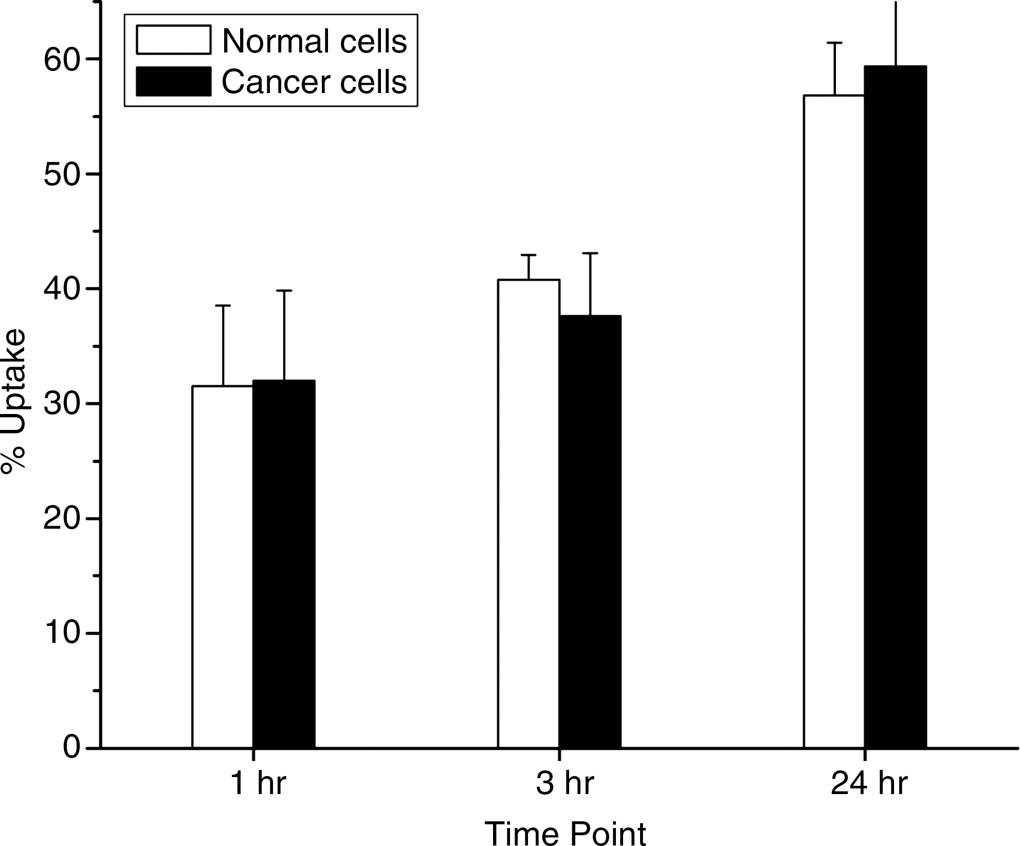
In vitro liver cell uptake pattern of 177Lu-oxine-lipiodol.
In vivo scintigraphic imaging studies
Scintigraphic studies showed the hepatic accumulation and retention pattern of 177Lu-oxine-lipiodol following its administration via the hepatic artery. Figure 2A and B depicts whole-body scintigraphic images of HCC-bearing Wistar rats recorded at 1 and 7 days postinjection (p.i.), respectively. At 1 day p.i., more than 90% of the injected activity was observed to be retained in the liver. However, in both normal as well as HCC-induced rats, the activity was observed to leach out of the liver gradually. Comparisons of loss of activity from the liver and whole body are depicted in Figure 3A and B, respectively. The leaching of 177Lu activity from the liver of normal rats was observed to be slightly more than that seen in the animals induced with hepatic cancers. Approximately 30% and 20% of the original injected activity was found to be associated with the livers of HCC-induced rats and normal rats, respectively, at 7 days p.i.

Whole-body scintigraphic images of HCC-bearing Wistar rat injected with 177Lu-oxine-lipiodol in hepatic artery at
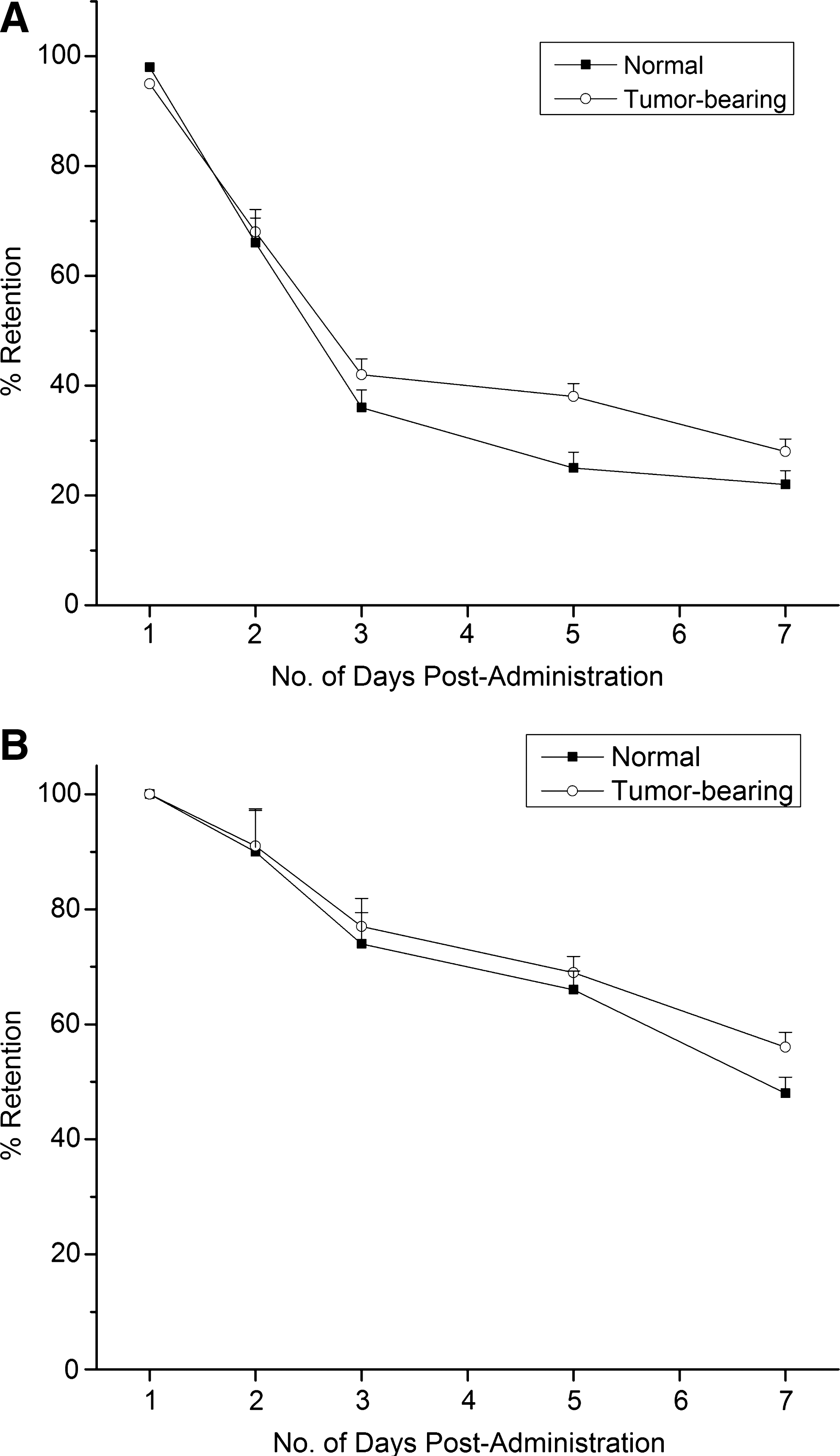
In vivo retention patterns of 177Lu-oxine-lipiodol in
Conclusions
177Lu-labeled lipiodol was prepared in good yield by dispersion of 177Lu-oxine complex in lipiodol medium. The radiolabeled preparation showed satisfactory in vitro stability in normal saline. In vitro cell uptake studies showed appreciable uptake in both normal and cancer origin liver cells. In vivo studies with Wistar rats showed good initial accumulation of the injected activity in liver. However, significant subsequent leakage of the preparation was observed from the liver region, with accumulation of 177Lu activity in the skeleton. Thus, 177Lu-oxine dispersed in lipiodol may not be suitable for the therapy of HCC owing to the poor retention of the radiotracer in the liver.
Footnotes
Acknowledgments
The authors are grateful to Dr. S.V. Thakare and Mr. K.C. Jagadeesan of Radiopharmaceuticals Division for their valuable help in carrying out irradiation of lutetium targets. The help rendered by the staff of the Animal House Facility of BARC is also gratefully acknowledged.
Disclosure Statement
The authors disclose that there are no financial or other conflicting interests in this work.