




Abstract
The imaging of the distribution of β-amyloid plaques in the brain is becoming an important diagnostic modality in Alzheimer's disease. The present study reports the synthesis of novel benzothiazole derivatives. The final products were characterized by spectral techniques such as FTIR, 1H NMR, and electrospray ionization–mass spectrometry. The structure–activity relationship of these benzothiazole derivatives is also reported. The K i values of these derivatives were evaluated by competitive binding assay studies. The analogs were labeled with 99mTc for the potential diagnostic imaging of Alzheimer's disease using stannous chloride as a reducing agent. The radiochemical stability was found to be ≥ 90% for both the compounds and they were stable for 10–12 hours in human serum. Biodistribution studies of the 99mTc complex in normal mice were performed after intravenous injection through the tail vein. The data showed high initial brain uptakes at 2 minutes (2.2% ± 0.1% ID/g), and brain activities washed out to 0.3% ± 0.02% ID/g at 6 hours. In conclusion, benzothiazole derivatives showed excellent binding affinities for β-amyloid aggregates and high initial brain uptakes in normal mice.
Introduction
Alzheimer's disease is a common neurodegenerative disorder, and the formation and accumulation of β-amyloid (Aβ) plaques in the brain are major factors in its pathogenesis. 1 Thus, in vivo assessment of Aβ plaques in the brain would be imperative for early diagnosis and monitoring of disease progression. 2,3
Several studies have been conducted to evaluate the structure–activity relationships of Aβ-aggregate–specific ligands, and lipophilic analogs of thioflavin-T have been reported to be potential imaging agents. 4 –8 In particular, benzothiazole analogs that contain five-membered sulfur-containing heterocyclic rings have shown high affinities for Aβ aggregates and high initial brain uptake in normal mice. 4,9,10 Benzoxazole and benzofuran derivatives, which contain heterocyclic oxygen rather than sulfur in a five-membered ring, have also shown similar properties. 6,7 Radiolabeled heterocyclic molecules give promising results and technetium-99m offers optimal characteristics as a radionuclide for nuclear imaging purposes. Our group has designed a molecule based on benzothiazoles and azomethine to generate a new series of radiopharmaceuticals, which can also be utilized as fluorescent sensors based on photon-induced electron transfer (PET) mechanism, which is exploration of photoelectronic transition of molecules.
Materials and Methods
All reagents and solvents were purchased from Aldrich. Column chromatography was carried out using silica (mesh size: 60–200 μm) and 1:9 MeOH:CHCl3 solvent system. TLC was done on aluminium sheets coated with silica gel 60 F254 (Merck). 1H and 13C NMR spectra were recorded on a Bruker Avance 400 MHz spectrometer. Electrospray ionization mass spectrometry was performed on Agilent 6310 system with ion trap detection in the positive and negative modes. Radiocomplexation and radiochemical purity of the synthesized compounds were checked by instant thin layer chromatography with ITLC-SG (Gelman Sciences).
QSAR analysis
The QSAR investigation was carried out by the linear free energy relationship model. In the present study, parameters for benzothiazole series (electronic, hydrophobic, and steric) were selected and considered as consistent. Geometries of all compounds were completely optimized by MM2 studies using Chem3D 6.0 software according to our previous work. 11,12
A classical Hansch multivariate regression analysis using the least-square method was chosen to derive QSAR equations for the dataset. The level of significance of each coefficient was judged by statistical procedures such as F tests and statistical analysis was carried out by employing the method of least squares with stepwise selection and elimination.
Synthesis of benzothiazoles
The condensed products were synthesized by mixing a methanolic solution (10 mL) of 2-aminobenzothiazole (0.01 mol) with equimolar concentrations of 2-nitrobenzaldehyde and imidazole-2-carboxyldehyde, respectively. The mixture was refluxed for 7–8 hours at 70°C. The precipitate obtained was collected by filtration using a Buckner funnel, recrystallized from ethanol, and dried at room temperature with 82%–84% yield.
Spectral analysis
Compound [1] (Schiff base of benzothiazole and imadazole-2-carboxyldhyde)
Compound [2] (Schiff base of benzothiazole and nitrobenzoaldehyde)
UV and luminescence measurements
UV–vis spectra of the benzothiazole derivatives were obtained on a Cary Varian double beam spectrophotometer (Cary BIO 100, at 550–600 nm). Temperature dependence was measured over a wide range of temperature typically between 273 and 343 K. All the measurements were done as per reported literature. 12 Both the ligands showed absorption maxima at 272 and 286 nm, respectively.
Luminescence measurements were carried out on FS920 (Edinburgh Analytical Instruments) spectrofluorimeter equipped with a Xenon arc lamp as the light source. The temperature of the sample holder was regulated with a Peltier-cooled thermostat. Luminescence lifetime measurements were calculated using a customized integrated steady-state spectrofluorimeter and fluorescence lifetime instrument (FL900CDT; Edinburgh Analytical Instruments). The excitation source used was a nanosecond flash lamp nF900 filled with low-pressure hydrogen gas (0.4 bar) operating at a frequency of 40 kHz. The slit width for both excitation and emission monochromators was kept fully open. The intensity decay curves were obtained at the emission maximum and fitted as sum of exponentials as follows:
where τi and Ai represent the fluorescence lifetime and preexponential factor for ith decay component, respectively. All measurements were carried out in a solution containing 5 mM of the respective benzothiazole derivative in 10 mM KMOPS buffer (pH 7.2) at 25°C. The luminescence decay was recorded in a short phosphorescence lifetime mode and repeated at least five times under each condition. The luminescence lifetime was calculated from the monoexponential fitting of the average decay data.
Radiochemical studies
Radiolabeling of compounds was done using our previous approach 13,14 with some modifications, by taking 50 μL of 0.02 nM solutions of the compounds dissolved in DMSO in a shielded vial. Further, 30 μL of 1 × 10⁻² M SnCl2 · 2H2O (dissolved in N2-purged 1 mL of 10% acetic acid) was added, followed by freshly eluted saline solution of sodium pertechnetate (NaTcO4, 74 MBq, 100 mL). The pH of the reaction mixture was adjusted to 6.5 using 0.1 M NaHCO3 and shaken thoroughly to mix the contents. The vial was incubated at room temperature for 20–30 minutes. Percentage of labeling of the compound, radiochemical purity, as well as R f of the 99mTc-based complex were determined by ITLC-SG strips using 0.9% NaCl aqueous solution (saline) as developing solvent and simultaneously in acetone and PAW (pyridine, acetic acid, and water in 3:5:1.5 ratio). Each ITLC was cut into 0.1-cm segments and counts of each segment were taken.
Fresh human serum was prepared by allowing blood collected from healthy volunteers to clot for 1 hour at 37°C in a humidified incubator maintained with 5% carbon dioxide and 95% air. Then the sample was centrifuged at 400 rpm and the serum was filtered through a 0.22-μm syringe filter into sterile plastic culture tubes. The above freshly prepared technetium radiocomplexes were incubated in fresh human serum at physiological conditions, that is, at 37°C, at a concentration of 100 nM/mL and then analyzed by ITLC-SG at different time intervals to detect any dissociation of the complex. Percentage of free pertechnetate at a particular time point was estimated using saline and acetone as mobile phase and was represented as percentage of dissociation of the complex at that particular time point in serum.
Blood clearance study was performed in albino New-Zealand rabbits weighing ∼2.5–3.0 kg after administration of 10 MBq of the 99mTc-labeled compounds through the ear vein. At different time intervals, about 0.5 mL blood samples were withdrawn from the dorsal vein of other ear and radioactivity was measured in the gamma counter. The data from the experiment were expressed as percentage of administered dose at each time interval (Fig. 1).
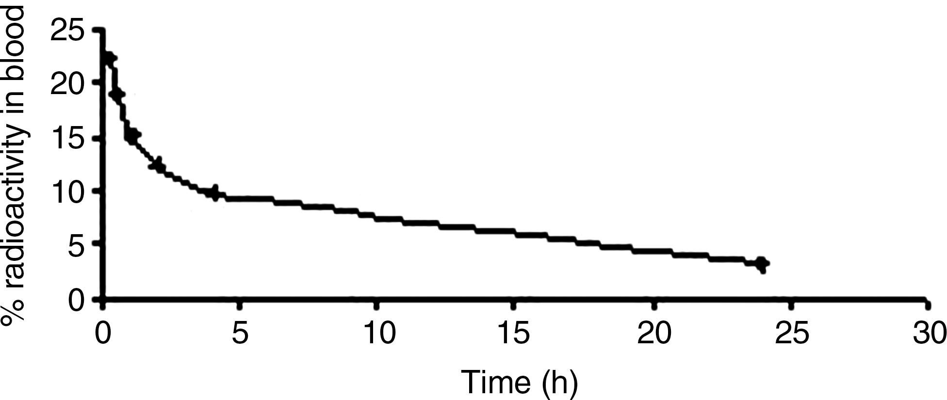
Blood kinetics of synthesized benzothiazole analog [1].
Balb/c mice (in triplicate) were used for the tissue distribution studies. Animal handling and experimentation were carried out as per the guidelines of the Institutional Animal Ethics Committee. An equal dose of 10 μCi of labeled test compound was injected in mice through the tail vein of each animal. At different time intervals, mice were sacrificed, blood was collected, and different tissues and organs were dissected and analyzed. The radioactivity was measured in a gamma counter. The actual amount of radioactivity administered to each animal was calculated by subtracting the activity left in the tail from the activity injected. Radioactivity accumulated in each organ was expressed as percentage of administered dose per gram of tissue weight. Total volume of the blood was calculated as 7% of the body weight.
In vitro binding assays
Binding studies were performed as described previously, with some modifications. 15 The K d values of compounds were evaluated using 20 nM Aβ aggregates. The binding affinities of benzothiazole derivatives to Aβ aggregates were evaluated by competitive binding assays as mentioned in literature. 5 One hundred microliters of Aβ aggregates (20 nM in the final mixture), 100 μL of benzothiazole derivatives (10−6 to 10−12 M in 50% ethanol containing 1 mM EDTA), 100 μL of 125I in 50% ethanol (0.1 nM in the final mixture), and 700 μL of phosphate-buffered saline (pH 7.2) were added to separate test tubes and incubated for 3 hours at room temperature for binding assay. Nonspecific binding was determined by incubation in the presence of 10 μM thioflavin-T. The reaction mixture was filtered through Whatman glass filters and washed twice with 3 mL of 10% ethanol aliquots. Filters were counted with a NaI well counter. The K i values of benzothiazole derivatives were then calculated from the results as shown in Figure 2.

Log P and binding affinity of fluorescent benzothiazoles for amyloid imaging.
Results
Benzothiazole derivatives are derived from condensation reaction of active hydrogen of amine and aldehyde groups. The progress of the reaction was monitored by TLC, which showed a single spot with an R f value different from the starting materials. The 1H NMR spectral data are reported along with the possible assignments in the experimental section. All the protons were found to be in their expected region and the proposed stoichiometry and structure were confirmed by comparing with the 1H NMR spectra of starting materials. Mass spectrometry spectra shows the presence of their molecular ions.
Preliminary complexation of novel synthesized compounds with 99mTc was found to give sufficiently stable complexes under physiological conditions. The in vitro serum stability of the radio complexes is a necessary parameter to measure the effectiveness of chelating moiety to coordinate the radiometal. In vitro serum stability of the complexes clearly indicates the good stability of the complex.
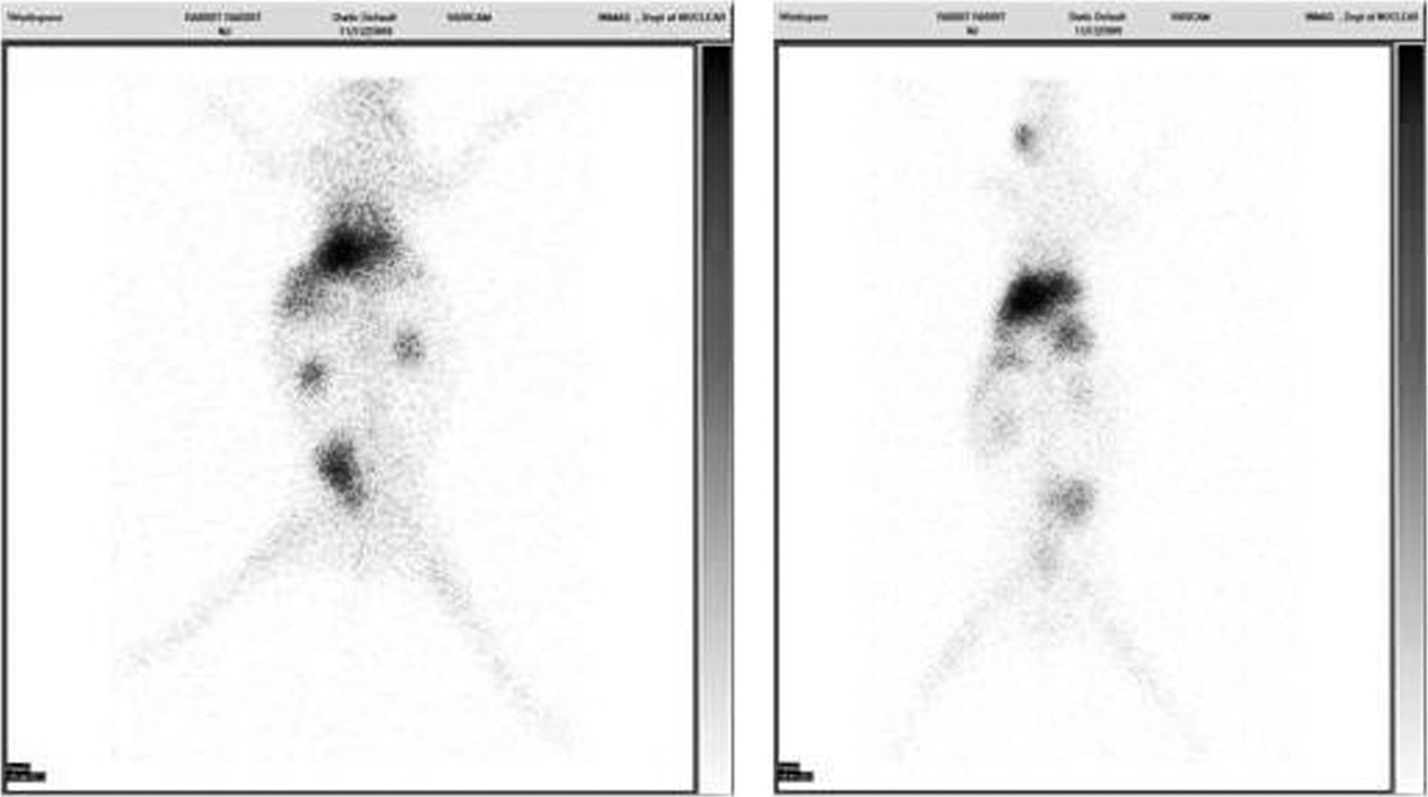
The octanol/buffer partition coefficients of 99mTc-labeled benzothiazole were found to be 150.82 (log P = 2.10) and 108.52 (log P = 2.08), respectively. These values are within the optimal range for passive diffusion over the BBB, which was also confirmed by the scintigraphic images (Figs. 3 and 4).
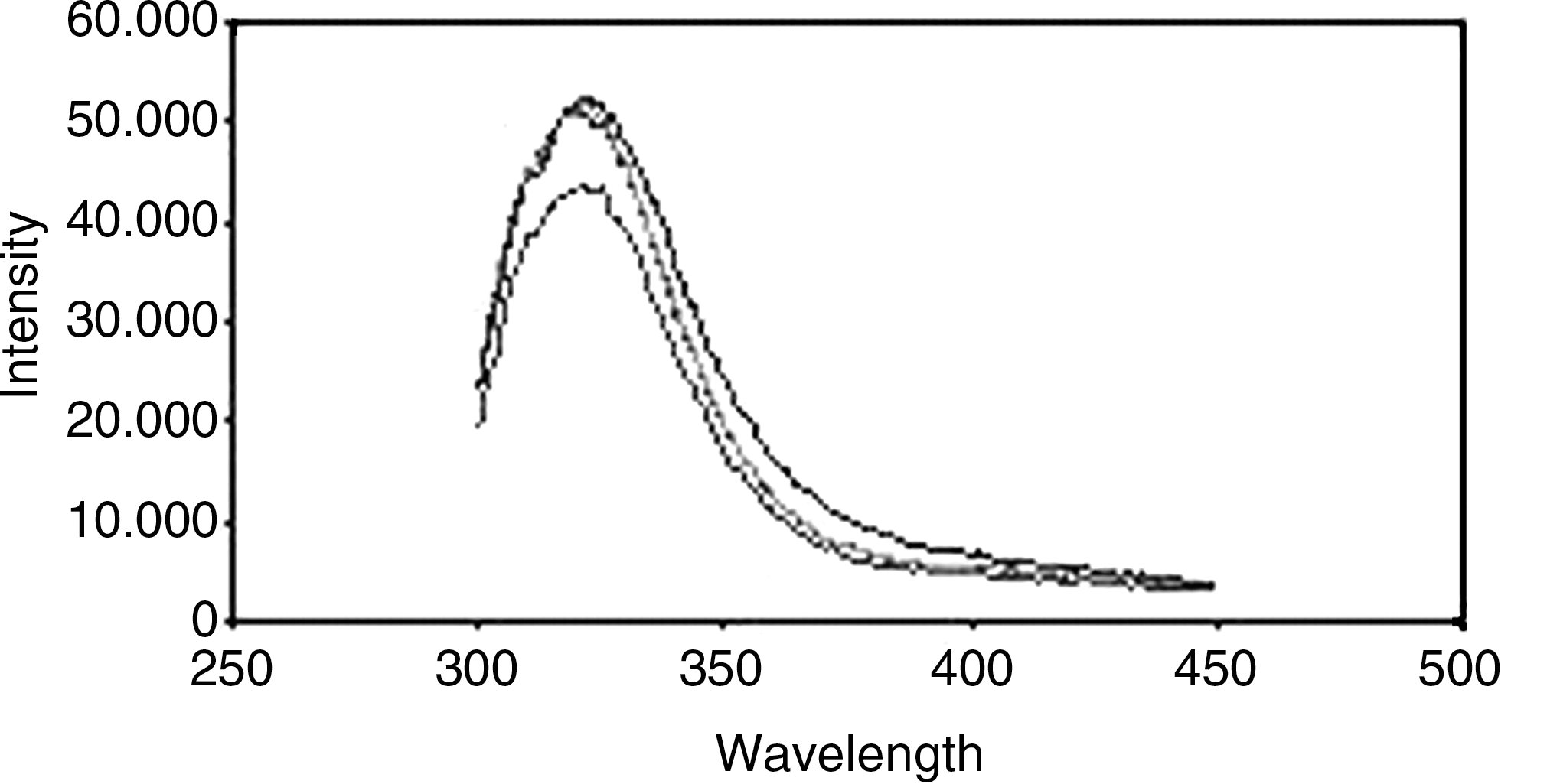
Emission spectra of benzothiazole analog [1].
Discussion
Table 1 shows the results of the biodistribution studies of novel analogs carried out in balb/c mice. The first requirement for a suitable tracer agent for detection of amyloid plaques in brain is the ability to cross the BBB. As both compounds show appropriate brain uptake, this important prerequisite is fulfilled. Excretion of the compounds proceeds mainly through the hepatobiliary pathway and, to a lesser extent, through the renal pathway, which is rather logical in view of the lipophilic nature of the compounds as indicated by the partition coefficient values as well. The relatively high uptake in the stomach suggests a partial reoxidation of the complexed technetium into pertechnetate in vivo.
n = 4 at each time point.
SD, standard deviation.
High-resolution UV–vis spectra were recorded using aqueous solution of the compounds. They show two bands whose intensity varied with temperature. The temperature dependence can be related to the existence of hydration equilibrium between the two species. Hydration numbers were determined by laser-induced luminescence, based on the difference in luminescence lifetimes measured in H2O and D2O solutions. The hydration numbers were calculated according to the revised equation of Beeby et al.:
where A′ is 1.15 ms and the correction factor for the contribution of the second and outer sphere water molecules is − 0.25 ms−1. The luminescence decay under all conditions strictly followed single-exponential decay as analyzed by the least square analysis. The luminescence lifetime and inner sphere water molecule measurements are given in Table 2. The noninteger numbers of q often imply the coexistence of differently hydrated states.
Absorption spectral measurements on HSA in the presence of a complex provide useful information related to the nature of interaction between a ligand and HSA to define the in vivo nature of molecule. In the present study, HSA solution (10 μM) was titrated against the compounds in 0.005 mM phosphate buffer at pH 7.2. The emission maxima (( max) was observed at 348 and 389 nm. For carrying out further studies, HSA was dissolved in H2O (2 mg/mL) containing phosphate buffer 10 and 2.5 mM of sodium hydroxide solution. The protein concentration was determined spectrophotometrically using extinction coefficient of 36,500 M−1 cm−1 at 280 nm.
The fluorescent spectrum of the compound recorded in aqueous solution displayed an emission band centered at 348 and 389 nm. The emission was observed to be strongly dependent on pH, “switching on” as the pH decreased, with no noticeable change in wavelength. This is typical of PET sensors, where basic functionalities form part of the receptor. A plot of fluorescent intensity against wavelength for both of the compounds is shown in Figures 4 and 5. The fluorescent enhancement (intensity in the range of 104) of these compounds was found as a result of an increase in the oxidation potential of the group upon protonation, removing the thermodynamic driving force for PET and switching the fluorescence “on” using a plot of − log(F MAX − F)/(F − F MIN) against pH (where F MAX is the maximum fluorescence intensity, F MIN the minimum fluorescence intensity, and F the measured fluorescence intensity).
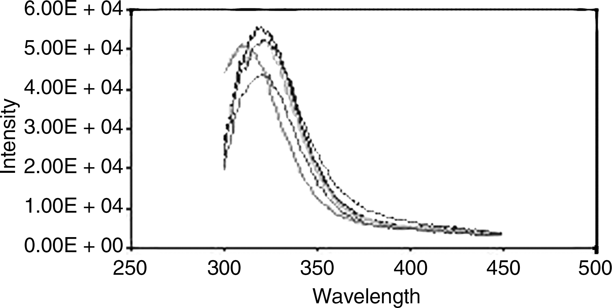
Emission spectra of synthesized benzothiazole analog [2].
Conclusions
In summary, the present study reports the facile synthesis of benzothiazole derivatives, which fulfill the basic requirements of luminescent probes that can be used as optical agents, namely good hydrophilicity, high kinetic inertness in aqueous solutions, long luminescence lifetime, and an efficient cation emission. The 99mTc-labeled benzothiazoles showed high brain uptake, indicating their potential application in imaging Aβ plaques in the brain. The therapeutic efficacy of these complexes can be further extended by applying them in different animal models and cell lines. Preliminary studies with these novel ligands are encouraging to carry out further in vivo experiments for targeted imaging of human subjects.
Footnotes
Acknowledgments
The authors thank Dr. R.P. Tripathi, Director of INMAS, for providing necessary facilities. This work was supported by Defence Research and Development Organization, Ministry of Defence, under R&D project INM-306.
Disclosure Statement
No competing financial interests exist.