
Abstract
The development of antisense oligonucleotides suitable for tumor targeting applications is hindered by low stability and bioavailability of oligonucleotides in vivo and by the absence of efficient and safe vectors for oligonucleotide delivery. Stabilization in vivo has been achieved through chemical modification of oligonucleotides by various means, but effective approaches to enhance their intracellular delivery are lacking. This study reports on the characterization in vitro of a fully phosphorothioated 20-mer oligonucleotide, complementary to p21 mRNA, radiolabeled with fluorine-18 using a thiol reactive prosthetic group. The potential of two novel synthetic block copolymers containing grafted polyamines on their hydrophobic blocks for vector-assisted cell delivery was studied in vitro. Extensive cellular uptake studies were performed in human colon carcinoma cell lines with enhanced or deficient p21 expression to evaluate and compare the uptake mechanism of naked and vectorized radiolabeled formulations. Uptake studies with the two novel biodegradable vectors showed a moderate increase in cell uptake of the radiofluorinated antisense oligonucleotide. The two vectors show, however, promising advantages over conventional lipidic vectors regarding their biocompatibility and subcellular distribution.
Introduction
Antisense oligonucleotides (AsODNs) are being intensively studied for possible applications in cancer therapy and as diagnostic tools to be used for the personalization of treatment approaches in individual cancer patients. AsODNs developed for antisense targeting are generally 18–20 bases in length and undergo Watson–Crick hybridization to targeted mRNA. The specificity for the target mRNA and ease of preparation make AsODNs ideal agents for molecular targeting purposes even though major challenges still hinder their effective use in vivo. 1
In the present study, the authors are interested in developing a probe for antisense targeting of the cyclin-dependent kinase inhibitor p21, which modulates apoptosis, regulates transcription of genes important in cell cycle progression and senescence, affects DNA repair processes, and potentially also functions as an oncogene. The status of p21 expression shows a potential clinical relevance in chemotherapy of various tumors, including hematologic malignancies, 2 prostate cancer, 3 breast cancer, 4 and nonsmall cell lung cancer. 5 This provides the rational for targeted gene therapies that focus on p21. Downregulation of p21 also shows potential in improving radiocurability. 6 A noninvasive method to monitor the p21 status in vivo could be predictive of the outcome of cancer therapy. Successful antisense imaging of p21 at the mRNA level has been already reported using an 111In-labeled anti-p21 ODN in animals bearing human breast cancer cells that were induced to overexpress p21 by intratumoral injection of epidermal growth factor. 7 The aim of the present study was to develop a fluorine-18–labeled AsODN probe against p21 mRNA for positron emission tomography to permit a more sensitive and quantitative detection. 8
Natural phosphodiester ODNs are known to exhibit low bioavailability 9 and low stability in vivo. 10 Because of the negative charge, these polyanions cannot cross the cell membrane effectively in vitro. 11 Moreover, ODN can bind to undesired targets, including proteins, such as cell membrane proteins or human serum albumin, and nontarget mRNA. To avoid degradation due to nuclease activity, AsODNs with a phosphorothioate backbone (PS-AsODNs) have been developed and have already shown suitability for therapeutic applications. 12 Still, effective delivery systems are required to overcome physical barriers and to enhance the intracellular delivery while decreasing nonspecific interactions. Synthetic vectors, such as cationic lipids and polyamines, are frequently used for this purpose.
In the present study, a 20-mer PS-AsODN, complementary to p21 mRNA, with a phosphorothioate monoester at the 5′ end conjugated to the radiofluorinated thiol-reactive prosthetic group 2-bromo-N-[3-(2-[18F]-fluoropyridin-3-yloxy)propyl]acetamide ([18F]FPyBrA) has been described. The major aim of this study was to evaluate the vector-assisted delivery of this radiolabeled probe using different vectors. Two novel biodegradable synthetic vectors, consisting of micelle-assembling block copolymers, were compared with a commercially available liposome formulation (Lipofectin). The novel block copolymers are based on poly(ethylene oxide)-block-poly(ɛ-caprolactone) (PEO-b-PCL) and are grafted with polyamines on their hydrophobic blocks. Cellular uptake studies with naked and vectorized 18F-labeled AsODN on human colon carcinoma cell lines with induced or deficient p21 expression
13
were performed to evaluate the cell uptake mechanism of the naked and vectorized radiolabeled formulations. These studies also evaluated the effects of excess levels of AsODN or random ODN (RdODN), incubation at 4°C, and coincubation with poly-(
Materials and Methods
Chemicals with high analytic purity and materials for cell culture with certified analysis were obtained from various commercial suppliers. A 20-mer DNA with a fully phosphorothioated backbone and a phosphorothioate monoester at the 5′ end complementary to p21 mRNA with the sequence 5′-TGT-CAT-GCT-GGT-CTG-CCG-CC-3′ (PS-AsODN) and the random sequence 5′-CCG-GTG-AAC-GAG-CGA-GCA-CA-3′ (PS-RdODN), 6 with a purity of >95%, were purchased from the University Core DNA Services. [18F]-Fluoride was produced on a TR-19/9 cyclotron (Advanced Cyclotron Systems) by the 18O(p,n)18F nuclear reaction.
The two recently described biodegradable polyamine-grafted PEO-b-PCL–based copolymers, grafted with spermine (SP) and tetraethylenepentamine (TP), with molecular weights of 8300 and 8500 g/mol, respectively, were prepared as described previously 14 and are presented in Scheme 1. A commercially available liposome formulation based on a 1:1 mixture of the cationic lipid N-[1-(2,3-dioleoyloxy)propyl]-N,N,N-trimethylammonium chloride and the fusogenic lipid dioleoyl phosphatidylethanolamine (Lipofectin reagent) was purchased from Invitrogen Corporation.

Biodegradable polyamine-grafted PEO-b-PCL–based copolymers, grafted with spermine and tetraethylenepentamine used for vectorization. PEO-b-PCL, poly(ethylene oxide)-block-poly(ɛ-caprolactone); SP, spermine copolymer; TP, tetraethylenepentamine copolymer.
Radiofluorination of PS-AsODN
For the preparation of the 18F-labeled prosthetic group, [18F]FPyBrA, the N-Boc–protected nitroprecursor, [3-(2-nitropyridin-3-yloxy)propyl] carbamic acid tert-butyl ester was synthesized according to Kuhnast et al. 15 The synthesis was adapted for the fully automated preparation in a commercially available modular synthesis system (Modular Lab, Eckert & Ziegler). 16 The preparation of [18F]FPyBrA including online normal-phase high-performance liquid chromatography (HPLC) purification was performed within 65 minutes, achieving 10%–20% decay-corrected yield with 18F starting activities of 3–5 GBq.
Conjugation of [18F]FPyBrA (100–300 MBq) with the PS-AsODN (200–500 μg) was carried out in a 250 μL mixture of 0.1 M phosphate-buffered saline (PBS) pH 7 and methanol (1/4, v/v) in a sealed V-vial at 120°C for 30 minutes. The reaction mixture was purified by gel filtration on an NAP-10 column (Sephadex™ G-25 DNA grade; GE Healthcare). The radiochemical purity of the 18F-labeled PS-AsODN was analyzed by reverse-phase HPLC on a Beckman Coulter chromatography system with a Model 126 analytical dual pump, Model 168 Diode Array variable UV detector, and ACE Mate™ Single Channel Analyzer radiometric detection, equipped with a Phenomenex Luna C18 column (Semi/Prep; 250 × 10.0 mm, porosity 10 μm), using a gradient system of 0.1 M triethylammonium acetate (pH 7) and acetonitrile (ACN) with a flow rate of 3 mL/minute and increasing levels of ACN: 0–20 minutes, 10%–50%; 20–21 minutes, 50%–70%; 21–25 minutes, 70%; 25–26 minutes, 70%–10%; 26–30 minutes, 10%. The PS-AsODN concentration was determined from the UV trace at 254 nm using standard curves and confirmed by UV spectroscopy at 260 nm using a Beckman DU 7400 spectrophotometer.
Preparation and characterization of vectorized radiolabeled PS-AsODN
Assessment of AsODN binding capacity and stability
The nonradiolabeled PS-AsODN (2 μg) was incubated with SP and TP (1–64 μg) at different vector-to-ODN weight ratios (0:1, 0.5:1, 1:1, 2:1, 4:1, 8:1, 16:1, 32:1) in a total volume of 40 μL, at 37°C for 30 minutes. Complexation of PS-AsODN was analyzed by agarose gel electrophoresis (2% agarose gel containing 0.05 mg/mL ethidium bromide) at 130 mV for 15 minutes, after addition of 10 μL of a solution containing 50% glycerol, 1% bromophenol blue, and 1% xylene cyanol FF in TBE buffer. The resulting gels were photographed under UV illumination. The digitized pictures were analyzed with Scion image analysis software to determine the density of ODN bands. The binding percentage was calculated based on the intensity of free PS-AsODN in the presence of vector with respect to the intensity of free PS-AsODN in the absence of vector. The stability of the different complexes prepared at optimal vector-to-ODN ratios was determined by challenge with the competing polyanion heparin in increasing heparin-to-ODN weight ratios (0:1, 0.025:1, 0.05:1, 0.1:1, 0.25:1, 0.5:1, 1:1, 5:1) at 37°C for 1 hour by gel electrophoresis as described earlier. The stability of naked and vectorized PS-AsODN in PBS containing 1% or 25% fetal bovine serum (FBS; v/v) after 4-hour incubation was investigated by comparing the density of the ODN bands with incubation in PBS alone. ODN binding ability, heparin challenge, and stability in PBS/FBS were also studied for Lipofectin as the reference complexing agent.
Preparation of radiolabeled vectorized PS-AsODN formulations
For the reference vectorized product, the Lipofectin reagent was diluted with Optimem medium to a concentration of 100 μg/500 μL and preincubated at room temperature (RT) for 45 minutes. Following addition of 500 μL of a 3 μM solution of [18F]PS-AsODN, the solution was incubated for another 15 minutes at RT. For vectorized formulations with SP and TP, 500 μL of a 3 μM solution of [18F]PS-AsODN was incubated with either 16 μL of a 5 μg/μL TP solution in water or 32 μL of the corresponding SP solution at 37°C for 30 minutes.
Evaluation of cellular uptake
Cell uptake studies were performed on two different human colon carcinoma cell lines: (1) HCT116 cells, with transcriptional transactivation of p21 through activation of the tumor suppressor TP53 protein and its translocation to the nucleus (induced by ionizing radiation); and (2) 80S4 cells, a derivative cell line of HCT116 cells, in which both p21 alleles have been deleted through homologous recombination.
13
Cells were cultured in DMEM/F12 nutrient medium supplemented with
For cell uptake experiments, cells were washed twice with ice-cold DMEM/F12 medium supplemented with 1% FBS (v/v). Cells were then supplied with fresh medium and incubated with naked and vectorized radiolabeled [18F]PS-AsODN formulations, which were diluted to reach a 50 nM ODN concentration in the assay and incubated at 37°C for 2 and 4 hours in triplicates. For naked [18F]PS-AsODN, cellular uptake at a lower concentration (5 nM) and blocking of the cellular uptake by addition of excess PS-AsODN and PS-RdODN (1 μM) were studied. Cell uptake was interrupted by the removal of the supernatant and two rapid rinses with ice-cold PBS. The cells were recovered from the plates by adding 1 N NaOH. All collected fractions were counted in a Wallac 1840 Wizard 3 automatic gamma counter (Perkin Elmer Life Sciences) and the cell uptake was calculated. The cell-associated radioactivity was expressed in relation to the total activity added (% of total activity). For statistical analysis, the independent t-test (significance level of 0.05) was used.
Inhibition of cellular uptake was evaluated by uptake studies at 4°C, a nonpermissive temperature for endocytosis. The role of cell surface proteoglycans in the cellular uptake was explored by coincubation with either 1 μM heparin sulfate, a free glycosaminoglycan known to inhibit the interaction with proteoglycans, or 300 nM poly-(
Two different cell membrane treatments were used to desorb the fraction of naked or vectorized [18F]PS-AsODN bound to membrane proteins. For this purpose, after removal of the supernatant and two rinses with PBS, the cells were additionally treated twice with either 0.04 N sodium acetate adjusted to pH 4.5 or PBS containing 10% FBS (v/v) at RT for 5 minutes before lysing the cells with 1 N NaOH. The relative retained cell uptake was calculated comparing the retained cell-associated radioactivity of treated versus untreated cells.
The cytotoxicity of the PS-ODNs and of the different vectors against HCT116 and 80S4 cells was evaluated using 3-(4,5-dimethylthiazol-2-yl)-2,5-diphenyltetrazolium bromide (MTT) assay. HCT116 and 80S4 cells in DMEM/F12 medium supplemented with 10% FBS (v/v) were seeded in 96-well plates at a density of 4,000 cells per well. After 24-hour incubation, the medium was aspirated and replaced by 200 μL fresh medium supplemented with 5% FBS (v/v) containing either PS-RdODN, PS-AsODN, Lipofectin, SP, or TP. Each component was added separately in triplicate at different concentrations (25, 5, 2.5, 0.5, 0.25, 0.025 μg/mL). After 48-hour incubation, 20 μL MTT in PBS was added to each well and the plate was incubated for 3 hours at 37°C. The medium was removed, DMSO (200 μL) was added, and the optical absorbance was measured at 570 nm using a FLUOstar OPTIMA microplate reader (BMG Labtech) to assess cell viability. The percentage of cell growth in relation to the control containing cell culture medium without any of the components was calculated.
Results
Radiofluorination of PS-AsODN
Radiolabeling of the PS-AsODN at the phosphorothioate monoester at the 5′ end was carried out through alkylation with [18F]FPyBrA, achieving a radiolabeling yield of 51.4%–74.4% as assessed by radio-HPLC. The 18F-labeled PS-AsODN was separated from unreacted [18F]FPyBrA by NAP purification, achieving a radiochemical purity of >99%. Quantification of the PS-AsODN amount by HPLC and UV spectroscopy resulted in a specific activity of 0.77–4.44 GBq/μmol (21–120 Ci/mmol) of the final product. The radiochromatogram and UV trace at 254 nm after purification are shown in Figure 1.
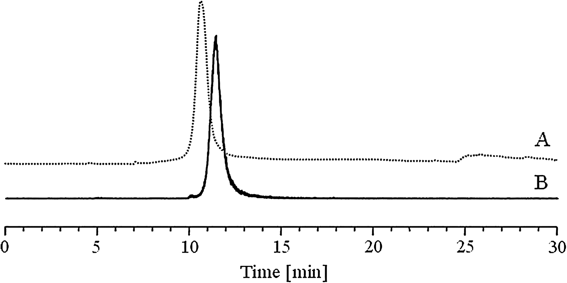
HPLC profile of [18F]PS-AsODN after NAP purification.
Preparation and characterization of vectorized [18F]PS-AsODN
The ability of SP and TP for micelle formation was assessed by gel electrophoresis using nonradiolabeled PS-AsODN. Disappearance of free ODN bands in the agarose gel was used as an indicator for complete complexation (Fig. 2). A vector-to-ODN ratio of 8:1 was sufficient to obtain >95% complexation of Ps-AsODN with TP, whereas for SP a ratio of 16:1 was needed. Also for Lipofectin complexes, quantitative PS-AsODN complexation was confirmed.

Binding capacity of nonradiolabeled PS-AsODN by gel electrophoresis with increasing vector-to-ODN weight ratios: 0:1 (1), 0.5:1 (2), 1:1 (3), 2:1 (4), 4:1 (5), 8:1 (6), 16:1 (7), and 32:1 (8).
The analysis of the PS-AsODN release from the different nonradiolabeled vectorized formulations by incubation with competing polyanionic heparin revealed complete release of the PS-AsODN at a heparin-to-polymer ratio of 0.5:1 for SP and TP, whereas for Lipofectin an incomplete release was observed even at a heparin-to-polymer ratio of 5:1. Gel retardation analysis on complexes incubated in PBS containing 1% and 25% FBS for 4 hours revealed a high stability of Lipofectin complexes with values >99%. For naked PS-AsODN, a densitometric intensity of >99% in PBS/1% FBS in comparison with PBS alone was found, whereas in PBS/25% FBS a lower intensity of 84.7% was found. For complexes with SP and TP, a similar stability was found at different PBS/FBS concentrations, with values ranging between 84.8% and 94.2%. Results are summarized in Figure 3.
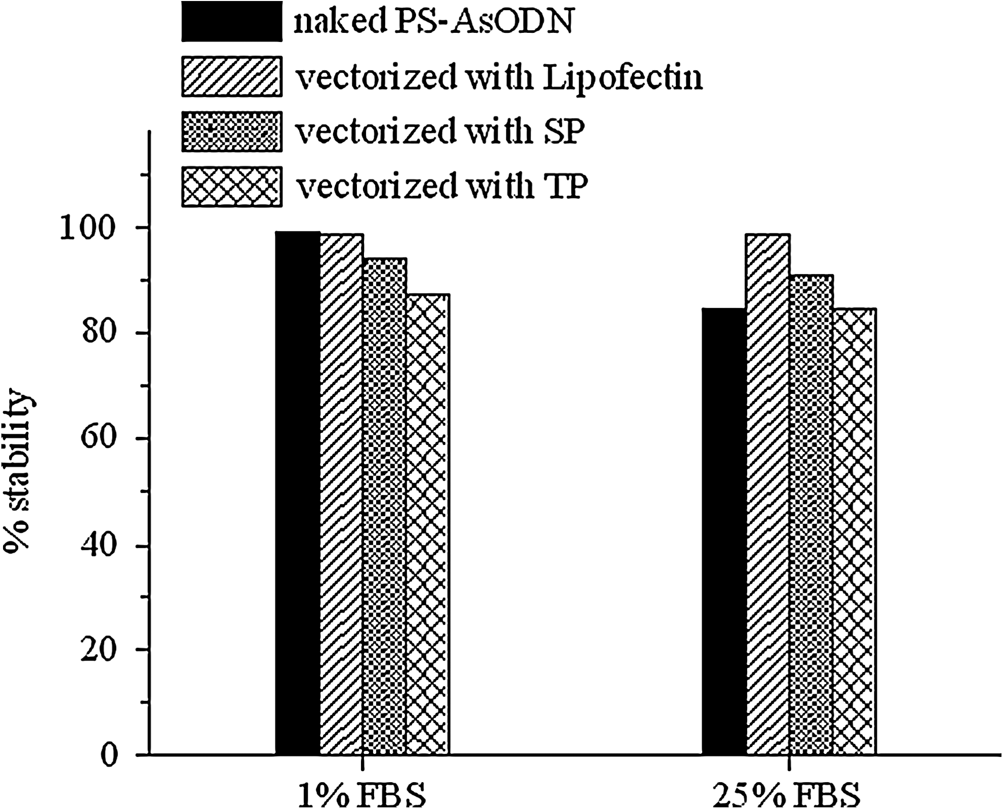
Stability based on gel electrophoresis of naked nonradiolabeled PS-AsODN and the different vectorized formulations after 4-hour incubation in PBS containing 1% and 25% FBS. SP, spermine; TP, tetraethylenepentamine; FBS, fetal bovine serum; PBS, phosphate-buffered saline.
Evaluation of cellular uptake
The immunofluorescence analysis at 24 hours after gamma irradiation confirmed induction of p21 expression in HCT116 cells, but not in 80S4 cells. Representative immunofluorescence images captured using a DAPI-FITC fluorescence filter combination show Alexa Fluor 488 detection in correspondence to DAPI-stained nuclei for HCT116 cells (Fig. 4). Initial cell uptake studies with different concentrations of naked [18F]PS-AsODN in HCT116 cells showed similar uptake at a concentration of 5 nM (1.50% ± 0.10%; n = 3) and 50 nM (1.42% ± 0.01%; n = 3). Uptake of [18F]PS-AsODN by HCT116 cells in the presence of excess levels of nonradiolabeled PS-RdODN and PS-AsODN was reduced by 83% and 84%, respectively. A similar uptake was found for 80S4 cells with 1.28% ± 0.14% (n = 3) at 50 nM, which was reduced by 84% and 89% in the presence of excess nonradiolabeled PS-RdODN and PS-AsODN, respectively (Fig. 5). The prosthetic group [18F]FPyBrA alone showed a very low cell uptake of less than 0.5% (data not shown). The uptake of naked [18F]PS-AsODN increased to some extent when the incubation time was raised from 2 to 4 hours in both cell lines, but still remained at values below 2%.
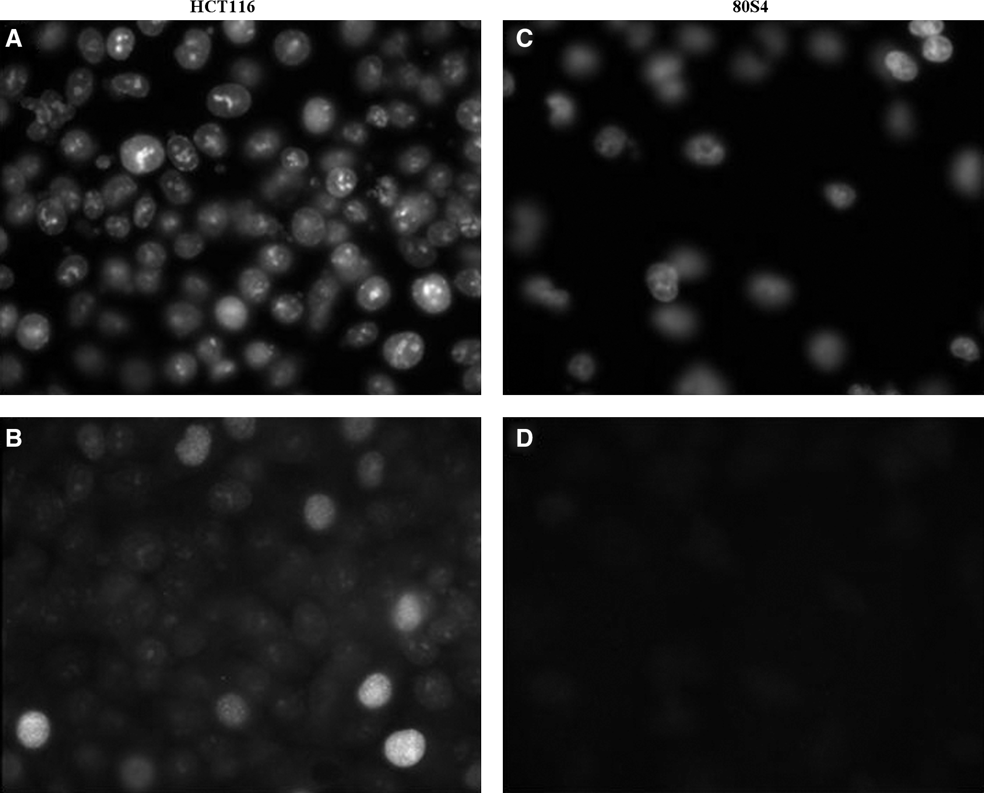
Immunofluorescence analysis showing enhanced p21 expression in HCT116 cells (
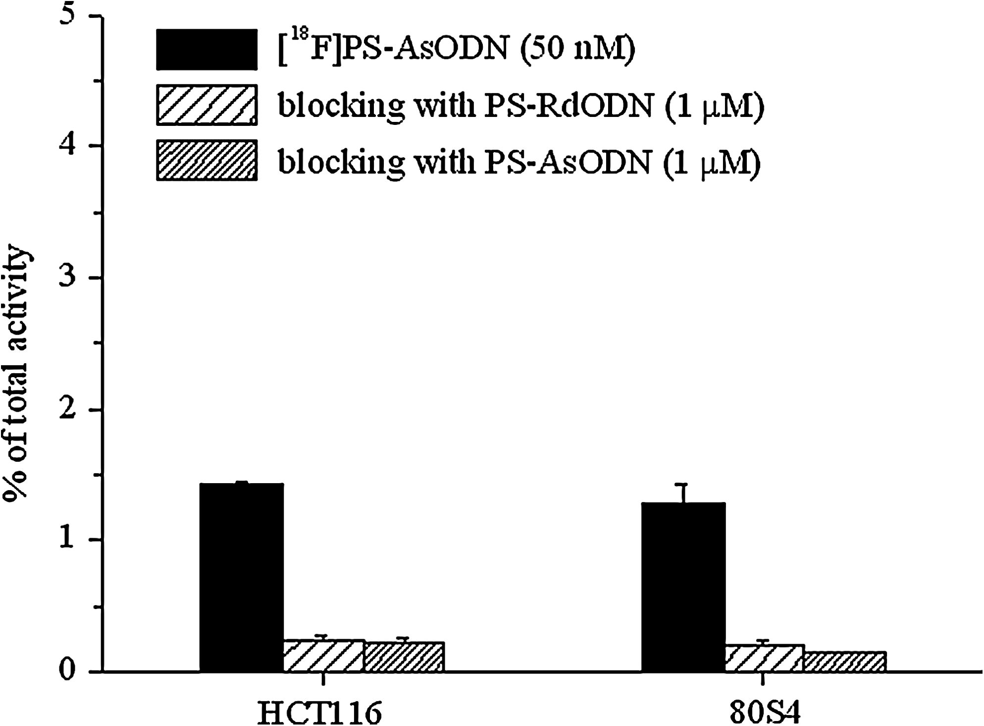
Cell uptake of naked [18F]PS-AsODN by HCT116 and 80S4 cells and blocking of uptake with nonradiolabeled PS-RdODN and PS-AsODN (mean ± standard deviation, n = 3).
Among the different vectorized [18F]PS-AsODN formulations, the highest uptake was found for Lipofectin complexes, showing 15.8% ± 0.51% and 15.7% ± 0.20% uptake in HCT116 and 80S4 cells after 2-hour incubation, respectively. The cell uptake increased significantly after 4-hour incubation, reaching 28.3% ± 0.50% and 29.7% ± 0.70% in HCT116 and 80S4 cells. A much lower uptake was observed for complexes with SP and TP. Vectorization with SP resulted in 5.29% ± 0.32% and 4.26% ± 0.10% for HCT116 and 80S4 cells, respectively, after 2-hour incubation. Under the same condition, vectorization with TP provided 5.70% ± 0.22% and 3.94% ± 0.19% uptake in the two cell lines, respectively. The cellular uptake at 4 hours after incubation did not increase significantly in any of the cell lines under study (Fig. 6). For vectorized formulations with SP and TP, a statistically significant higher uptake in HCT116 versus 80S4 cells was found at 2 and 4 hours after incubation.
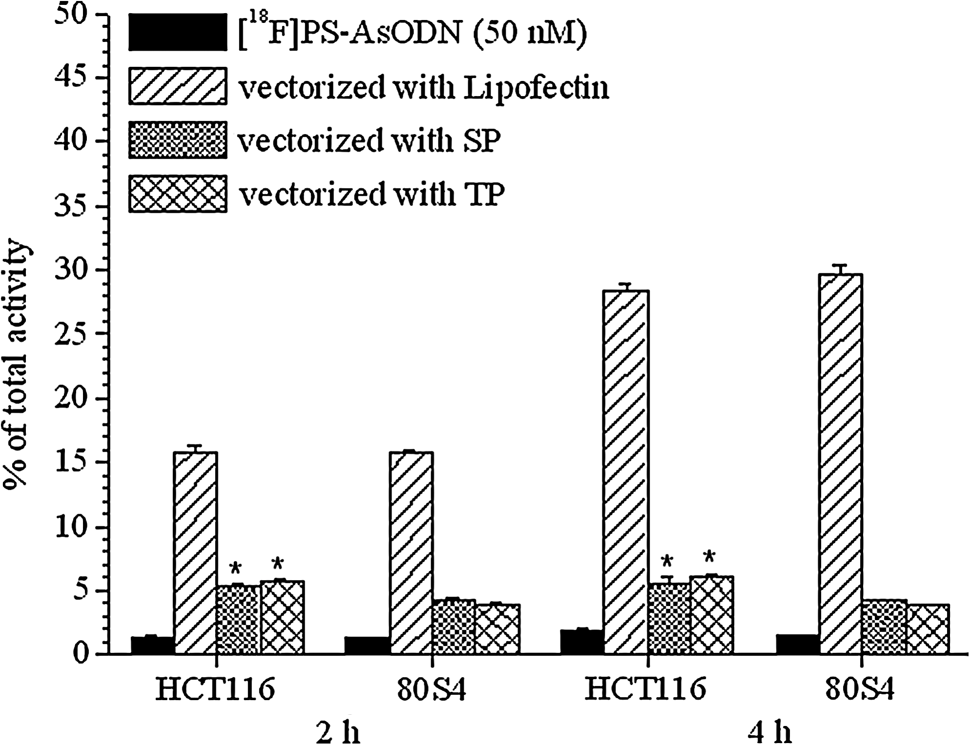
Cell uptake of naked and vectorized [18F]PS-AsODN by HCT116 and 80S4 cells at 2 and 4 hours after incubation (mean ± standard deviation, n = 3; *significantly increased versus 80S4 cells, p < 0.05).
Uptake studies performed at 4°C indicated a temperature-dependant uptake of naked [18F]PS-AsODN. At 4°C, a relative cell-associated radioactivity of only 52.9%–53.5% when compared with incubation at 37°C was found (Table 1). Temperature had a minor effect on the uptake of vectorized [18F]PS-AsODN, where a relative cell-associated radioactivity of 77.3%–90.7% was observed. Heparin sulfate caused a dramatic decrease in uptake for the three vectorized formulations in both cell lines (Table 1). This effect was more pronounced for vectorization with SP and TP in comparison with Lipofectin complexes. A relative cell-associated radioactivity of 21.7%–24.9% was observed for coincubation with heparin sulfate for vectorized formulations with SP and TP versus 37.9%–44.4% found for Lipofectin. Coincubation with poly-(
Data are expressed as percentage of uptake in relation to the cell-associated radioactivity at 37°C without coincubation with heparin sulfate (mean ± SD, n = 3). Absolute values used for reference are presented in Figure 6.
SP, spermine; TP, tetraethylenepentamine.
Cell membrane treatments with either 0.04 N sodium acetate (pH 4.5) or PBS/10% FBS resulted in a reduction of the cell-associated radioactivity in all cases (Table 2). There were slight differences between the cell lines, and in general, the treatment with PBS/10% FBS was more effective in reducing the cell-associated radioactivity than the treatment with sodium acetate. The reduction of the cell-associated radioactivity was higher for naked [18F]PS-AsODN and vectorized formulations with SP and TP, resulting in a relative retained cell uptake of 66.2%–84.9% with sodium acetate and of 31.6%–47.4% with PBS/10% FBS. In contrast, for Lipofectin complexes the relative retained cell uptake was always >80%.
Data are expressed as percentage of radioactivity relative to the cell-associated radioactivity without membrane treatment (mean ± SD, n = 3). Absolute values used for reference are presented in Figure 6.
SP, spermine; TP, tetraethylenepentamine; FBS, fetal bovine serum; PBS, phosphate-buffered saline.
No in vitro cytotoxicity was noted for PS-RdODN, PS-AsODN, and the various vectorization agents at similar concentrations to that used in cellular uptake studies.
Discussion
A number of prosthetic groups have been described for the radiolabeling of ODN with 18F. 15,18 –25 To date, alkylating agents binding to a phosphorothioate moiety have shown the most promising results. 26,27 In the present study, these literature precedents guided the choice of the alkylating agent [18F]FPyBrA for the radiolabeling of PS-AsODN complementary to p21 mRNA. Recently, the fully automated preparation of this prosthetic group in a commercially available modular synthesis unit has been described. 16 Radiolabeling of the PS-AsODN proceeded with 50%–75% yield as indicated by radio-HPLC of the crude product. The radiolabeling mixture was purified by size-exclusion chromatography, resulting in a pure product approaching 5 GBq/μmol. The slightly longer retention time of [18F]PS-AsODN compared with the unlabeled PS-AsODN precursor, as shown in Figure 1, suggests that a material of much higher effective specific activity could be isolated, but for the present cell-based study the specific activity was sufficiently high.
The preparation of vectorized [18F]PS-AsODN formulations under optimized conditions resulted in stable ODN binding of >90% as confirmed by gel electrophoresis. No cytotoxic effects were observed with the polymer amounts used for cellular uptake studies. All formulations were stable in PBS containing 1% FBS up to 4 hours and this FBS concentration was used for cell studies. Heparin challenge indicated that polyamine-grafted PEO-b-PCL micelles were more prone to dissociation in comparison with Lipofectin complexes.
The cell uptake of naked ODN, particularly PS-ODN, is known to take place by adsorption to cell-surface proteins, followed by receptor-mediated endocytosis, although it is recognized that other PS-ODN binding proteins are also present on the cell surface. 28 As expected, uptake of [18F]PS-AsODN in the absence of vectorization occurred to a very limited extent (<2%). This limited uptake was efficiently blocked by coincubation with excess nonradiolabeled PS-RdODN or PS-AsODN and considerably reduced at 4°C (Fig. 5 and Table 1). This is in accordance with a saturable uptake mechanism by receptor-mediated endocytosis 9 and suggests that on the surfcace of the cell membrane there are proteins capable of binding ODN. The percentage of uptake in relation to the total activity added was similar when incubating the cells with a 5 and 50 nM dilution of naked [18F]PS-AsODN, and therefore, all uptake studies were performed with vectorized formulations at 50 nM ODN concentration. Similar results have been reported in other studies 29 and saturation seems to occur only at higher concentrations. 30
For vectorized [18F]PS-AsODN, an increase of the uptake up to 20-fold with Lipofectin complexes and up to 3-fold with SP and TP was found, compared with naked PS-AsODN (Fig. 6). Energy depletion at 4°C had little influence on the uptake of vectorized formulations (Table 1), suggesting that other energy-independent uptake mechanisms in addition to electrostatic adsorption followed by endocytosis, such as fusion with or destabilization of the cell membrane, are involved. 31 An increase of the uptake at a later timepoint of 4 hours when compared with 2 hours was observed only for naked [18F]PS-AsODN and Lipofectin complexes, whereas no significant changes in the uptake were observed for vectorization with SP and TP (Fig. 6). This finding could potentially be explained by an early equilibrium or saturation mechanism at work with these novel vectors or a slowdown of the endocytosis process in the presence of the PEO shell.
Anionic cell surface proteoglycans, especially heparin and heparan sulfate proteoglycans, have been proposed to be involved in the electrophilic attraction of vector cations on the cell membrane.
32
Therefore, coincubation with poly-(
Antisense targeting for therapeutic and imaging purposes using AsODNs requires hybridization to the target mRNA following internalization, and thus, a cytosolic rather than nuclear delivery pathway for the AsODN is required. It is known that naked ODNs remain sequestered in endosomal and lysosomal vesicles after endocytosis, where they may undergo degradation by nucleases or cell elimination by exocytosis 34 and only a very limited amount might be able to access the target in the cytosol. Vectorization, therefore, not only aims to increase cellular uptake, but also to modify the intracellular distribution after internalization. Synthetic polymers, such as cationic polymers and cationic lipids, are being increasingly investigated for their potential for site-specific delivery of ODN to tumor cells. 35
Vectorization of ODN with cationic lipids and polymers leads to the formation of complexes with high affinity to cell membranes. Cell entry takes place by nonspecific interaction with the membrane, enhancing adsorptive endocytosis. Endosomal escape of the ODN is accomplished by destabilization of endosomes or lysosomes at acidic pH. For Lipofectin complexes, destabilization is accomplished by the “helper lipid” dioleoyl phosphatidylethanolamine, and after escape, N-[1-(2,3-dioleoyloxy)propyl]-N,N,N-trimethylammonium chloride directs the ODN preferentially to the nucleus. 31 However, if the AsODN remains complexed with the cationic lipids, hybridization to target mRNA in the cytosol will be compromised. When cationic block copolymers, such as SP and TP, are used for ODN delivery, endosomal escape takes place by the so-called “proton sponge effect,” the buffering capacity of the polymers causing increased osmotic pressure and, therefore, swelling and rupture or leakage of the vesicle with release of their cargo into the cytosol. 36 Especially, for the novel vectors used in this study, an efficient endosomal escape and a specific cytosolic delivery pattern has been shown. 14 Because of the higher propensity for dissociation, more “free” AsODN might be available to hybridize to the target mRNA, enhancing the potential for antisense targeting.
In the cellular uptake studies, besides slightly increased uptake of naked [18F]PS-AsODN in HCT116 versus 80S4 cells at a later timepoint after incubation, a higher cell uptake of [18F]PS-AsODN in HCT116 versus 80S4 cells when delivered with SP and TP was observed. This finding could potentially reflect the better endosomal escape and cytosolic delivery of the AsODN by these vectors compared with others, but has to be confirmed by further experiments, comparing the uptake of naked and vectorized radiolabeled AsODN and RdODN in the two cell lines.
Although cationic lipids, such as Lipofectin, are very effective for transfection in vitro, they have limiting disadvantages in vivo related to cytotoxicity, inhibition of the delivery in the presence of serum, and size-limiting cellular membrane permeability. 31 In contrast, biodegradable block copolymers may prove to be better candidates for delivery of AsODN for molecular targeting purposes because of their lower toxicity, relatively small particle size of <100 nm, and especially, more efficient ODN delivery into the cytosol. 14 The availability of a radiolabeled probe additionally provides the possibility to monitor and quantify the fate of the AsODN in vivo. Future investigations in the field of ODN delivery will have to elucidate the influence of the delivery agent on the pharmacokinetics and biodistribution and, especially, the intracellular trafficking of ODN. 37 One major challenge will be to overcome the accumulation of ODN in endosomes and inefficient release to the cytosol. In this perspective, the described novel vectors or an improved generation holds high promise to overcome some of the obstacles currently encountered with ODN delivery.
Conclusions
Initial studies in vitro with radiofluorinated PS-AsODN demonstrated improved cellular uptake by using novel synthetic vectors for delivery when compared with naked PS-AsODN. The results of the present study support further investigations, which are ongoing, comparing the uptake of radiolabeled AsODN and RdODN in the two cell lines, as well as studying the subcellular distribution and efflux of naked and vectorized PS-AsODN labeled with fluorescent probes. Even though a lower vector-assisted PS-AsODN uptake was observed in vitro with SP and TP in comparison with Lipofectin, these novel block copolymers still promise advantages in terms of safe administration in vivo, serum integrity, and improved subcellular distribution with high potential for application for antisense targeting approaches.
Footnotes
Acknowledgments
The authors thank the radiochemistry staff at the Edmonton PET Center (radiochemistry), Xiao-Bing Xiong from the Faculty of Pharmacy and Pharmaceutical Sciences (block copolymer synthesis), and Bonnie Andrais and Geraldine Barron from the Division of Experimental Oncology, Faculty of Medicine and Dentistry (cell studies and fluorescent microscopy). Elisabeth von Guggenberg was funded by an Erwin Schrödinger Fellowship of the Austrian Science Fund. Research funding from the Alberta Cancer Board and the Alberta Cancer Foundation is gratefully acknowledged.
Disclosure Statement
No financial conflicts of interest exist.