
Abstract
The present investigation was focused on developing Technetium-99m (99mTc)–labeled poly(
Introduction
In an increasing number of malignancies, metastatic spread of the disease through the lymphatic system is considered as a major factor in the prognosis of the disease. 1 The sentinel lymph node (SLN) is the first to receive lymphatic drainage from a cancerous lesion and is therefore a potential trap for cancer cells moving from the primary tumor into the circulatory system with consequent metastatic effects. 2 Especially in cancers of the breast, 3 head and neck, 4 skin, 5 and gynecological origin, 6 where the surrounding lymphatic network is well mapped, there is an increasing call for use of SLN detection (SLND) and biopsy to accurately assess the disease, while minimizing the burden of side-effects to patients, because of large-scale axillary lymph node excision, and also reducing the number of biopsies for medical workers.
The earliest attempt at identifying sentinel nodes was by Morton et al., 7 who used colloidal gold scintigraphy and blue dye mapping to track the pathway of lymphatic drainage in melanoma patients. Although the nodes can be successfully isolated by visual identification, the rapid spread of the dye to adjacent lymph nodes necessitates injection during surgery and considerable surgical skill to identify and isolate the sentinel node(s). To alleviate this issue, a more convenient class of SLND radiotracer, Technetium-99m (99mTc)–labeled diagnostic radiocolloid preparations, which could be injected beforehand and detected during surgery with a hand-held gamma probe, came into practice. The 140 keV γ emissions of the 99mTc label also make it convenient to localize the sentinel node by scintigraphic imaging prior to surgery. Recent reviews have suggested that a combination of radiocolloid and blue dye approaches provides the best results in terms of sensitivity and accuracy. 8
The working principle of colloidal SLND agents is that they pass through tissue interstices at the site of injection and are drained into the adjacent lymph node where they are retained long enough for convenient detection. Depending on the choice of material and mode of preparation, particle diameter of radiolabeled colloids may vary from 20 to 1000 nm, which reflects appropriately on their mobility from the site of injection and retention in the sentinel node. Among the 99mTc-labeled colloids, filtered sulfur colloid and human serum albumin nanocolloid are commonly used for SLND in the current scenario. 9 With sulfur colloid, there may be a significant variation in the particle size, which negatively affects its suitability. Also, in many cases, 99mTc labeling of sulfur colloids is in situ, followed by a filtration process in which significant loss of radioactive label occurs. Human serum albumin nanocolloid gives a more uniform particle size range (100–200 nm) and convenient radiolabeling, but is a product of biological origin with possible allergy reactions and requires handling of blood products. Thus, there is a requirement for a synthetic nonallergenic biocompatible nanoparticle preparation with narrow particle size range and adequate shelf-life that can be conveniently labeled with 99mTc for use as an SLND agent.
Among various polymeric systems used in clinical applications, poly(
In the present work, PLGA nanoparticles were obtained by emulsion solvent evaporation method. Formulation optimization studies were carried out and the optimized product was characterized with respect to its size and morphology. The formulation was subjected to freeze drying during which the suitability of several cryoprotectants and poloxamers as a stabilizer was investigated. 99mTc labeling of the nanoparticle preparation was performed and optimized, and stability of the labeled product was studied. Assessment of the in vivo suitability of 99mTc-labeled PLGA nanoparticles was carried out in Wistar rat models by biodistribution studies as well as scintigraphic imaging studies.
Materials and Methods
Materials
PLGA (50/50) was procured as a gift from PURAC Asia Pacific Pvt. Ltd;
Saline used for 99mTc labeling, purification, and in vivo biological evaluation studies was prepared using ultrapure water from Aquamax-Ultra 370 water purification system (Young Lin) and purged with nitrogen gas (Inox Chemicals) before use to minimize oxygen content. NaCl and SnCl2 (AR grade) were obtained from Sigma. 99mTc was obtained as pertechnetate (99mTcO4 −) by elution with saline from an in-house 99Mo-99mTc generator at the Radiopharmaceuticals Division, BARC. PD-10 columns (GE Healthcare) containing Sephadex G-25 resin were employed for labeled product separation. Orbital shaker-incubator used for radiolabeling reaction was from Biosystem Scientific Co. NaI(Tl) counter from Electronics Corporations of India Limited was used for radioactivity measurement studies, and ultraviolet (UV) absorption studies were conducted on the V-530 UV–vis spectrophotometer (Jasco). Scintigraphic imaging studies were carried out with the Millennium MPS gamma camera system (Wipro-GE Medical Systems).
Methods
Preparation of nanoparticles and optimization studies
The PLGA nanoparticles were synthesized by emulsion solvent evaporation method. PLGA solution in acetone (22 mg in 5 mL) was emulsified with 0.05% (w/v) solution of Vit. E TPGS in double-distilled water (20 mL) using Ultraturrax T25 at 17,500 rpm for 5 minutes. The emulsion formed was stirred using a blade-type stirrer at 2000 rpm for 8 hours to evaporate the organic phase, which resulted in the formation of nanoparticles. The process was optimized with respect to the effect of different nonionic surfactants and effect of surfactant concentration. The effect of these parameters on the particle size and size distribution of the resulting product was studied.
Particle size analysis of nanoparticles
During the optimization of formulation, the average particle size and polydispersity index (PI) of the formulations was determined in duplicate by PCS. Measurements were carried out at an angle of 90° at 25°C. Each sample was diluted 10-fold with double-distilled water immediately before measurement and filtered through a 0.45-μm membrane filter, which enabled the nanoparticles to pass through and simultaneously retained any external impurities possibly present in the water used for sample dilution, which could affect the particle size determination. It has been reported that dilution of the sample does not alter the particle size and distribution. 11 The mean particle diameter and PI of the optimized formulation was further assessed by DLS at 25°C. The light source was argon ion laser operated at 514.5 nm with a maximum output power of 2 W. Measurements were carried out at scattering angles of 50°, 70°, 90°, 110°, and 130° and the correlation functions were analyzed by the method of cumulants.
Freeze drying of nanoparticles
The samples were subjected to lyophilization using a laboratory freeze-dryer (n = 3). The shelf temperature was initially reduced to −40°C and the sample was held therein for 8 hours. The temperature was then increased from −40°C to −15°C at 0.3°C/minute, with the chamber pressure maintained at 0.08 mbar, for primary drying. These conditions were maintained for 6 hours. The secondary drying was executed by further increasing temperature to 25°C at 0.5°C/minute, with chamber pressure maintained at 0.08 mbar for 4 hours. During the entire process, the temperature of the cold-trap was maintained at −52°C. Different concentrations (percentage by weight of solid content of the nanoparticle dispersion) of cryoprotectants and stabilizer were added to the formulation to assess optimum concentration. The final samples were stored at 4°C until analysis.
Reconstitution of freeze-dried products
After freeze drying, the products were reconstituted by the addition of double-distilled water. Rehydration was observed visually after gentle manual agitation of the samples, leading to the formation of aqueous liquid. Mean particle diameter and PI of the reconstituted product were determined by PCS as earlier described (n = 3).
Particle morphology
The shape and surface morphology of the freeze-dried nanoparticles were determined by AFM using a scanning probe microscope. Rectangular cantilevers of silicon nitride having a force constant of 3 N/m were employed for measurement. A drop of the nanoparticle formulation was mounted on the silicon chip, air-dried, and scanned with the microscope. The procedure was conducted in triplicate.
99mTc labeling of PLGA nanoparticles
PLGA nanoparticles reconstituted with saline from the lyophilized form were labeled with 99mTc by direct reduction technique with SnCl2, adapting the procedure from the protocol reported by Tafaghodi et al. 12 Briefly, to 5 mg of lyophilized PLGA preparation, 3.7–37 MBq of 99mTc (as 99mTcO4 −) in 1 mL saline was added, and the nanoparticle suspension was reconstituted in that mixture. SnCl2 (2 mg/mL in saline) was added to this and the mixture was mixed vigorously on a vortex mixer. The labeling reaction was then carried out with shaking (80 rpm) in an orbital shaker-incubator. The reaction was optimized with respect to SnCl2 content, reaction temperature, and time. In the standardized protocol, the reaction was carried out using 250 μg SnCl2 for 60 minutes at 37°C.
Purification and stability
99mTc-labeled PLGA nanoparticles were separated by size exclusion chromatography on a PD-10 Sephadex G-25 column. Saline was used as the eluant and 1 mL fractions were collected. 99mTc radioactivity was determined using a narrow well-type NaI(Tl) counter. The labeling yield was estimated by measurement of 99mTc activity in the eluted fraction that corresponded to PLGA nanoparticles. The elution pattern of PLGA nanoparticles on PD-10 column was previously determined using an in-house devised assay for studying PLGA absorbance at 220 nm on a UV–vis spectrophotometer. Stability of the labeled product in saline was ascertained by repeated column elution up to 12 hours. The nanoparticles were also tested for any changes in particle size and dispersion characteristics under conditions of 99mTc labeling.
In vivo radioactivity distribution studies
Wistar rat (female, 200–250 g) was used as the animal model for performing in vivo studies of biological efficacy of 99mTc-labeled PLGA. The procedures followed herein were in concurrence with the institutional guidelines governing conduct of animal experiments. The animals were anesthetized with xylazine:ketamine (1:10, i.p.). Fifty (50) microliters of the 99mTc-labeled PLGA preparation (∼120 KBq 99mTc) was administered to each animal subcutaneously via the footpad. Gentle massage of the administered region was performed using absorbent pad for ∼1 minute. Rejection criteria followed was the observation of any bleeding at the site of injection or measurement of >0.5% of administered dose on the absorbent pad. The animals were then kept in separate sets for incubation periods of 15 minutes, 1 hour, and 3 hours. Five (5) minutes prior to the end of the respective incubation periods, animals in each set were administered with 50 μL of Patent Blue dye solution (1%, w/v, in saline) in the same region using a protocol identical to administration of 99mTc-labeled PLGA. At the end of incubation, the animals were sacrificed and the relevant organs and tissues, including the popliteal (sentinel) and iliac nodes, were excised for determination of in vivo distribution of 99mTc activity. Radioactivity measurement was done on a flat bed-type NaI(Tl) detector. Activity retained in each organ/tissue was expressed as a percentage of the total injected dose (% ID).
Scintigraphic imaging of SLN
SLN scintigraphic imaging studies were again performed in Wistar rat model. Using the same amount of nanoparticles as for the in vivo radioactivity distribution studies, the activity administered per animal was ∼1.2 MBq. The protocols followed for anesthesia of animals and administration of 99mTc-labeled PLGA nanoparticles were the same as for the above-referred biodistribution studies, but without administration of blue dye. The animals were placed with their dorsal side facing the camera and scintigraphic images were acquired at various time-points with the Genie Acq Image Acquisition software (Release 3.0). Acquisition parameters were as follows: matrix 256 × 256, pixel size ∼2.2 mm, zoom 1.33, acquisition time 5 minutes. During acquisition the site of injection was masked with lead shielding. Image processing was achieved with the Xeleris image-processing software (Version 1.0272).
Results and Discussion
Formulation optimization studies
The PLGA nanoparticle formulation was optimized with respect to various surfactants and surfactant concentrations. The results are depicted in Figure 1 (n = 3). From the figure, it is evident that Vit. E TPGS resulted in a low particle size and narrow distribution under all the experimental concentrations. A decrease in particle size and PI was observed as the concentration was increased from 0.025% to 0.05% (w/v). However, with a further increase in the concentration, only a marginal decrease in the particle size was observed. Hence, the concentration of 0.05% (w/v) was finalized. Use of the surfactants Pluronic F127 and Pluronic F68 resulted in comparable particle size, when used at the same concentration levels. However, these surfactants gave a very nonhomogeneous size distribution, as indicated by the high PI values. The results obtained with Vit. E TPGS may be attributed to its superior emulsification ability, which ultimately governs the particle size as the solvent evaporates. This in turn may be due to the structural attributes of this molecule, that is, the presence of polyethylene glycol chain along with the tocopherol succinate portion, both of which are of a bulky nature and increase the interface area during emulsification. 13

Effect of surfactants and surfactant concentrations on the particle size and PI of the nanoparticles prior to freeze-drying (n = 3, mean ± SD). PI, polydispersity index; SD, standard deviation.
Particle size analysis
The mean particle diameter and PI obtained by analyzing the sample at the various scattering angles are illustrated in Figure 2 (n = 6). The particle size of the nanoparticles does not vary significantly with the change in the scattering angle, which indicates the homogeneity of the formulation. The low PI also indicates the narrow particle size distribution.
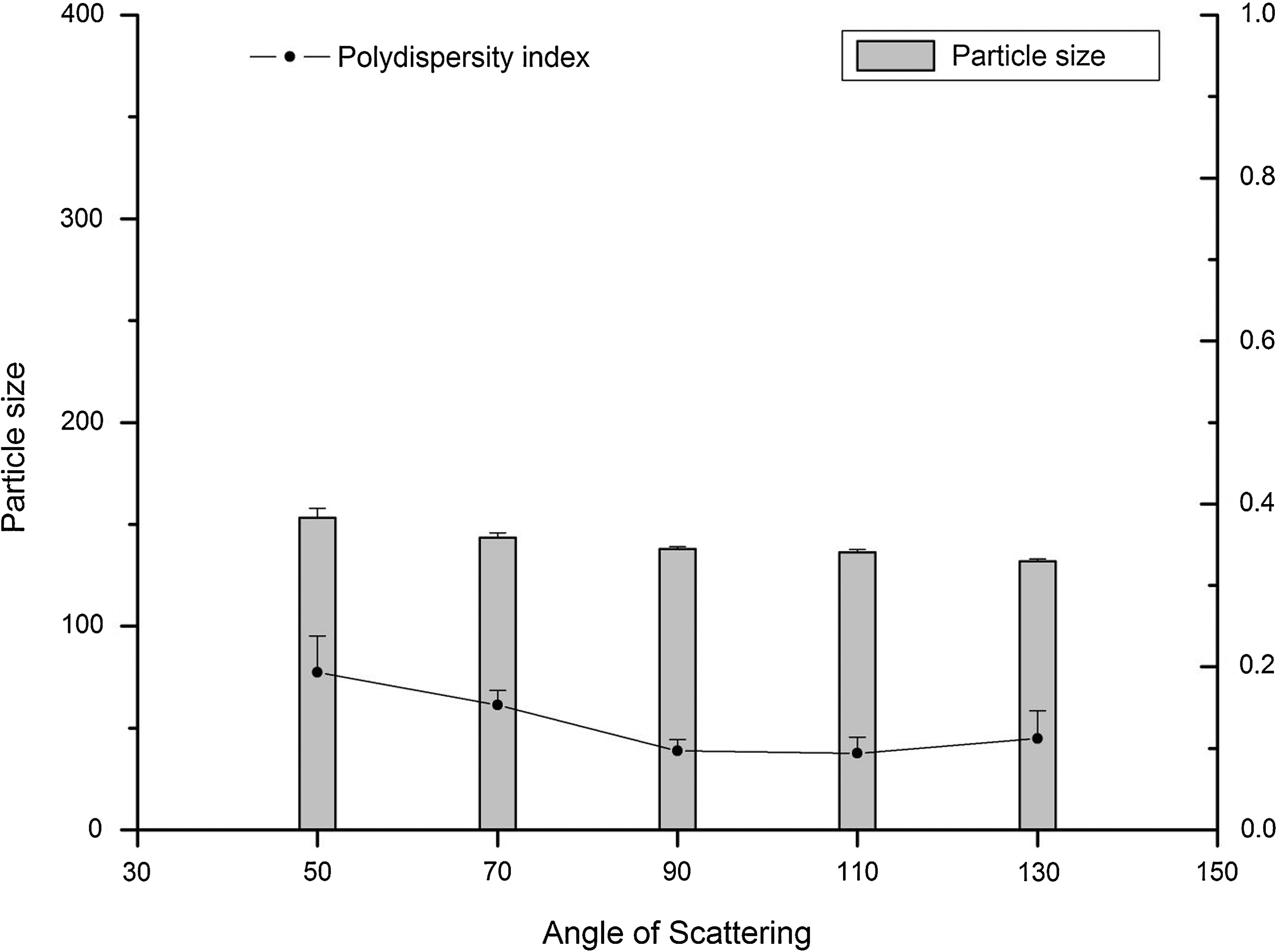
Effect of scattering angle on the particle size and PI of the nanoparticles during dynamic light scattering prior to freeze-drying (n = 6, mean ± standard deviation).
Freeze drying of nanoparticles
The various concentrations of the cryoprotectants either alone or in combination with Poloxamer 188 and their effects on the characteristics of the freeze-dried nanoparticles are listed in Table 1. The particle diameters and the PI of the reconstituted products are demonstrated in Figure 3. The solid sugar or sugar–stabilizer mixture, used as the cryoprotectant, was dissolved in the final nanoparticle formulation before being subjected to the drying process. Redispersion of the freeze-dried products in purified water was studied by manual shaking. The velocities of reconstitution were also different with different cryoprotectants (Table 1). Trehalose and dextrose formulations exerted a rapid hydration velocity at higher concentrations (<1 minute); the lyophilized products possessed excellent features and could be very easily and rapidly rehydrated. It was difficult to reconstitute the mannitol formula uniformly and the particles exhibited agglomeration.
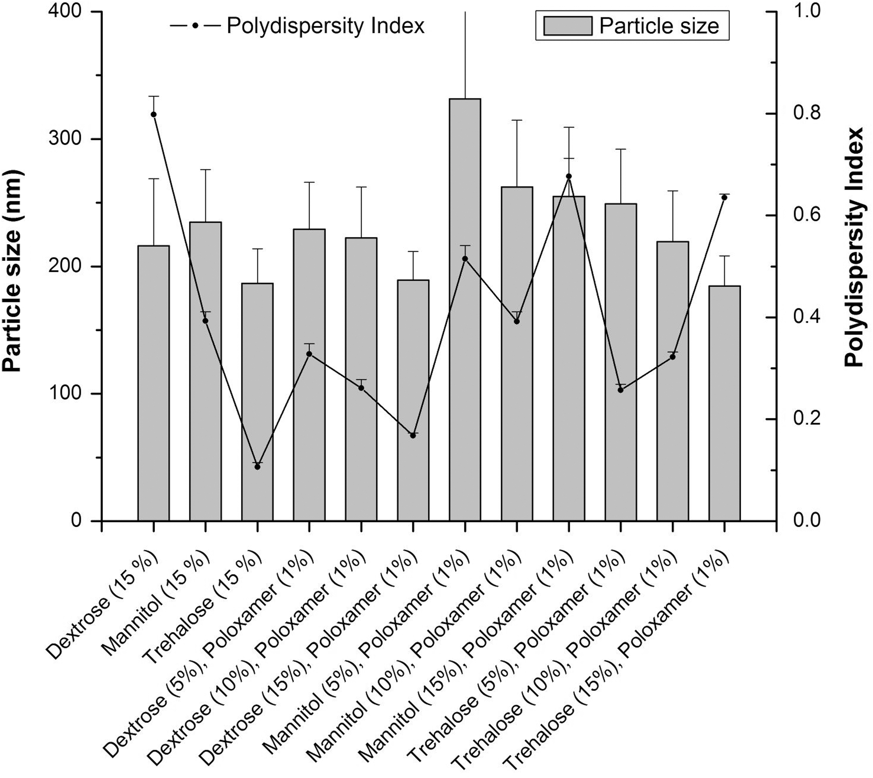
Particle diameters and the PI of the reconstituted nanoparticles post–freeze-drying (n = 3, mean ± standard deviation).
++, Best: brittle and snow-like, smooth full; +, better: a little shrinkage and rugged; -, bad: collapse, rather porous; --, worse: serious shrinkage leading to cake formation.
The particle size analysis of the reconstituted products showed that the particle diameter results were in accordance with the physical nature of the freeze-dried cake and the reconstitution velocity. Formulations with mannitol alone or in combination with the stabilizer showed increased particle size, which might be due to the higher energy input (ultrasonication) required for solubilizing this poorly water-soluble sugar. The results obtained with trehalose alone at a concentration of 15% and in combination with 1% Poloxamer as well as those obtained with a combination of 15% dextrose and 1% Poloxamer were similar. However, the combination of 15% trehalose and 1% Poloxamer resulted in a wider particle size distribution as indicated by the PI of the sample. The distributions with 15% trehalose and 15% dextrose with 1% Poloxamer were comparable. In this respect, the later combination would be preferred at an industrial scale, in event of scale-up of nanoparticles, because of the more economic cost of dextrose.
Particle morphology
AFM images depicting the three-dimensional morphology of the nanoparticles are depicted in Figure 4A and B. The figures reveal the spherical nature of the formulated PLGA nanoparticles. This morphology is especially important for easy movement of the nanoparticles through the interstices of the blood vessels, facilitating a superior image during diagnosis. The particle size deduced from these images was in accordance with the size range obtained by PCS and DLS. Further, the images also showed a smooth surface without any pores, which is characteristic of PLGA nanoparticles. 14

Spherical morphology
99mTc labeling of PLGA nanoparticles
Following the absorbance at 220 nm, PLGA nanoparticles were found to be eluted in the void volume with saline elution on PD-10 column. Using the optimized labeling procedure, ∼70% of the added 99mTc was associated with the PLGA nanoparticles and the labeled product could be easily separated on a PD-10 size exclusion column (Fig. 5). The effect of reaction time and temperature on 99mTc-labeling yield is shown in Figure 6. Under reaction conditions of 60 minutes at 37°C, an increase of SnCl2 content in the reaction mixture from 250 to 500 μg improved the labeling yield by <5% (Fig. 6); hence, it was decided to use the lower SnCl2 concentration. The labeled product was found to be stable (>90% retention of activity in PLGA fraction) for up to 12 hours when stored at 4°C in saline. From particle size analysis it was observed that exposure to conditions of 99mTc labeling did not significantly alter the particle size distribution.
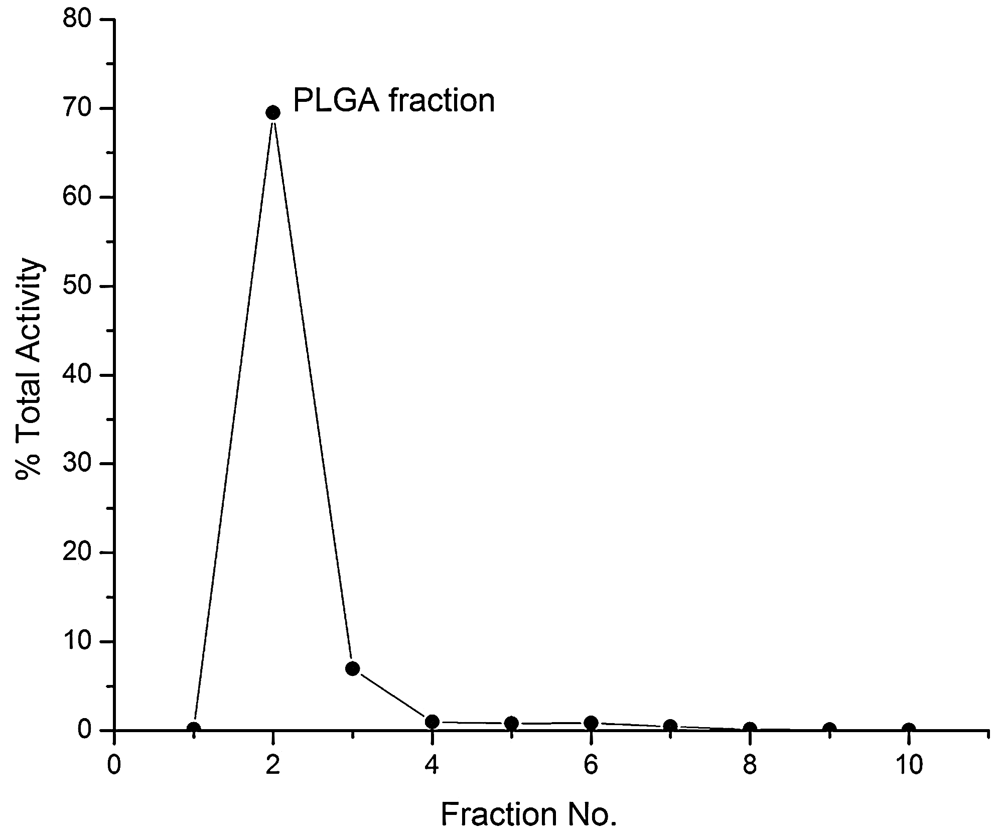
Elution profile of 99mTc-labeled PLGA on PD-10 column based on radioactivity profile of elution in saline.

Effect of reaction conditions on 99mTc-labeling yield of PLGA (n = 3; columns are associated with lower abscissa [time] and lines are associated with upper abscissa [SnCl2 concentration]).
In vivo radioactivity distribution and scintigraphic imaging
In vivo distribution pattern of 99mTc-labeled PLGA nanoparticles as observed from the measurement of retained activity in excised organs/tissues is given in Table 2. Scintigraphic image examples showing localization of 99mTc-labeled PLGA nanoparticles in the sentinel node at 15 and 60 minutes are shown in Figure 7 (n = 3). 99mTc activity appears to be extracted in significant amounts from the site of injection. 99mTc-PLGA shows accumulation in the popliteal and iliac nodes, which appears to be stable over the tested period of 3 hours. As the popliteal node serves as the sentinel node in this procedure, popliteal extraction (PE) is an indicator of the suitability of a preparation as an agent for SLND. It is calculated using the following formula:
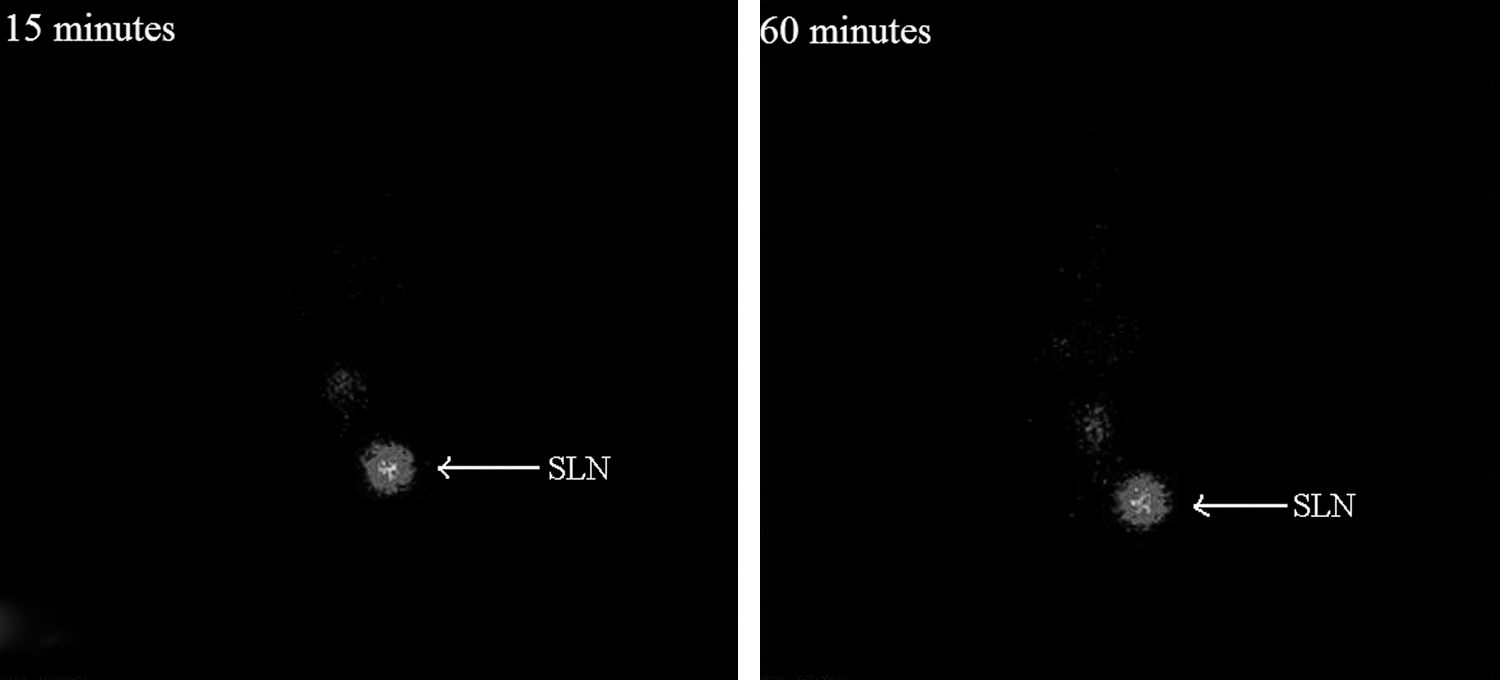
Scintigraphic imaging of sentinel lymph node in Wistar rat model at different time points.
All values are expressed as percentage of total injected dose per organ/tissue, except where indicated (n = 4, mean ± standard deviation).
The PE is good in the initial time period (15 minutes) and competitive with the result obtained with commercially available sulfur colloid kit Nanocis (PE ∼81%). The scintigraphic image is also reflective of the favorable properties of the labeled preparation. The added advantage of a ready-to-use cold kit with convenient labeling would make it more logistically suitable than sulfur colloid at the clinical end. PE value is lower at the 60 minutes mark, but after this it remains stable even at 180 minutes p.i. The absolute node uptake is lower than that observed for 99mTc-labeled Nanocis (∼4.88% at 180 minutes in popliteal node) but that would appear to be related more to the stability of the 99mTc label than as a characteristic of the labeled nanoparticle preparation. Apart from the nodes, radioactivity is observed to accumulate in the blood, liver, stomach, and intestine. As there is no observed bleeding during subcutaneous administration in any of the animals included in the study, this pattern of retention indicates that the 99mTc label may be getting separated in vivo from the PLGA nanoparticles. It may then go into the circulation and subsequently accumulate in the above-listed organs. Repeated radiolabeling and in vivo studies were conducted 6 months after freeze-drying the formulation, giving similar results. This clearly indicates the storage stability of the nanoparticles during this time and their ability to be reconstituted into a nanoparticulate suspension for their radiolabeling and subsequent animal administration.
From the above observation it may be hypothesized that Tc in reduced form is adsorbed as a layer onto the surface of the PLGA nanoparticles. This may be getting reoxidized to some extent in vivo and thereby separated from the nanoparticles. Further work is necessary to modify the radiolabeling protocol to ensure a more stable association of 99mTc activity with the PLGA nanoparticles and study the impact of that on the biological efficacy of the preparation as an SLND agent. Loading the PLGA nanoparticles with a suitable ligand for complexation with 99mTc may provide a more optimal radiotracer.
Conclusions
PLGA nanoparticles with a sub-200 nm particle diameter suitable for SLN imaging and a narrow and homogenous size distribution were formulated and developed in freeze-dried form. The developed particles were rigorously tested for the effect of various parameters on the particle size and related characteristics, and the data were used to refine and optimize the final preparation. The data suggest that the developed systems would pass through the tissue interstices at the site of injection and drain into the adjacent lymph node. The nanoparticles were labeled with 99mTc and purified by size exclusion chromatography. In vitro stability of the labeled preparation was ascertained. The system was then investigated using biodistribution and gamma scintigraphy studies to study the in vivo localization and assess the biological suitability as an SLND agent. The labeled preparation was observed to go into the sentinel node. Although there are issues with the need for purification of labeled product and in vivo stability of current labeling protocol, the present work provides a sufficient proof-of-concept for the heretofore unreported application of PLGA-based nanoparticles labeled with 99mTc as a means of SLND. Further work needs to be done to modify the labeling route to obtain a product with greater suitability for the clinical application.
Footnotes
Acknowledgments
The authors are thankful to PURAC Asia Pacific Pvt. Ltd., BASF India Ltd., and Gangwal Chemicals Pvt. Ltd. for the gift samples of the excipients. Prajakta Dandekar is thankful to University Grants Commission, Delhi, India, for providing the fellowship necessary for executing the work.
Disclosure Statement
The authors declare that there are no financial or other conflicting interests in the present work.