
Abstract
The current study was aimed at synthesizing a glucuronide derivative of D-penicillamine (D-PA) to be used for imaging purposes. First of all, D-PA-glucuronide (D-PA-Glu) was synthesized by experimental treatments starting with uridine 5′-diphospho-glucuronosyltransferase enzyme rich microsome preparate. Then, the synthesized compound was labeled with technetium (99mTc) by using a reduction method with stannous chloride. Quality controls were performed by using high-performance liquid chromatography and thin-layer radio chromatography (TLRC). Radiolabeling yield of 99mTc-D-PA-Glu was more than 98% according to TLRC results. In vitro evaluations of radiolabeled complexes were investigated on PC-3 human prostate cancer cells. 99mTc-D-PA-Glu exhibited more accumulation on PC-3 cells versus 99mTc-D-PA at 240 minutes. In order to determine its radiopharmaceutical potential, biodistribution studies were carried out in male Albino Wistar rats. The biodistribution results of 99mTc-D-PA-Glu, showed the highest uptake in prostate at 120 minutes postinjection with the main excretion route being through kidneys and bladder. 99mTc-D-PA-Glu and 99mTc-D-PA have exhibited different biodistribution results.
Introduction
Glucuronides are common soluble conjugates formed as a step in the metabolism and excretion of many toxins and drugs, such as phenols and alcohols. Uridine 5′-diphospho-glucuronosyltransferase (UDP-glucuronosyltransferase or UGT) enzymes produce products that are more water soluble, usually less biologically active, and more readily excreted than the parent compounds. The glucuronides can be excreted by renal and biliary elimination. 1 Due to the deglucuronidation of a glucuronide conjugate on the cell membrane by β-glucuronidase, they have become interesting as targeting entities in cancer research. 2 –4 Fishman et al. reported that β-glucuronidase was a component of the tumor cells and that some types of normal cells (such as fibroblasts and leukocytes) also contained the enzyme. 5 In other words, cancer cells have the enzyme of β-glucuronidase at an elevated ratio. The compounds of different glucuronide derivatives when conjugated with toxic aglycons can be used as prodrugs for cancer therapy. 6 Glucuronide-based prodrugs can be used in prodrug monotherapy for selective cancer therapy. Therefore, the glucuronide prodrugs are useful for application in the antibody-directed enzyme prodrug therapy strategy such that β-glucuronidase can be targeted to tumor cells by administration of antibody- β-glucuronidase conjugates. 7
Ertay et al. studied a peptide glucuronide, Exorphin C glucuronide, labeled with technetium (99mTc) to image opioid receptor-expressing tissues and tumor cells containing higher enzyme β-glucuronidase activity. 3 They reported that the radiolabeled peptide is rapidly cleared via the urinary system, thus showing much lower liver uptake. Müftüler et al. synthesized an antiestrogen glucuronide compound radiolabeled with 99mTc, 99mTc-TOR-G, as a new anti estrogen glucuronide imaging agent for ovary tumors. 8
It is known that 99mTc is one of the most important radioisotopes used in diagnostic imaging. Nowadays, radiolabeled compounds labeling with 99mTc have been used for bone scintigraphy as well as myocardial perfusion scintigraphy, and they have been started to be used with the aim of tumor imaging in recent years. In addition, radiolabeled amino acids can be used for tumor diagnostic labeling or radiotherapy. To this end, small and biologically active amino acids and peptides have been labeled with radioelements. 9 –11
Penicillamine can be considered an amino acid, because it is a dimethyl derivative of cysteine. All natural amino acids have D- and L- configurations. It is known that amino acid drugs have significant differences between the pharmacological and toxicological properties of drug enantiomers. 12 –15
There are limited studies regarding labeling of penicillamine with 99mTc, and no research has been found related with glucuronidation of penicillamine. In the light of this information, the current study is aimed at synthesizing a compound, via a glucuronidation reaction, by using a D-amino acid derivative with a glucuronic acid to form a product that is selectively hydrolyzed by β-glucuronidase enzyme, known to be rich in the tumor cells.
Experimental
Materials
All reagents were commercially available and of analytical grade. Na99mTcO4 was obtained from a 99mTc/99Mo generator (Monrol). D-penicillamine (D-PA) was purchased from Aldrich, and other chemicals were supplied from Merck Chemical Co. Cellulose–coated, plastic thin-layer chromatography plates were purchased from Merck (Merck 5555). Bidistilled water was supplied by using a Millipore (Milli-Q Gradient A-10; Millipore S.A.) water purification instrument. Radioactivities were counted by using a Cd(Te) detector equipped with an RAD 501 (Işın Electronic Ltd.) single-channel analyzer. All animal experiments using the test article were carried out according to the relevant Institutional Animal Review Committee of Ege University.
Glucuronide conjugation of D-PA
The microsomal enzyme preparation was prepared according to the procedure previously described by Zihnioglu. 16
Glucuronidation reaction
The glucuronidation reaction was performed similar to the Biber et al. report. 2 The microsomal enzyme preparation (6 mg protein/0.5 mL) was added to 5 mL of 50 mM Tris buffer (pH 8.0) containing 6 mM CaCl2, 10 mM uridine diphosphate glucuronic acid, and 1 mM dithiothreitol. The reaction mixture (total volume 5 mL), containing UGT was stirred at 37°C in a water bath for 10 minutes. The contents were then sonicated in an ultrasonic bath for 30 seconds to disperse the microsomes, and the reactions were started by the dropwise addition of 1 mg/0.1 mL D-PA in water, with stirring. Slow stirring at 37°C was continued for 18 hours. The reaction was terminated after 18 hours by addition of 0.5 mL acetonitrile, and the precipitated protein was removed by centrifugation at 1500 rpm for 30 minutes by using a microcentrifuge. 4,17,18
High-performance liquid chromatography
High-performance liquid chromatography (HPLC) system for the analysis of the compounds consisted of a Shimadzu quaternary gradient pump (Model LC-10Atvp Shimadzu), an automated syringe injector (20 μL loop) on a 5 μm particle size reverse-phase column (RP-C-18 column [250×4.6 mm I.D.] Macherey- Nagel; Shimadzu) eluted with acetonitrile: water (80:20) at 1 mL/min. The column oven was set to 25°C. The eluted components were detected at 214 nm and 280 nm by a UV detector SPD- 10A/V (Shimadzu).
Liquid chromatography–mass spectrometry/mass spectrometry analysis of D-PA-glucuronide
The instrument used in liquid chromatography–mass spectrometry/mass spectrometry (LC-MS/MS) measurements was Agilent LC-MSD SL ion trap (Agilent HPLC 1100 binary pump, degasser, autosampler, and column oven). LC-MS/MS spectra were collected at Ege University, at the Research and Application Center of Drug Development and Pharmocokinetics.
NMR spectroscopy
Both the 1H/13C NMR spectra were obtained by using the facility of Erzurum Ataturk University, Faculty of Science, Department of Chemistry. The 1HNMR and 13CNMR experiments were carried out on a Varian-Gemini 200 MHz at 50.34 MHz spectrometer at 23°C. Dimethylsulfoxide-d6 and Water (D2O) were used as the solvent for 13C NMR and 1H NMR, respectively.
Radiolabeling procedure of D-PA-glucuronide with 99mTcO4 −
Radiolabeling of D-PA-glucuronide (D-PA-Glu) was carried out with 99mTc by using the stannous chloride (SnCl2) reduction method. First, 25 μg SnCl2 in 25 μL water was added into a glass vial containing 374 μL (100 μg) of synthesized D-PA-Glu. Second, this solution was labeled with 55.5 MBq (1.5 mCi) 99mTcO4 −and reacted at pH 7 for 30 minutes at room temperature.
Thin-layer radio chromatography
Thin-layer radio chromatography (TLRC) was used to determine the R f values of the radiolabeled product(s). A mixture of 0.2 M potassium phosphate, 2 mM mercaptoethanol, and 0.4% tritonX100 (pH=7) was used as a TLRC solution. Each TLRC sheet was counted by using a Bioscan AR-2000 Imaging Scanner. The R f values and radiolabeling yield were determined.
High-performance liquid radio chromatography
High-performance liquid radio chromatography (HPLRC) system for the analysis of the compounds consisted of an RAD-501 single-channel analyzer eluted with acetonitrile (0.01% TFA): water (0.01% TFA) (60:40) at 1 mL/min. The column oven was set to 25°C. The eluted components were detected by a radioactivity detector.
Lipophilicity (partition coefficient)
The lipophilicity (logP) of complexes was measured. The partition coefficient was calculated as the ratio of count per second (CPS)/g of octanol to CPS/g of water. Specifically, 100 μL of 99mTc-D-PA-Glu sample was taken in a centrifuge tube, and 200 μL n-octanol and phosphate buffer (pH 7) were added. The reaction mixture was vortexed at room temperature for 1 minute and centrifuged at 2500 rpm for 30 minutes. Aliquots of 0.1 mL of sample from the octanol and aqueous layers were transferred into other tubes. Finally, each phase was counted in a Cd(Te) detector equipped with an RAD 501 single-channel analyzer. The measurement was repeated thrice and averaged.
Stability study in serum of 99mTc-D-PA-Glu
In vitro stability of 99mTc-D-PA-Glu in serum was determined by incubating 100 μL of the labeled compound with 300 μL of serum at 37°C. The aliquots were then analyzed at time intervals of 0, 30, 60, and 300 minutes after incubation.
Cell culture
Human prostate cancer (PC-3) cell lines was obtained from American Type Culture Collection. Cell lines were cultured in Dulbecco's modified Eagle medium low glucose supplemented with
PC-3 cells were seeded in 24-well plates at 1.0×10⁶ cells per well and cultured to confluence. Monolayers were washed thrice with phosphate-buffered saline (PBS), and [99mTcO4]− labeled samples (10 μCi [8μg]/mL in a culture medium) were then added to the cells. Additionally, 0.5 mL of labeled samples in a culture medium was used as a control group for radioactivity measurement. Due to time-dependent incorporation of the radiolabeled compounds, the cells were incubated for 30, 60, 120, and 240 minutes. After incubation, the cells were washed thrice with PBS, and radioactivity measurement was performed on a Cd(Te) detector equipped with an RAD 501 single-channel analyzer. To investigate concentration-dependent incorporation of [99mTc-D-PA] and [99mTc-D-PA-Glu] compounds, concentrations of 4, 8, 20, and 60 μg/mL radiolabeled compounds (10 μCi) were sequentially added to neighboring wells on the culture plate. Cells were incubated for 240 minutes, and the same procedure just explained was repeated.
Statistical analysis
Statistical significance was assessed with one-way analysis of variance and linear regression by the GraphPad program. Probabilities of (p-values) <0.05 were considered statistically significant.
Biodistribution studies on male Albino Wistar rats
All animal experiments using the synthesized compound were carried out according to the relevant instructions set by the Institutional Animal Review Committee of Ege University. The percentage of injected radioactivity per gram of tissue for selected organs is given as the mean value of the measurements for 3 rats. Biodistribution studies for the complex were performed in male Albino Wistar rats (weighing approximately 130–180 g). After sterilization by passing through a 0.22 μm membrane filter, the 99mTc labeled compound was injected into the tail vein of the animals (2 μg/rat). The activity was approximately 11.1 MBq/rat. The rats were sacrificed at 30, 120, and 300 minutes postinjection with a ketamine+xylazine cocktail (75–100 mg/kg and 5–10g/kg, resp.) by intraperitoneal injection. Organs of interest were excised, and blood samples were taken. All organs were wet weighed and counted. The percent of radioactivity per gram of wet tissue weight (% injected activity/g tissue) was determined.
Statistical analysis
Differences in the mean values of the measured activities were statistically evaluated by the SPSS 13 program (Univariate Variance Analyses and Pearson Correlation). Probability values <0.05 were considered significant. Pearson correlation was carried out between different organs for 99mTc-D-PA-Glu.
Scintigraphy studies on rats
Animal care and use was performed in compliance with the approval of The Animal Research Ethic Committees of the Ege University and in accordance with Principles of Laboratory Animal Care. Two (2) rats (120–130 g weight) were used for scintigraphic imaging. Each rat was anesthetized with a cocktail of ketamine/xylazine injected intramuscularly on the brachial muscle. 99mTc-D-PA and 99mTc-D-PA-Glu (9.25 MBq/100 μL), respectively, were injected in the tail vein of rats. Static (256×256 matrix, 1 zoom) images were performed until 4 hours. An LEHR collimator was used to acquire images with a 1-mm pixel size. Static images of 500,000 counts were obtained at 10 distinct time points (5 minutes to 4 hour) by using the supine position. To compare the radiolabeled complex accumulation, regions of interest (in counts per pixel) were determined.
Results and Discussions
HPLC results
The HPLC assay was developed to efficiently separate D-PA and D-PA-Glu. D-PA and D-PA-Glu eluted at retention times of 1.85 and 2.59 minutes, respectively, as shown by HPLC (Fig. 1). D-PA-Glu was synthesized in reference to the procedure just given to provide a high yield of 98%. The high yield was also confirmed with HPLC chromatograms at room temperature (Fig. 1).
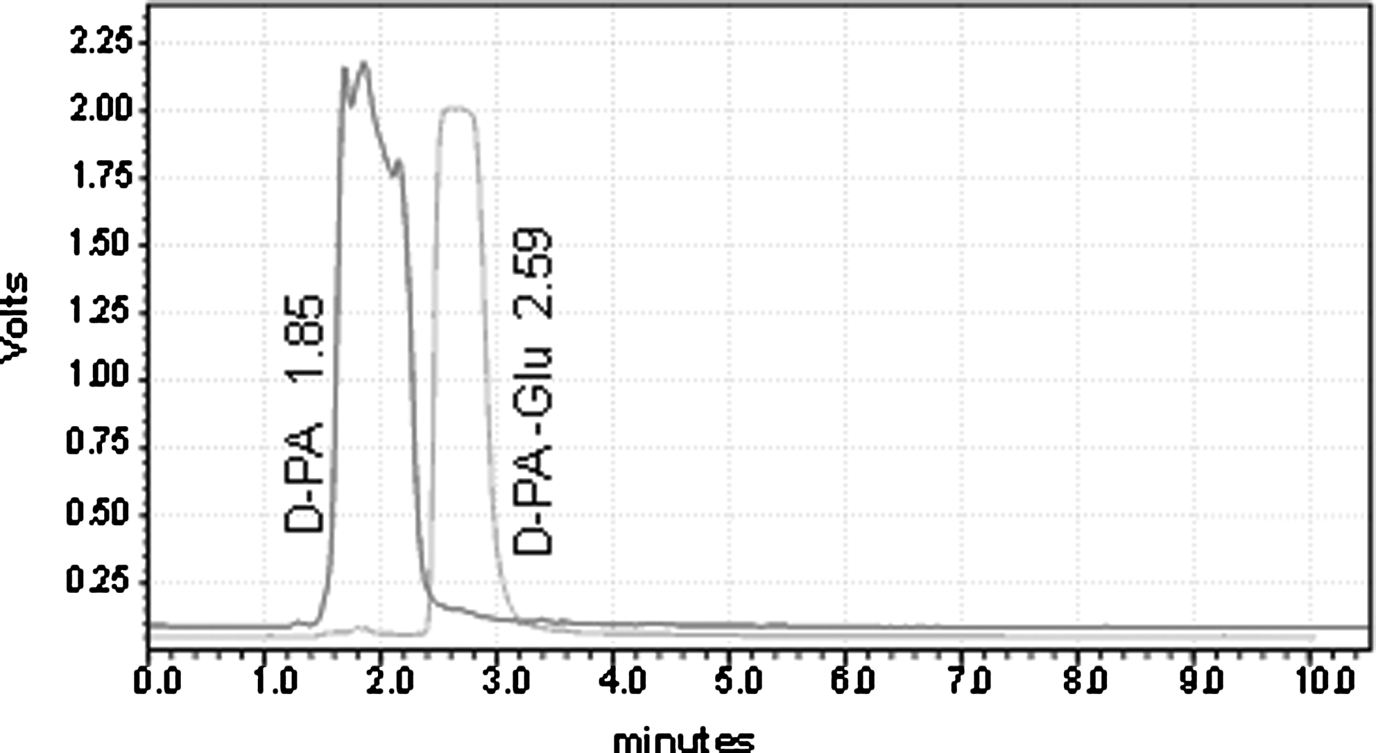
Ultraviolet-visible high-performance liquid chromatograms of D-PA and D-PA-Glu. D-PA-Glu, D-penicillamine-glucuronide.
LC-MS/MS analysis of D-PA-Glu
The negative ion MS/MS spectra (Table 1) showed abundant product ions formed by loss of glucuronide moiety with or without loss of one or two water molecules. The negative ion MS/MS spectra of D-PA-Glu showed intense product ions derived from the deprotonated D-PA-Glu [D-PA-Glu–2H]2−, and losses of two water [D-PA-Glu–2H2O]− (Table 1). The spectra also showed product ions formed by the abstraction of two protons [Glu-2H]2− and losses of two water [Glu–2H2O]−. The positive and negative ion MS/MS spectra indicate clearly the presence of the glucuronide moiety. However, the MS/MS data do not provide unambiguous information on the site of glucuronidation. These conclusions agree with Hintikka et al.'s study. 19
D-PA-Glu, D-penicillamine-glucuronide.
NMR spectroscopy
1H NMR and 13C NMR were used to determine the position of the glycosidic linkage. D-PA contains three possible sites for glucuronidation, namely hydroxyl, amine, and thiol functional groups in positions 1, 2, and 3. Comparison of the spectrum with its glucuronide-conjugated product spectrum showed that the H7 signal shifted to 7.9 ppm and on the other hand, the H10 signal did not shift, which suggests that the glucuronide is indeed an N-glucuronide. Theoretical and experimental δ (ppm) values of 1H/13C NMR spectral for D-PA-Glu are given in Table 2. He et al., studied the N-glucuronide of some pyrolizidine alkaloids. 20 According to information of NMR results, the proposed structure of D-PA-N-Glu was drawn and numbered by using ACD/LogP Algorithm Computer Programme (Fig. 2).
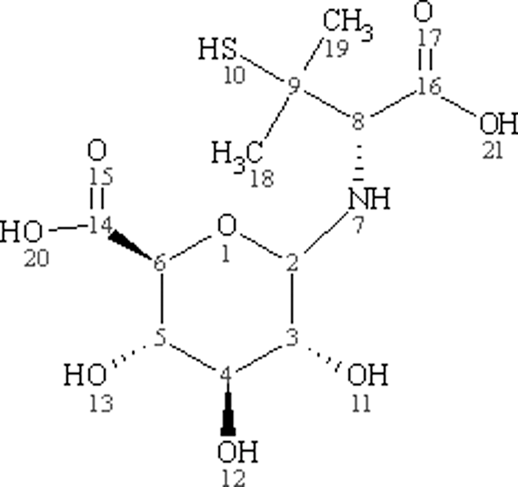
Proposed structure of D-PA-N-Glu.
TLRC and HPLRC results
The D-PA-Glu complex was easily labeled with 99mTc by SnCl2 reduction. The result of TLRC studies is given in Table 3. A mixture of potassium phosphate, mercaptoethanol, and tritonX100 is the most suitable developing solution to establish R f values. The best labeling yield obtained was 97.6%±1.2% when the pH value was set to 7. In addition, 99mTc-D-PA-Glu and 99mTcO4 − eluted at retention times of 2.31 and 3.03 minutes, respectively, as shown by HPLRC (Fig. 3).

High-performance liquid radiochromatograms of 99mTc-D-PA and 99mTcO4 −.
TLRC solvent: 0.2 M potassium phosphate, 2 mM mercaptoethanol, 0.4% tritonX100 (pH=7).
TLRC, thin layer radio chromatography.
Stability study in serum of 99mTc-D-PA-Glu
The stability of the complex in serum was investigated by the TLRC method at intervals of 0, 30, 120, and 300 minutes after radiolabeling. The results demonstrated that approximately 94.2%±1.8% of 99mTc-D-PA-Glu existed as an intact complex in serum up to 300 minutes as seen in Table 4.
Lipophilicity (partition coefficient)
The logP of D-PA has been theoretically calculated for the uncharged molecule. The N-glucuronidation reaction increased the theoretical logP value for D-PA (Table 5). On the other hand, experimental results showed that 99mTc-D-PA-Glu for N-glucuronidation display more lipophilicity than 99mTc-D-PA. Leu et al. also reported that the glucuronidation of 10-hydroxycamptothecin exhibits high liphophilicity when compared with the main compound. 7 High liphophilicity may retard compound excretion and, hence, result in prolonged exposure at the tumor site. Thus, development of new prodrugs with a high liphophilic nature may be advantageous. 7
Cell culture
Biological activity of wells having certain specific radioactivity of [99mTcO4]−, [99mTc-D-PA], and [99mTc-D-PA-Glu] complexes were investigated in vitro by means of seeded PC-3 cells in 24-well plates.
Incorporation values of the [99mTcO4]− labeled compounds on PC-3 were determined at certain time intervals. In vitro cellular uptake using the human prostate adenocarcinoma cell line showed that there was a markedly increased uptake of [99mTc-D-PA-Glu] as a function of time compared with [99mTc-D-PA] control group. The data points were calculated as a percentage of uptake. As seen in Figure 4, the highest incorporation was observed at 240 minutes for both radiolabeled complexes; however, 99mTc-D-PA-Glu exhibited fivefold more accumulation on PC-3 cells versus 99mTc-D-PA. It is known that cancer cells have the enzyme of β-glucuronidase at an elevated ratio, and glucuronide compounds were selectively hydrolyzed by β-glucuronidase enzyme. 6 This high accumulation of 99mTc-D-PA-Glu in PC-3 cells can be explained by this approach. When the incorporation values of radiolabeled compounds were statistically discussed, there is a significant correlation between [99mTc-D-PA-Glu] and [99mTc-D-PA] (p<0.001). In addition, there is a significant correlation (p<0.01) between incorporation values and time for [99mTc-D-PA-Glu] and [99mTc-D-PA].

Incorporation of radiolabeled compounds on PC-3 cells as a function of time. PC-3, human prostate cancer cell lines.
The antitumor activities of several glucuronide derivatives of molecules have been tested in vitro against two human tumor-cell lines and reported by several researchers. The glucuronide methylesters of podophyllum derivatives are some of the most active compounds. In addition, long-term administration of the O-glucuronide methylester of 5-fluorouracil (5-FU), to mice bearing slow-growing tumors, has been shown to be remarkably effective compared with its parent compound 5-FU. 21 Medine et al., studied the in vitro examination of 125I radiolabeled glucuronide derivatives of uracil on Hutu-80, Caco-2, and Detroit 562 cell lines. They concluded that radiolabeled glucuronide derivatives of uracil have more binding affinity compared with their parent compound. 22
The results of concentration-dependent incorporation of [99mTc-D-PA] and [99mTc-D-PA-Glu] compounds are given in Figure 5. The incorporation of [99mTc-D-PA-Glu] at 20 μg/mL was significantly different from other concentrations. In addition, the high incorporation value for [99mTc-D-PA] was 8 μg/mL. Incorporations of [99mTc-D-PA-Glu] were higher than those of [99mTc-D-PA] at all concentrations tested.

Incorporation of radiolabeled compounds on PC-3 cells as a function of concentration.
Biodistribution studies on male Albino Wistar rats
When the biodistribution results of 99mTc-D-PA-Glu were examined, the prostate showed a high uptake at 120 minutes, and then, the uptake of activity decreases with time. Fishman et al. reported that β-glucuronidase enzyme is localized mainly in human tumor versus normal tissues. The tumors reported were of the jejunum, sigmoid, rectosigmoid, uterus, adrenal, prostate, and thymus. The location of the enzyme in each of these tumors was mainly in the cytoplasm of the neoplastic cells. 5 In addition, it is well known that liver is the main source of glucuronidation. Besides the liver, glucuronidation is also known to occur in organs such as the lung, kidney, gastrointestinal mucosa, prostate, and olfactory epithelium. 23 In our study, exhibition of high uptake in prostate could be explained due to the prostate being rich in β-glucuronidase enzyme. According to Ünak et al.'s study, they found that the prostate and the pancreas have also considerable β-glucuronidase levels. 24 In addition, Jenab and Thompson reported that the increased β-glucuronidase activity could also affect hormone-related cancer, such as prostate cancer in men. 25
The therapeutic response was also observed in patients with prostate cancer and renal cancer having high β-glucuronidase activity, but not in patients of other cancers, especially breast tumors, due to their intracellular location of β-glucuronidase. 21
In the current study, the main excretion route of 99mTc-D-PA-Glu was through kidneys and bladder. Thus, renal excretion was observed, as characteristic for glucuronides. 26 The report of Ertay et al. supports our results. In their study of scintigraphic imaging of 99mTc- exorphin C glucuronide in rabbits, the main excretion route of their radiolabeled peptide-glucuronide was found as renal. 3 As a result, the 99mTc-D-PA-Glu was cleared via the urinary system, showing much lower liver uptake and, thus, a more favorable extrahepatic biodistribution.
99mTc-D-PA-Glu and 99mTc-D-PA showed a different biodistribution route. 99mTc-D-PA displayed a high uptake in the liver and kidneys, as previously reported. It was cleared by renal and hepatobiliary excretion, which differs from 99mTc-D-PA-Glu. 27 This could be due to the lipophilicity differences between D-PA and D-PA-Glu complexes; that is, the 99mTc-D-PA-Glu and 99mTc-D-PA complexes have different activity localization mechanisms and clearance (Fig. 6).
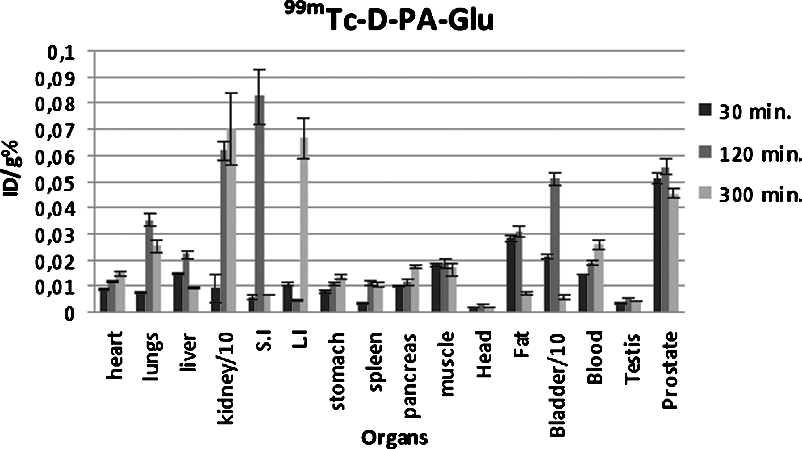
ID/g% biodistribution scheme for 99mTc-D-PA-Glu.
Statistical analyses (univariate analysis of variance) demonstrated that there were significant differences in uptake between the kidneys and spleen, the kidneys and small intestine, head and liver, and head and kidneys for the 99mTc-D-PA-Glu complex (p<0.05).
Scintigraphy studies on rats
The scintigraphic images (Fig. 7) obtained at 5 minutes after injection of 99mTc-D-PA-Glu in rat clearly show a higher uptake of the radiocomplex in the kidneys and bladder than the liver and heart. Gamma scintigraphy of 99mTc-D-PA-Glu also showed that the radiolabeled complex had a high accumulation in the kidneys, which suggests that the radiolabeled complex was cleared primarily through the renal route and was not retained in the liver and spleen. In addition, 99mTc-D-PA accumulates in the liver and bladder at 5 minutes after injection (Fig. 8). Over time, 99mTc-D-PA clears via the hepatobiliary route of the intestinal system. Scintigraphy data, thus, were in accordance with the data obtained from biodistrubiton. Further, low uptake in the thyroid and stomach reveals no 99mTc reoxidation for the two radiolabeled complexes.

Scintigraphic images of 99mTc-D-PA-Glu on male Wistar Albino rats.

Scintigraphic images of 99mTc-D-PA on male Wistar Albino rats.
Conclusions
99mTc-D-PA-Glu was obtained in high radiochemical purity and high yield. 99mTc-D-PA-Glu has displayed a different biologic behavior relative to 99mTc-D-PA. In vitro evaluations of radiolabeled complexes were investigated in PC-3 cells. 99mTc-D-PA-Glu exhibited fivefold more accumulation in PC-3 cells versus 99mTc-D-PA at 240 minutes. In addition, compared with its parent compound, 99mTc-D-PA-Glu displays different uptake and clearance kinetics of distribution as can be seen in both scintigraphic and biodistribution studies. However, further studies should be performed to investigate its biological behavior, specifically the in vivo stability.
Footnotes
Disclosure Statement
The authors declare that they have no conflict of interest.