
Abstract
Silver nanoparticles were prepared from silver nitrate using a vitamin C derivative, 6-palmitoyl ascorbic acid-2-glucoside (PAsAG), via a sonochemical experiment. The resultant golden yellow solution that contained silver nanoparticle–PAsAG complex (SN-PAsAG) of about 5 nm particle sizes was explored for its potential to offer protection to DNA from γ-radiation–induced damages. The presence of SN-PAsAG during irradiation inhibited the disappearance of covalently closed circular (ccc) form of plasmid pBR322 with a dose modifying factor of 1.78. SN-PAsAG protected cellular DNA from radiation-induced damage as evident from comet assay study on mouse spleen cells, irradiated ex vivo. When orally administered with SN-PAsAG at 1 hour prior to whole-body radiation exposure, cellular DNA was found protected from radiation-induced strand breaks in various tissues (spleen cells, bone marrow cells, and blood leucocytes) of animals. Also, SN-PAsAG could enhance the rate of repair of cellular DNA in blood leucocytes and bone marrow cells when administered immediately after radiation exposure. The studies, under in vitro, ex vivo, and in vivo radiation exposure conditions, showed effective radiation protection.
Introduction
Human exposures to ionizing radiation has become a concern on account of the rapid technological advancement with wider application of nuclear energy in diverse fields such as medicine (therapeutic and diagnostic applications), food preservation, and space travels. Ionizing radiation in the cellular milieu generates several reactive oxygen species such as superoxide, hydrogen peroxide, and hydroxyl radical, which react with cellular macromolecules such as DNA, lipids, and proteins resulting in damage to genomic DNA, peroxidation of membrane lipids, protein oxidation, and altered gene expression causing activation of cytotoxic and cytoprotective cellular signaling pathways. 1,2 There is a need to protect man and animals from the deleterious effects of ionizing radiation and this is one of the major challenges in radiation biology. 3 Nuclear accidents, nuclear terrorism, space program, and industrial and medical use of radiation and radioisotopes have created a global awareness for finding safer radioprotectors for human application. 4 Various herbal extracts, synthetic drugs, phytoceuticals, and nutraceuticals were analyzed for their radioprotective efficacy 5,6 and reports show varying degrees of success in laboratory-bench level.
Recently, it has been reported that metal oxide nanoparticles such as cerium oxide (CeO2) prevent the harmful effects of radiation exposure to normal tissues by scavenging reactive oxygen species. 7,8 Because of the electron clouds that surround them, nanoparticles could have high reactivity with free radicals. 9 In the present work, silver nanoparticles prepared using a vitamin C derivative was explored for radiation protection properties. Silver nanoparticles are currently used in several pharmaceutical products for topical application because of their antimicrobial activity. 10,11 Silver nanoparticles also have applications in catalysis, electronics, and optoelectronics. 12 Challenges associated with the synthesis of capped or coated silver particles/powder by solution method are tremendous in terms of dispersion and crystallinity of the particles. Fish oil, which contains carboxylate and amine functional groups, can act as a reducing agent as well as surfactant for the synthesis of silver nanoparticles. 13 In the present work, the possibility of preparing silver nanoparticles using a vitamin C derivative, 6-palmitoyl ascorbic acid-2-glucoside, which has various functional groups such as OH and C = O, with a long chain fatty acid (palmitic acid), was explored and the complex obtained was analyzed for its potential to offer protection against γ-radiation–induced DNA damages.
Ascorbic acid is a natural product having the ability to protect cells from radiation-induced damages. It plays an important role as the first defense against reactive oxygen species (ROS) such as hydroxyl radical (OH•), super oxide anion (O2 −), hydrated electron (eAq−), hydroperoxy radical (HO2), hydrogen peroxide (H2O2), and hydronium ion (H3O+) and reduces cellular damage produced by ionizing radiation and H2O2 in vivo 14 and in vitro because it scavenges ROS before they attack cellular macromolecules. 15 Its susceptibility to thermal and oxidative degradation has led to development of derivatives with increased stability and in vivo activity. 6-acyl ascorbic acid-2-glucoside, 6-palmitoyl ascorbate, etc., are stable ascorbate derivatives and exhibit inherent vitamin C activity of radical scavenging in vivo 16 and have improved antioxidant activity and bioavailability. 17,18 6-Palmitoyl ascorbic acid-2-glucoside (PAsAG) has been reported to possess radioprotecting activity. 19
In this study, silver nanoparticles were prepared as a complex with PAsAG and evidence is presented to show that silver nanoparticles in combination with PAsAG (SN-PAsAG) protected DNA, the genetic material and the principal biological target for radiation injury, under in vitro, ex vivo, and in vivo conditions of radiation exposure.
Materials and Methods
Animals
Swiss albino mice (8–10 weeks old) weighing 22–25 g were obtained from the Small Animal Breeding Section, Mannuthy, Thrissur, Kerala. They were kept under standard conditions of temperature and humidity in the centre's Animal House Facility. The animals were provided with standard mouse chow (Sai Durga Feeds and Foods) and water ad libitum. All animal experiments in this study were carried out with the prior approval of the Institutional Animal Ethics Committee and were conducted strictly adhering to the guidelines of committee for the purpose of Control and Supervision of Experiments on Animals constituted by the Animal Welfare Division of Government of India.
Chemicals
PAsAG was obtained from Dr. V.T. Kagiya, Health Research Foundation, Kyoto, Japan. Bovine serum albumin and high- and low-melting-point agarose were obtained from Sigma Chemical Company. pBR 322 DNA was obtained from Bangalore Genei. Analytical-grade silver nitrate was obtained from Qualigen India Ltd. All other chemicals were of analytical grade and procured from reputed Indian manufacturers.
Sonochemical preparation of silver nanoparticle by 6-palmitoyl ascorbic acid-2-glucoside
One percent solution of silver nitrate and 1% solution of PAsAG were prepared in triple glass distilled water. The solutions were mixed in different proportions and sonicated at 25°C in dark for 20 minutes. The appearance of golden yellow color and absorption at 415 nm indicated the formation of silver nanoparticles. It was found that when the mixture was having equimolecular concentrations of silver nitrate and PAsAG, the golden yellow color developed was stable. This solution was designated as SN-PAsAG, which contained 12 mM PAsAG, that is, 7.5 mg PAsAG/mL. Ultrasonication was carried out at a frequency of 20 kHz at 250 W.
Characterization of SN-PAsAG
The SN-PAsAG complex was characterized by absorption spectrum, X-ray diffraction (XRD), and transmission electron microscopy (TEM). UV–visible absorption spectrum was recorded in JASCO V570 model UV-VIS-NIR spectrophotometer. XRD was recorded as dried powder in a miniflex Regaku model using Cu Kα as source of radiation. TEM was done using copper grid on Technai G20-STWIN (200 kV).
Exposure to γ-radiation
Irradiation was carried out using a 60Co-Theratron Phoenix teletherapy unit (Atomic Energy Ltd.) at a dose rate of 1.88 Gy/min.
Alkaline single-cell gel electrophoresis or comet assay
The DNA strand breaks in mouse tissues were measured using alkaline single-cell gel electrophoresis performed using the method by Singh, 20 with minor modifications. Microscopic slides were coated with normal-melting-point agarose (1% in PBS containing 0.8% NaCl, 0.02% KCl, 0.14%Na2HPO4, and 0.02% NaH2PO4), immediately coverslipped, and kept at 4°C for 10 minutes to get the agarose solidified. After removal of the coverslip, 200 μL of 0.8% low-melting-point agarose containing 50 μL of treated cells was added onto the slide, coverglasses were placed immediately, and the slides were kept at 4°C. After solidification, the coverglasses were removed and the slides were immersed in prechilled lysing solution containing 2.5 M NaCl, 100 mM Na2EDTA, 10 mM Tris-HCl (pH 10), 1% DMSO, and 1% Triton X and kept for 1 hour at 4°C. After lysis, the slides were drained properly and placed in a horizontal electrophoretic apparatus filled with freshly prepared electrophoresis buffer containing 300 mM NaOH, 1 mM EDTA, and 0.2% DMSO (pH ≥13). The slides were equilibrated in buffer for 20 minutes and electrophoresis was carried out for 30 minutes at 20 V. After electrophoresis, the slides were washed gently with 0.4 mM Tris-HCl buffer (pH 7.4) to remove alkali. The slides were again washed with distilled water and kept at 37°C for 2 hours to dry the gel and then silver staining was carried out. 21 Exposure of cells to radiation results in fragmentation of cellular DNA, and during comet assay, these DNA fragments under the electric field moves faster from the nucleus than the DNA from unirradiated cells. Upon staining the DNA, the irradiated cells form a comet-like image with fragments constituting the tail, whereas a normal cell will have a circular, disc-like appearance. The images of cells after comet assay were visualized using Olympus BX-41 microscope and more than 50 comet images were captured and analyzed using the software CASP that measures the various comet parameters such as % DNA in tail, tail length, tail moment, and olive tail moment, which directly corresponds to the extent of cellular DNA damage. The parameter tail moment is the product of tail length and % DNA in tail, and olive tail moment is the product of the distance between the center of the head and the center of the tail and % DNA in tail. 22 Results are given as mean ± standard deviation.
Protection of plasmid pBR322 DNA by SN-PAsAG
The plasmid DNA (100 ng) in phosphate buffer (0.1 M, pH 7.4) was exposed to γ-irradiation (0–15 Gy) in the presence and absence of SN-PAsAG (1.2 mM PAsAG in complex with nanosilver) on ice. After irradiation, DNA was electrophoresed on 0.8% agarose at 55 V for 2 hours and the DNA damage was analyzed by Digital Gel Documentation and Analysis Software (Biotech R&D Laboratories). The reduction in the covalently closed circular (ccc) form of plasmid DNA is directly related to the radiation dose and the dose-modifying factor (DMF) was calculated. The ratio of the doses of radiation that have the same extent of DNA damage in the presence of protector and in its absence is taken as DMF, which was determined from the relation between the percentage of DNA remaining versus the radiation dose.
Protection of cellular DNA by SN-PAsAG in mouse splenocytes, ex vivo
Spleen tissue was excised from Swiss albino mice and single-cell suspensions of splenocytes were prepared in 0.1 M phosphate buffer. The cell suspension (106 cells/mL) was exposed to 6 Gy γ-radiation in the presence or absence of SN-PAsAG (1.2 mM PAsAG in complex with nanosilver) and comet assay was performed.
Protection of cellular DNA by SN-PAsAG in mouse tissues, in vivo
Swiss albino mice were divided into four groups and treated as follows: 0.2 mL distilled water + Sham irradiation 0.2 mL distilled water + 6 Gy 60Co-γ-rays 0.2 mL SN-PAsAG + Sham irradiation 0.2 mL SN-PAsAG + 6 Gy 60Co-γ-rays
Animals were administered with 0.2 mL of SN-PAsAG (the animal received 60 mg PAsAG/kg body weight) or distilled water. One hour after administration, the animals were exposed to whole-body 6 Gy γ-irradiation. Immediately after irradiation, the animals were sacrificed; blood, spleen, and bone marrow were collected; and alkaline single-cell gel electrophoresis or comet assay was performed.
Effect of SN-PAsAG on repair of γ-radiation–induced cellular DNA damage
To study the repair of γ-radiation–induced DNA lesions in vivo, blood and bone marrow were collected by sacrificing whole-body 6 Gy γ-irradiated animals at different time intervals. The treatment group of animals received 0.2 mL SN-PAsAG (60 mg PAsAG/kg body weight) immediately after irradiation and these animals were also sacrificed at various time intervals. Alkaline single-cell gel electrophoresis or comet assay was performed on these cells.
Statistical analysis
Results are presented as mean ± standard deviation of the studied groups. Statistical analyses of the results were performed using analysis of variance with Tukey–Kramer multiple comparisons test. The treated groups were compared with the respective control groups.
Results
Sonochemical preparation of silver nanoparticle by palmitoyl ascorbic acid-2-glucoside
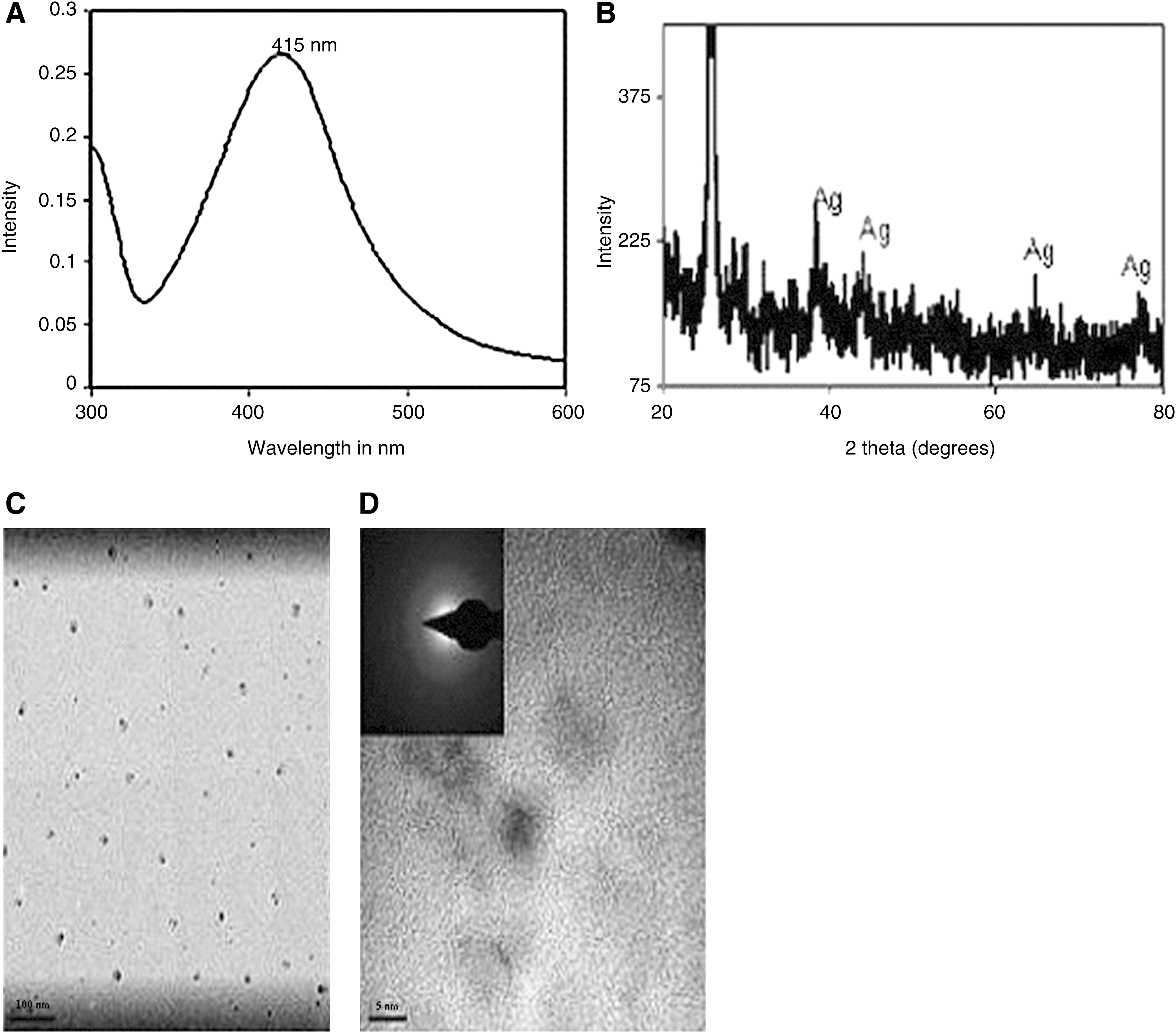
PAsAG (Fig. 1) has various functional groups such as OH and C = O, with long aliphatic carbon chain. It is a suitable surfactant to complex with nanoparticles. PAsAG reduced silver nitrate under ultrasonication and resulted in the formation of SN-PAsAG. The absorption spectrum of the complex obtained by sonochemical treatment is presented in Figure 2A, which shows an absorption maximum at 415 nm, characteristic of the surface plasmon resonance of silver nanoparticles.
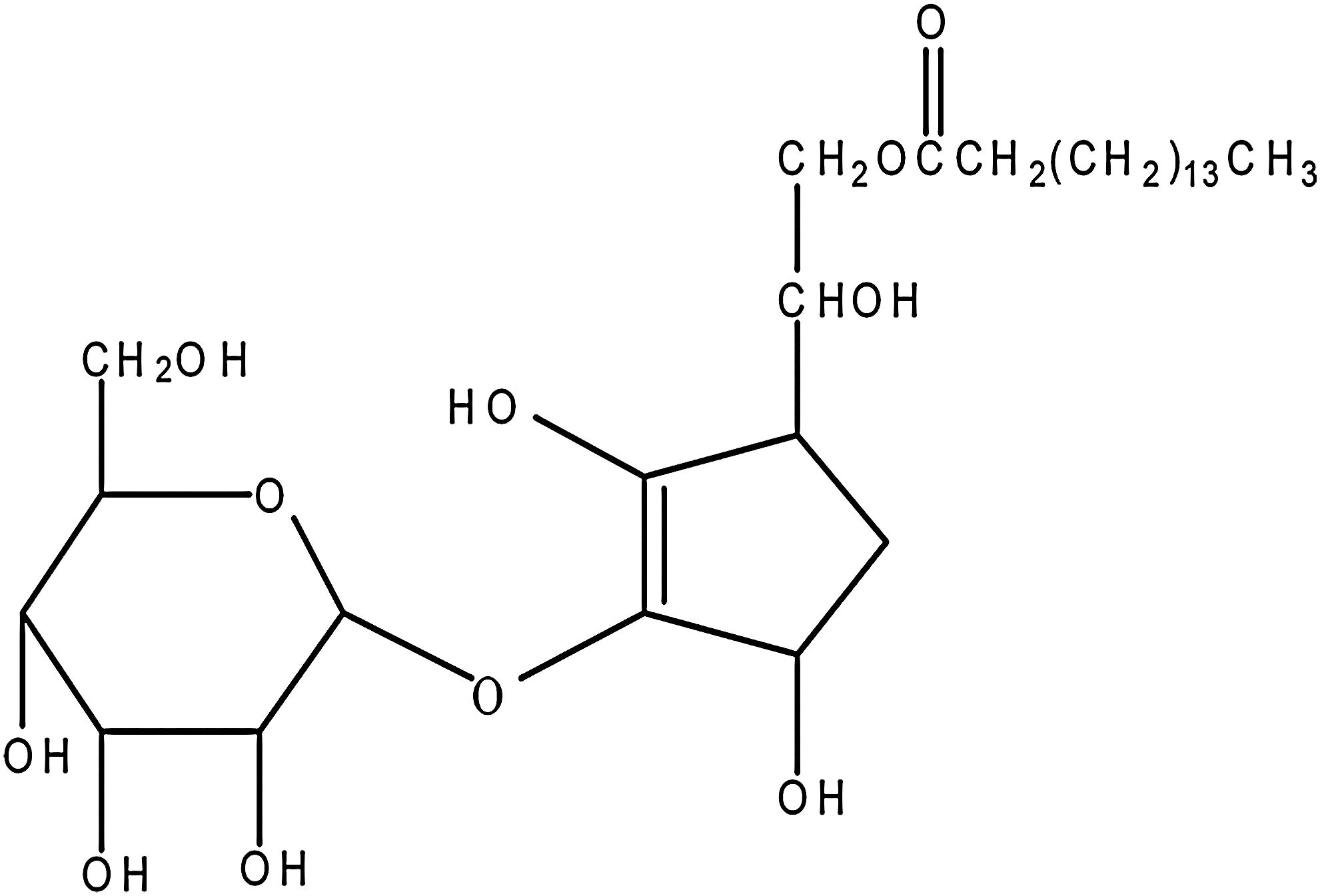
Structure of 6-palmitoyl ascorbic acid-2-glucoside (PAsAG).
Characterization of silver nanoparticle 6-palmitoyl ascorbic acid 2-glucoside (SN-PAsAG).
The XRD and TEM profile of the complex are shown in Figure 2B and C and D, respectively. Surface plasmon resonance (SPR) was obtained in all experiments conducted under similar conditions involving silver salt and the PAsAG molecule. The reaction mixture in all cases turned golden yellow to dark brown depending on the concentration of the PAsAG and the silver nitrate as well as the duration of the sonication. UV–visible absorption spectrum of the sample showed sharp SPR bands at about 415 nm, as shown in Figure 2A. The ratio between the drug molecule and the silver nitrate played a major role in controlling the particle size and SPR wavelength of the nanoparticles. The preparations by varying the concentration between the silver salt and the drug showed broad SPR because of condensation of dimeric silver complexes or because of interparticle clustering, which is known to result in low-energy SPR band or broad SPR. 23,24 Such events for silver nanoparticles obtained from silver myristate in DMF have been already reported. 25 As the prepared silver-bonded drug solutions were confirmed to possess silver nanoparticles, it was directly used for further testing with no external reducing reagents or surfactants.
The XRD pattern of the preparation (Fig. 2B) revealed peaks at 111, 200, 220, 311, and 222 crystal planes for face-centred cubic crystal structure of bulk silver metal. The broad and noisy XRD pattern suggested the nanoparticles to be poorly crystalline, meaning that they may be close to amorphous nature because of the extremely small diameter as well as their surface being surrounded by the drug molecule. The crystallite size was estimated using Scherer formula from the line broadening at 111 peak (D = kλ/β cos 2θ (where D is the crystallite size; k is a constant, that is, 0.9, assuming that the particles are spherical; λ is the wavelength of the X-ray radiation (1.5406 Å); β is the full width at half of the maximum; and θ is the angle of diffraction). The calculations gave an average particle size of <5 nm, which matched well with the observation made using TEM.
TEM analysis (Fig. 2C and D) showed scattered particles that appeared to have organic layer around them, making them look as agglomerated nanosilver particles with spherical morphology. The agglomeration was considered to be the result of the presence of excess drug molecule, which was considered necessary for the scope of the work. The lattice fringes can be easily seen in a 5 nm particle, indicating crystalline nature of the nanoparticles. The electron diffraction pattern (inset in Fig. 2D) indicates very small particles owing to unclear relevant concentric rings for face-centered cubic silver. Normally, spherical particles are considered highly suitable for biological applications, such as antibacterial agents and in drug loading.
Protection of DNA by SN-PAsAG
The nanoparticle complex (SN-PAsAG) was analyzed for its ability to offer protection against γ-radiation–induced strand breaks in DNA under in vitro conditions with plasmid pBR322 DNA, under ex vivo conditions with mouse splenocytes, and under in vivo conditions using Swiss albino mice.
Protection of plasmid pBR322 DNA by SN-PAsAG
γ-radiation induces strand breaks in plasmid DNA, resulting in loss of the supercoiled or ccc form. The reduction in the ccc form of plasmid DNA can be taken as an index of the radiation-induced DNA damage, which can be measured using a Digital Gel Documentation and Analysis Software. As evident in Figure 3, the presence of SN-PAsAG (1.2 mM PAsAG complexed with silver nanoparticles) protected plasmid DNA from radiation-induced damage with a DMF of 1.78.

Protection of plasmid pBR322 DNA by SN-PAsAG against different doses of γ-radiation (0–15 Gy).
Protection of cellular DNA by SN-PAsAG in mouse splenocytes ex vivo
Comet assay was performed on single-cell suspensions of mouse splenocytes exposed to a dose of 6 Gy γ-radiation under in vitro conditions in the presence of SN-PAsAG.
Figure 4 depicts the comet parameters (% DNA in tail, tail length, tail moment, and olive tail moment) of splenocytes exposed to 6 Gy dose of γ-radiation. The comet parameters such as % DNA in tail, tail length, tail moment, and olive tail moment were increased from 0.33 ± 0.17 to 7.27 ± 0.53, from 3.3 ± 0.70 to 17.94 ± 5.13, from 0.03 ± 0.02 to 2.86 ± 0.63, and from 0.15 ± 0.07 to 5.74 ± 0.66, respectively, when the splenocytes were exposed to 6 Gy γ-radiation. When the cells were exposed to radiation in the presence of SN-PAsAG, the comet parameters such as % DNA in tail, tail length, tail moment, and olive tail moment were brought down to the levels of 3.09 ± 0.91, 4.14 ± 1.32, 0.23 ± 0.15, and 0.65 ± 0.21, respectively, and this decrease in the comet parameters is indicative of the ability of SN-PAsAG to offer protection to cellular DNA against radaition-induced strand breaks.
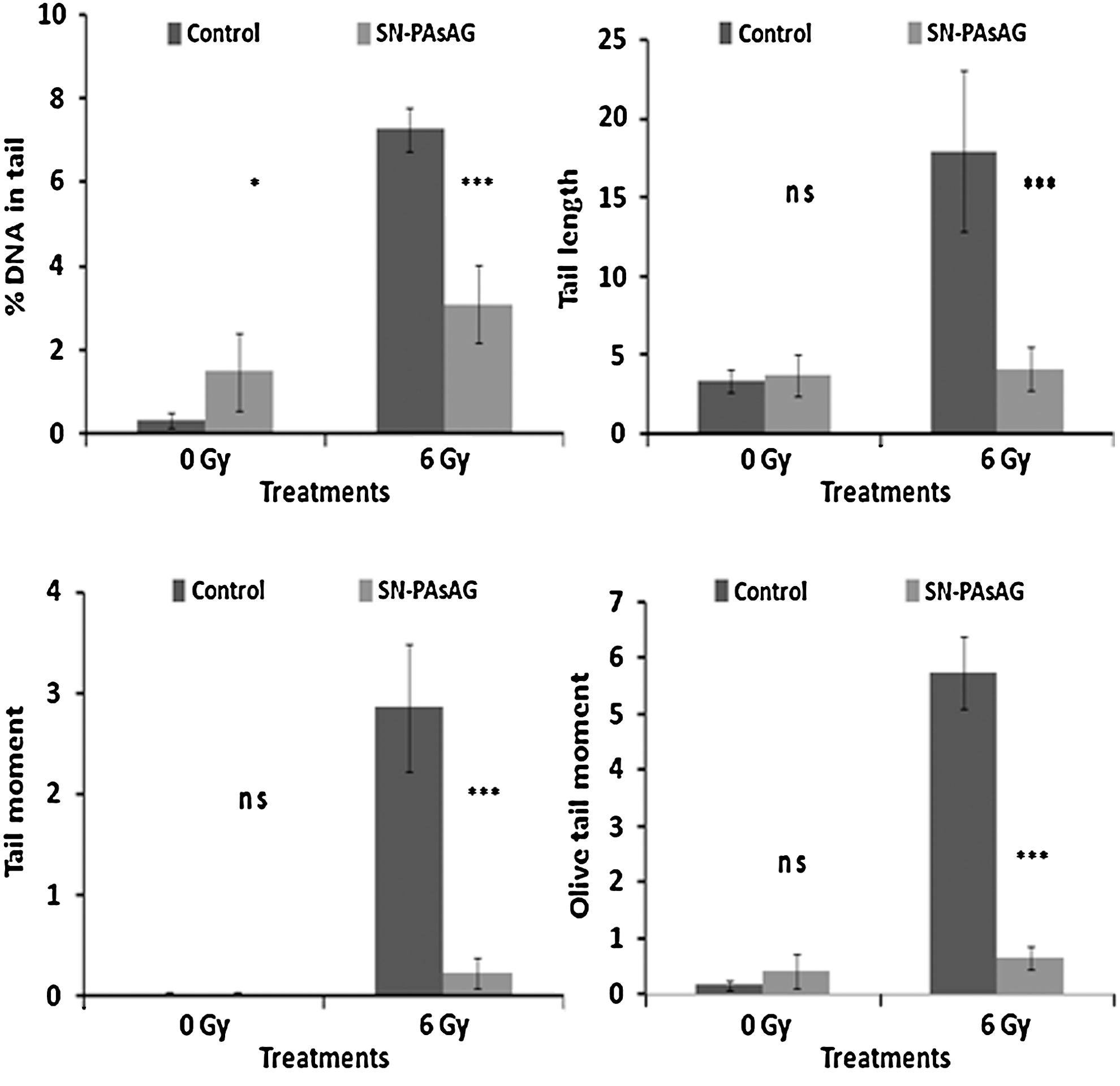
Effect of SN-PAsAG on DNA damage in murine splenocytes induced by exposure to γ-radiation, ex vivo (6 Gy), as measured by comet assay. Percentage of DNA in tail, tail length, tail moment, and olive tail moment are presented as mean ± standard deviation (SD) (*p < 0.05, ***p < 0.001 when compared with respective control; ns, not significant).
Protection of cellular DNA by SN-PAsAG in mouse tissues in vivo
For in vivo studies, SN-PAsAG was orally administered to Swiss albino mice at 1 hour prior to a 6 Gy whole-body γ-radiation exposure, and cellular DNA damage in various tissues such as spleen, bone marrow cells, and blood leucocytes was analyzed by performing comet assay. Figure 5A, B, and–C depicts the comet parameters of spleen cells, bone marrow cells, and blood leucocytes of mice exposed to whole-body 6 Gy γ-radiation. The comet parameters such as % DNA in tail, tail length, tail moment, and olive tail moment were increased from 0.93 ± 0.43 to 6.73 ± 1.26, from 3.29 ± 0.01 to 10.82 ± 2.87, from 0.03 ± 0.01 to 1.00 ± 0.22, and from 0.40 ± 0.18 to 2.44 ± 0.47 in spleen cells; from 1.06 ± 0.55 to 10.44 ± 3.01, from 3.13 ± 0.33 to 8.42 ± 3.73, from 0.13 ± 0.11 to 1.72 ± 0.76, and from 0.27 ± 0.12 to 2.6 ± 1.22 in bone marrow cells; and from 0.69 ± 0.14 to 7.38 ± 2.63, from 3.16 ± 0.55 to 7.31 ± 2.62, from 0.12 ± 0.05 to 0.93 ± 0.40, and from 0.59 ± 0.22 to 1.49 ± 0.61 in blood leucocytes, respectively. Oral administration of SN-PAsAG (60 mg PAsAG complexed with silver nanoparticles/kg body weight) to mice at 1 hour before 6 Gy whole-body radiation exposure significantly decreased these comet parameters down to levels of 0.52 ± 0.22, 3.20 ± 0.51, 0.02 ± 0.01, and 0.40 ± 0.14 in splenocytes; 1.00 ± 0.33, 3.25 ± 0.55, 0.14 ± 0.03, and 0.50 ± 0.18 in bone marrow cells; and 3.80 ± 1.49, 3.38 ± 0.84, 0.14 ± 0.05, and 0.83 ± 0.30 in blood leucocytes, respectively. These results suggested effective protection of cellular DNA from γ-radiation in these tissues by SN-PAsAG.
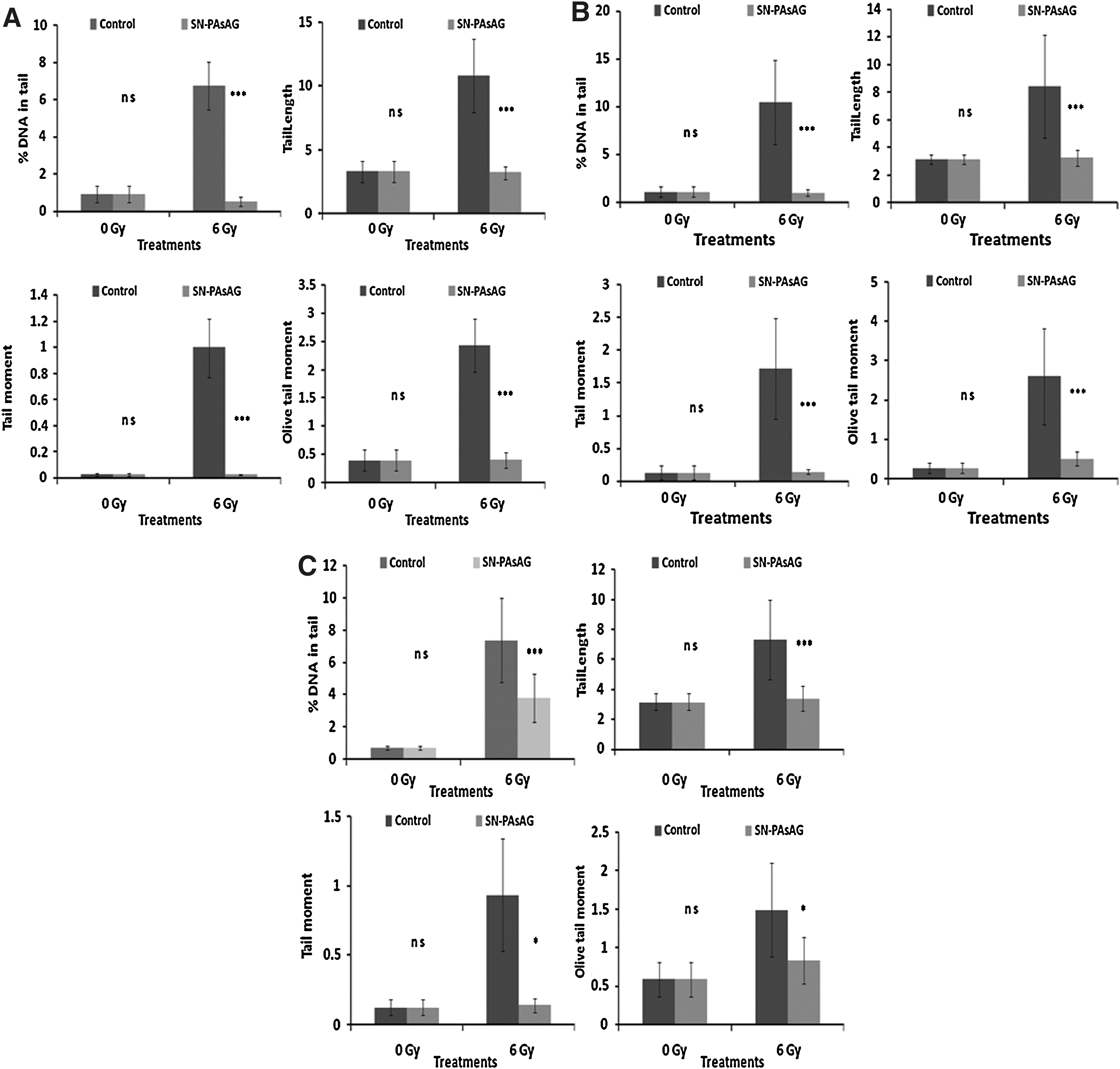
(
Effect of SN-PAsAG on repair of γ-radiation-induced cellular DNA damage
The effect of SN-PAsAG on DNA repair was determined by examining the comet parameters of the blood leucocytes and bone marrow cells of whole-body 6 Gy γ-irradiated mice at different postirradiation intervals, after postirradiation administration of SN-PAsAG (60 mg PAsAG/kg body weight). The exposure of animals to 6 Gy γ-radiation resulted in cellular DNA damage and the animals were administered with SN-PAsAG postirradiation to study the ability of SN-PAsAG to enhance the repair of radiation-injured cellular DNA. As can be seen from the results presented in Figure 6A, administration of SN-PAsAG to mice immediately after whole-body radiation exposure significantly enhanced the repair of cellular DNA in blood leukocytes because the various comet parameters such as % DNA in tail, tail length, tail moment, and olive tail moment were brought down to levels of 4.27 ± 1.7, 6.25 ± 0.72, 0.54 ± 0.20, and 1.25 ± 0.21, respectively, at 1 hour postirradiation, whereas these parameters remained at 6.98 ± 3.43, 8.73 ± 2.88, 0.95 ± 0.29, and 2.07 ±0.60 in control animals at the same time point. Similarly, these comet parameters were brought down at 1 hour postirradiation to levels of 4.41 ± 2.07, 8.42 ± 2.90, 0.55 ± 0.19, and 2.09 ± 0.76, respectively, in bone marrow cells of animals treated immediately postirradiation with SN-PAsAG, whereas at the same time point these levels remained at 6.98 ± 1.95, 13 ± 3.60, 1.1 ± 0.48, and 3.54 ± 1.23, respectively, in control animals, as shown in Figure 6B. There was a decrease in the comet parameters at a faster rate, starting at 15 minutes postirradiation, than in the control irradiated animals, and at 60 minutes most of the repair process was completed and the comet parameters were almost the same as those of the unirradiated controls in the SN-PAsAG–treated animals. Thus, the results indicated that postirradiation administration of SN-PAsAG to mice significantly enhanced the cellular DNA repair process in blood leukocytes and bone marrow cells.
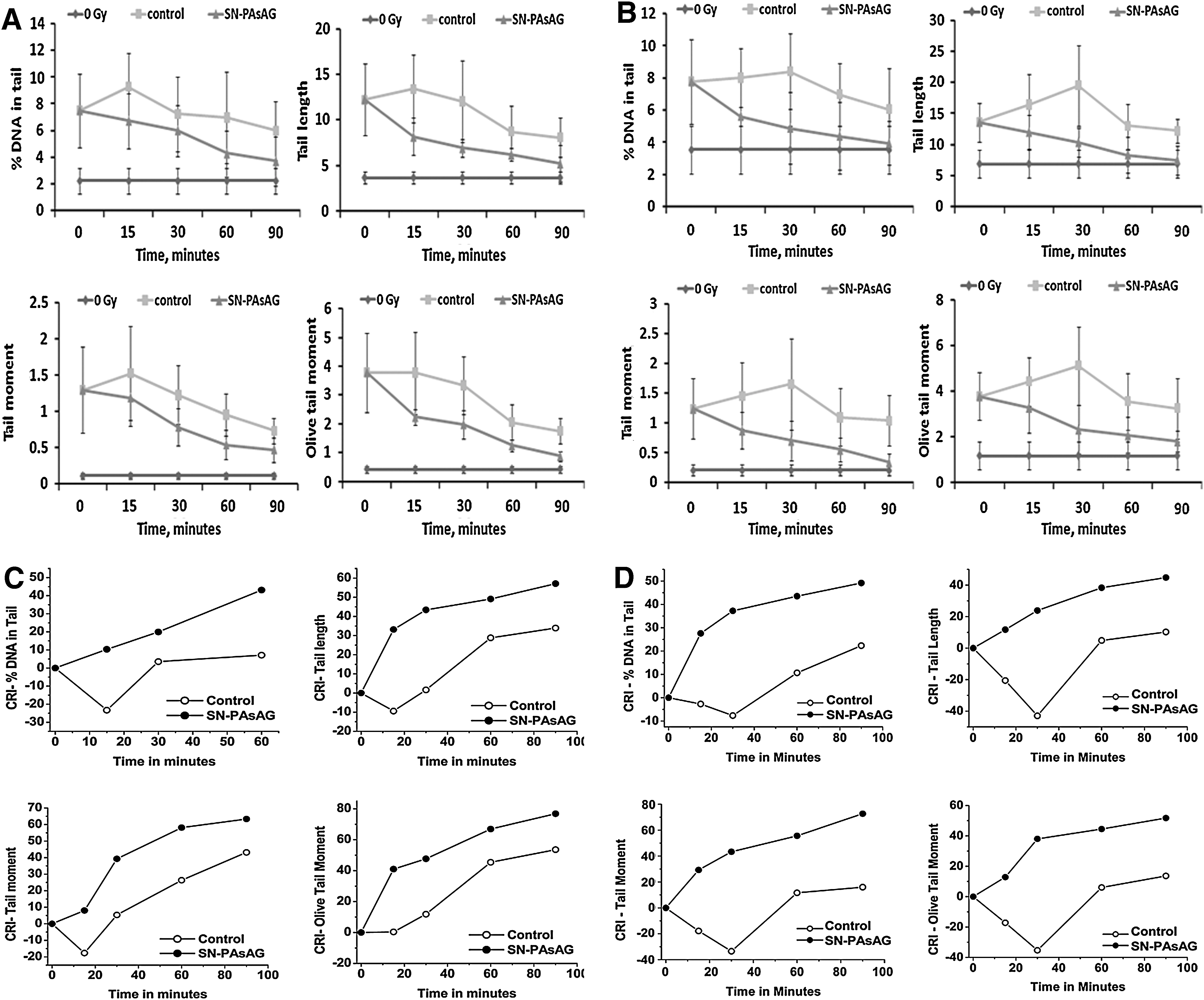
(
To quantify the efficiency of the cells to repair and rejoin strand breaks in DNA, a relation based on the comet parameters of the cellular DNA named cellular DNA repair index (CRI) can be used. CRI for a particular comet parameter is defined as the percentage of decrease from the initial value resulting from repair.
As the rate of decrease in the comet parameters is attributed to cellular DNA repair, the efficiency of the cells to repair DNA strand breaks following different treatments can be quantified by determining the CRI. The CRI increase reflects the disappearance of DNA strand breaks or DNA repair.
The data on CRI determined from the comet parameters of blood leucocytes and bone marrow cells of mice following 6 Gy in vivo γ-irradiation and postirradiation administration of SN-PAsAG are presented in Figure 6C and D. The repair capability of the cells following radiation could be understood from the data on CRI. Thus, SN-PAsAG significantly enhances cellular DNA repair efficiency as can be discerned from the data on all the comet parameters and CRI.
Discussion
γ-radiation induces damages in biological systems either by direct hit or indirectly through generating free radicals such as OH•, H•, eAq
In the present study, SN-PAsAG was prepared, characterized, and examined for its potential to offer protection against radiation-induced damages to naked or cellular DNA. The effect of this complex on DNA repair was also studied in an in vivo model.
SN-PAsAG (1.2 mM PAsAG complexed with silver nanoparticles) protected plasmid DNA from radiation-induced damage with a DMF of 1.78.
Cellular DNA is damaged by a variety of lesions such as single-strand breaks, double-strand breaks, DNA-DNA, and DNA–protein crosslinks, and nucleotide damages on exposure to γ-radiation. Alkaline comet assay is a powerful and sensitive technique to monitor DNA strand breaks and alkali-labile DNA lesions and is widely used to study genotoxicity, cellular DNA lesions such as single- or double-strand breaks, apoptosis, and DNA repair. 27,28 The results of ex vivo and in vivo alkaline comet assays performed in murine systems (splenocytes, bone marrow cells, and blood leucocytes) indicated that SN-PAsAG does not induce any DNA damage by itself and that it protected cellular DNA from radiation-induced damages when administered to animals at 1 hour prior to the radiation exposure.
The results of comet assay performed at different time intervals to study DNA repair showed that oral administration of SN-PAsAG to mice immediately after 6 Gy whole-body radiation exposure significantly enhanced cellular DNA repair process. In this experiment, to study the effect of SN-PAsAG on the repair of cellular DNA, the animals were exposed to whole-body γ-radiation to induce cellular DNA damage and were immediately administered with SN-PAsAG. Comet assay performed at various time intervals to analyze the extent of DNA damage showed a faster decrease in the comet parameters in the SN-PAsAG–treated animals. Also, the values of CRI were higher in the SN-PAsAG–treated group than in the untreated control. This fast decrease of comet parameters and the increased CRI are due to the enhanced repair of DNA strand breaks in the tissues of mice administered with SN-PAsAG postirradiation. The repair system involves removal of DNA lesions such as radiation-induced base modification through a process of incision and excision at the sites of damages in DNA strands. 29
Whole-body exposure to γ-radiation will cause damage to cellular DNA in various tissues of an animal. The whole-body γ-irradiation of mice is known to result in the depletion of hematopoietic organs owing to the intensive destruction of cells and the violation of their reproduction due to decreasing ability to proliferate. 30 Radiation-induced loss of viability of cells has been mainly attributed to unrepaired lesions in DNA. The results of in vivo alkaline comet assay in different tissues of γ-irradiated mice clearly indicated the efficiency of SN-PAsAG to offer radiation protection to cellular DNA of the hematopoetic system (spleen cells, bone marrow cells, and blood leucocytes). It is evident from the results that, apart from offering protection to cellular DNA against radiation, SN-PAsAG also enhanced the cellular DNA repair process.
Even though no previous reports on the radiation protection or free radical scavenging property of silver nanoparicles have been published, it has been already speculated by researchers at the Houston-based nanotechnology firm C Sixty that the electron clouds that surround buckyballs might soak up the free radicals generated during radiation therapy or chemotherapy. 9 Carbon nanoparticles such as fullerenes have been reported to display the potential to scavenge ROS, behaving as a “free radical sponge,” 31 including hydrogen peroxide, hydroxyl radical, hydroperoxy radicals, superoxide, 32 and nitric oxide, and also could inhibit nitric oxide synthase. 33,34 Carboxyfullerene and other water-soluble fullerenes have a potential to be cytoprotectors. 35 Cerium oxide nanoparticles are reported to possess anti-inflammatory, radioprotective, and longevity-enhancing capabilities. 8 The present work demonstrates the radiation protection potential of SN-PAsAG complex. The authors' earlier work has shown that there was significant decrease in antioxidant levels and increase in oxidative stress monitored as MDA in various tissues of mice exposed to 4 Gy whole-body γ-radiation. 19 In animals administered with SN-PAsAG and exposed to 4 Gy whole-body γ-radiation, the tissue antioxidant parameters and MDA levels were near the unirradiated control levels, indicating radioprotection (the authors' unpublished results). This complex was obtained by the reduction of silver nitrate by the antioxidant compound PAsAG to silver nanoparticles. The complex exhibited antimicrobial activity against Gram-positive and Gram-negative bacteria at 2 μM concentration onward in disc assay (the authors' unpublished observations). It offered protection to naked and cellular DNA from radiation-induced injuries, as evidenced by the plasmid relaxation assay and ex vivo and in vivo studies using comet assay in murine systems, and also enhanced the DNA repair process. The radioprotecting ability of SN-PAsAG may be attributed to the free radical scavenging property of both PAsAG and silver nanoparticles of the complex.
Conclusions
The antioxidant compound PAsAG itself reduced silver nitrate to silver nanoparticles, eliminating the need for an additional reducing and/or stabilizing agent. The SN-PAsAG complex obtained was found to be radioprotective because it protected plasmid DNA and cellular DNA from radiation-induced strand breaks and enhanced the rate of repair of cellular DNA from damages caused during radiation exposure. Other biological applications of the SN-PAsAG complex, such as in drug delivery and antimicrobial agents, are yet to be further explored.
Footnotes
Acknowledgments
C.K.K.N. expresses his gratitude to BRNS, Department of Atomic Energy, Government of India, for the financial support as a research grant awarded to him. P.K.K. thanks the Executive Director, C-MET and Department of Information Technology, Government of India, for financial support (grant no. 20(7) 2003VCND). D.K.C. thanks CSIR, Government of India, for the award of JRF.
Disclosure Statement
No competing financial interests exist.