




Abstract
CpG oligodeoxynucleotides (ODNs) are synthetic DNA sequences containing unmethylated cytosine–guanine motifs with potent immune modulatory effects. Via Toll-like receptor 9 agonists of dendritic cells and B cells, CpG ODNs induce cytokines, activate natural killer cells, and elicit vigorous T-cell responses that lead to significant antitumor effects. On the basis of these properties of CpG ODNs, a previous study has tested that they could enhance tumor response to single-dose radiotherapy. The present study extended this finding to the fractionated radiotherapy of the Lewis lung cancer and assessed the ability of CpG ODN 1826 to increase the immune function of mice and the effect of CpG ODN 1826 on the apoptosis of Lewis lung cancer. First, tumor growth delay was observed, and the enhancement ratio of CpG ODN 1826 was found to be 2.4; decreased tumor weight was found after combined treatments with CpG ODN 1826 and X-ray radiation compared with either treatment alone (p < 0.01). Second, enhanced cell apoptotic index was found after combined treatments with CpG ODN 1826 and X-ray radiation compared with either treatment alone (p < 0.01). Increased tumor necrosis factor-α and decreased interleukin-10 concentration in serum and enhanced spleen exponent were observed after combined treatments with CpG ODN 1826 and X-ray radiation compared with X-ray radiation treatment alone (p < 0.01). Results suggest that CpG-ODN1826 can increase the radiosensitivity of Lewis lung cancer, which may be associated with stimulation of immune system and enhanced cell apoptosis.
Introduction
Lung cancer is one common malignant tumor and the long-term survival in lung-cancer patients remains desperately poor. The majority of lung cancers are diagnosed at late stages, and they lose the chance of operation. Radiotherapy is one of the important therapies, but the therapeutic efficacy of partial patients is unsatisfactory. Tumor growth and its response to radiation are influenced by many factors related to tumor cells, tumor pathophysiology, and tumor host. The immune system is one of these factors. It has been recognized that conventional cancer therapies, including radiotherapy, may be more effective in tumor hosts whose immune system is intact, and their therapeutic efficacy can be increased by stimulation of innate or adaptive immunity. 1 –3
One immunotherapeutic approach has been to stimulate antitumor immunologic reactions of the tumor-bearing host by using bacteria or bacterial extracts, such as Bacillus Calmette-Guerin and Corynebacterium parvum. These bacteria or their extracts are potent elicitors or augmentors of many features of immunologic reactions, including activation of macrophages, induction of natural killer (NK) cell lytic activity, induction of antibody-dependent cell cytotoxicity, and production of various cytokines with antitumor activity, such as IFN-γ and tumor necrosis factor (TNF)-α. They were also shown to be very potent against various types of tumors in rodents and to improve the efficacy of chemotherapy and radiotherapy. 4 It was discovered that the immunostimulatory activity of bacteria resides in their DNA, 5 notably in unmethylated DNA CpG motifs prevalent in bacterial but not in vertebrate genomic DNA. 6 This discovery enabled synthesis of oligodeoxynucleotides (ODNs) containing unmethylated CpG motifs. 7 CpG ODNs preferentially induce Th1 immune response 8 through its receptor, TLR9, with the production of cytokines, such as TNF-α, IL-12, and IFN-γ, appropriate for the development of antitumor immunity. 9 In cancer immunotherapy, CpG ODNs have been used as monotherapy, 10,11 as adjuvant for vaccination, 12 and in combination with chemotherapy, 13 radiation treatment, 14 antibody therapy, 15 cellular vaccines, and others.
Increasing evidence in experimental animals shows that CpG ODNs exert antitumor activity against different types of tumors in both preventive and therapeutic settings. Treatment with CpG ODNs has also been reported to improve the outcome of surgery, chemotherapy, and, most recently, radiotherapy. 16 –18
In the present study, to assess therapeutically relevant interactions between CpG ODNs and radiotherapy, CpG ODN 1826 was combined with fractionated radiotherapy to treat mice with Lewis lung cancer cell implants. Tumor growth delay, cell apoptosis, and immunity index were measured. In addition, whether CpG ODN 1826 could enhance the radiation response of a tumor was tested.
Materials and Methods
Mice and tumors
C57BL/6J female mice bred and maintained in the authors' clean mouse colony were 6–8 weeks old, weighing 18–22 g at the beginning of the experiments, and housed 6 or 7 per cage. Animals used in this study were provided by Xipule-Bikai Ltd. Company. Lewis lung cell carcinoma used in this study was kindly supplied by the Life Science Institute of Academia Sinica in Shanghai. This animal study has been approved by the Ethics Committee of Jinshan Hospital, Shanghai, China. Solitary tumors were produced in the right anterior leg subcutaneously by the injection of 5–6 × 10⁶ cells. Tumor cell suspensions were prepared by mechanical disruption and digestion of nonnecrotic tumor tissue. Caliper measurements of tumor development and growth were documented at least every 3 days, and volumes were determined as width 2 × length × 0.52.
CpG oligodeoxynucleotide
ODNs, provided by Shanghai Sangon Biological Engineering Technology and Service Limited Company, were completely phosphorothioate modified. The sequence of CpG ODN 1826 (CG dinucleotides indicated) was as follows: 5′-TCC ATG ACG TTC CTG ACG TT-3′. ODNs were diluted with PBS to a concentration of 0.5 mg/mL and maintained at 4°C for up to a week. Injection was done intraperitoneally in a volume of 0.1 mL to achieve a dose of 50 μg per mouse. The experiment was begun when tumors were 8 mm in diameter. The mice that received CpG ODN 1826 as the only treatment were given 50 μg of the agent per mouse for six times: the 1st day, the 3rd day, the 5th day, the 8th day, the 11th day, and the 13th day; the total dose was 0.3 mg. When the agent was combined with tumor irradiation, the disposal was the same as that described above.
Tumor irradiation
Radiation was begun when tumors were 8 mm in diameter. Unanesthetized mice were tied to small boards, and tumors were centered in a 3 cm square field. Radiation was delivered to the tumor-bearing legs using accelerator linear X-ray unit at a single dose of 3 Gy for six times: at 1, 3, 5, 8, and 11 days; the total dose was 18 Gy. The radiation was carried out in the same day as that of CpG ODN. When radiation was combined with CpG ODN, radiation was given at 6 hours after administration of CpG ODNs.
Tumor growth delay
The effects of CpG ODN 1826 on tumor radioresponse were determined by tumor growth delay and sensitization enhancement ratio.
To obtain tumor growth curves, two orthogonal tumor diameters were measured at 1–3-day intervals with a Vernier caliper, and the mean values were calculated. Regression and regrowth were followed until the experiment reached day 5. Tumor growth delay was expressed as either the absolute or normalized growth delay (ATGD or NTGD). AGTD is defined as the time (in days) for tumors in the treatments (X-ray or CpG ODN1826) to grow from the original mean volume to four times volume minus the time in days for the tumors in the untreated control to reach the same size. NTGD was defined as the time for tumors in groups treated with a combination of X-ray and CpG ODN1826 to grow from the original mean volume to four times volume minus the time for tumors to reach the same size in mice with irradiation alone. Then, the sensitization enhancement ratio (SER) was defined as the difference between NTGD and ATGD divided by ATGD. Groups consisted of 8 mice each. Comparison of tumor growth delay was carried out by t-test.
Tumor weight
On day 5, tumor-bearing mice were killed by selecting eye globes, tumors were excised and weighed, and the mean values were calculated. Also, the tumor inhibitory rate was calculated as follows: tumor inhibitory rate = 1 − (the mean tumor weight of treated group/mean tumor weight of control group) × 100%.
Cell apoptosis analysis
The standard technique with TUNEL kit was used to detect tumor apoptosis in every group. Apoptosis features were by pyknosis and brown nodules deposited in the nucleus and gathered in the perimeter of the nucleus, but the nucleus of unapoptotic cells was blue. Two (2) pathology doctors counted 1000 cells in 10 high-power microscope fields of vision. Apoptosis index was calculated as follows: the pieces of apoptosis cells divided by all tumor cells.
Measurement of interleukin-10 and TNF-α
The immunomodulatory properties of CpG-containing DNA were assessed in vivo by studying the production of interleukin-10 (IL-10) and TNF-α in mice serum. At the fifth day of the experiment, the serum was collected and spleens and tumor tissues were stripped. The standard technique with Quantikine ELISA Kit from R&D Systems was used to measure murine IL-10 and TNF-α in serum.
Spleen exponent
Tumor tissues were stripped from mice, and spleens were also removed. The spleen exponent was calculated as follows: the weight of spleen divided by the weight of mouse whose tumor has been stripped. Then, the mean values were calculated.
Statistical analysis
Statistical analysis was done using SPSS12.0 (Novell). Data are expressed as means ± standard deviation (SD). Student's t-test was used to test the differences between groups. Statistical significance was considered as p < 0.05.
Results
Tumor growth delay
Mice bearing tumors were treated with CpG ODN 1826 six times, fractionated local tumor irradiation, or both CpG ODN 1826 and radiation. The results in Table 1 and partly in Figure 1 show that the total dose of 18 Gy radiation given in 3 Gy fractions produced an ATGD of 2.0 ± 0.5 days. CpG ODN 1826 alone delayed tumor growth for 2.2 ± 0.4 days (ATGD = 2.2 ± 0.4 days). In contrast, CpG ODN 1826 combined with X-ray radiation was strongly effective, and the ATGD of these tumors was 4.8 ± 1.2 days. This ATGD value was considerably higher than the sum of tumor growth delays caused by individual treatment. The enhancement factor of CpG ODN 1826 was 2.4.
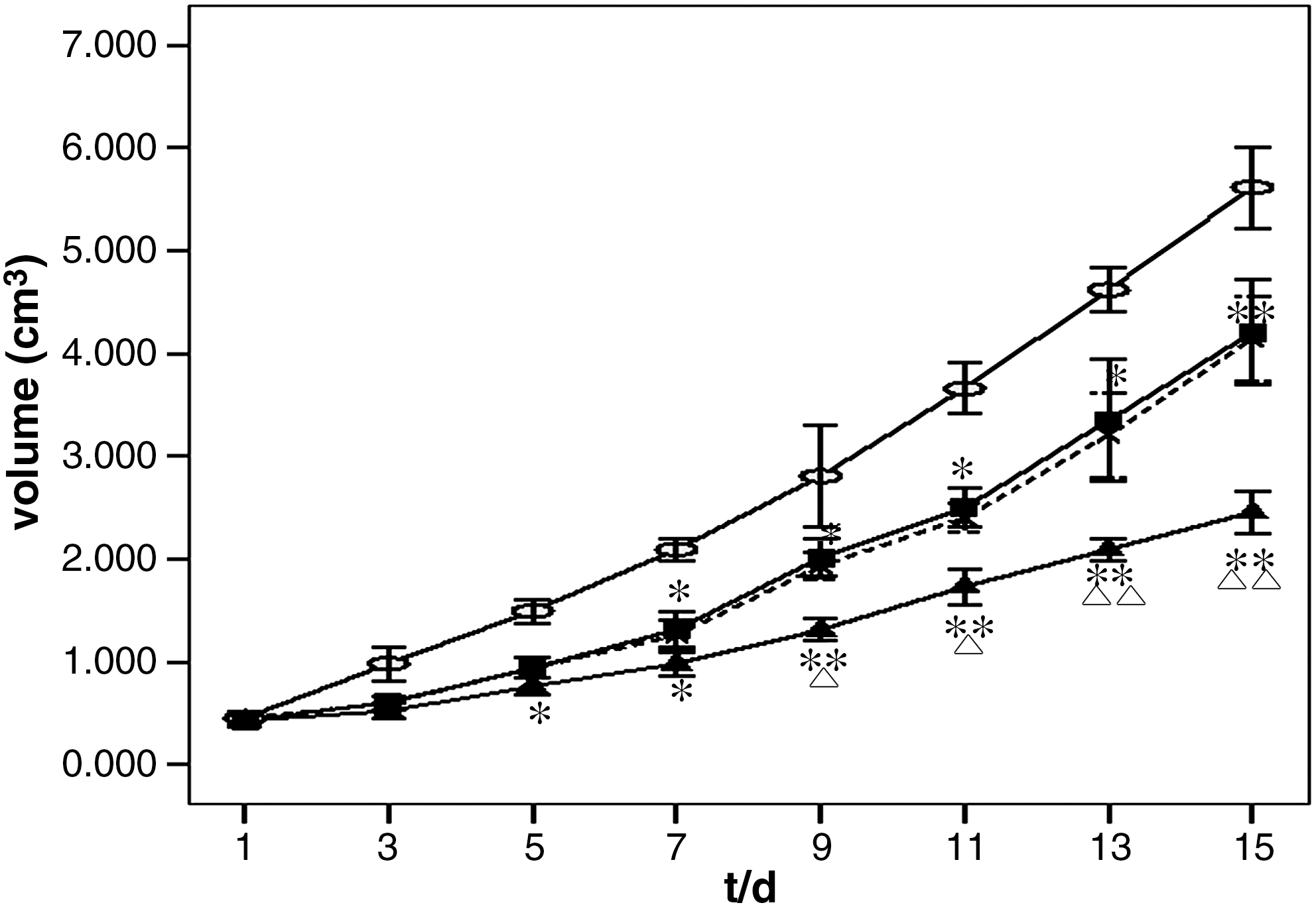
Effect of X-ray, CpG ODN1826, and CpG ODN1826 combined with X-ray on tumor growth. Mice bearing tumors were untreated (○, Control), treated with X-ray radiation only (3 Gy/F, on days 1, 3, 5, 8, 10, and 12) (▪, IR), treated with CpG ODN1826 only (0.05 mg on days 1, 3, 5, 8, 10, and 12) ( × , CpG ODN), or treated with CpG ODN1826 plus X-ray radiation (administered with CpG ODN at 6 hours before X-ray radiation) (▴, CpG ODN + IR). Experiments were begun on the same day when tumors were approximately 8 mm in diameter. *Significantly different from control at p < 0.05; **significantly different from control at p < 0.01; ▵significantly different from either IR or CpG ODN at p < 0.05; ▵▵significantly different from either IR or CpG ODN at p < 0.01. IR, irradiation; ODN, oligodeoxynucleotide.
Mice bearing tumors were untreated (Control), treated with X-ray radiation only (3 Gy/F, on days 1, 3, 5, 8, 10, and 12) (IR), treated with CpG ODN1826 only (0.05 mg on days 1, 3, 5, 8, 10, and 12) (CpG ODN), or treated with CpG ODN1826 plus X-ray radiation (administered with CpG ODN at 6 hours before X-ray radiation) (CpG ODN + IR). Experiments were begun on the same day when tumors were approximately 8 mm in diameter. Two orthogonal tumor diameters were measured at 1–3-day intervals with a Vernier caliper, and the mean values were calculated. Regression and regrowth were followed until experiment reached day 5. AGTD is defined as the time (in days) for tumors in the treatments (X-ray or CpG ODN1826) to grow from the original mean volume to four times volume minus the time in days for the tumors in the untreated control to reach the same size. NTGD was defined as the time for tumors in groups treated with a combination of X-ray and CpG ODN1826 to grow from the original mean volume to four times volume minus the time for tumors to reach the same size in mice with irradiation alone. SER was defined as the difference between NTGD and ATGD divided by ATGD, and the SER of CpG ODN1826 was 2.4.
SD, standard deviation; SER, sensitization enhancement ratio; ATGD, absolute tumor growth delay; NTGD, normalized tumor growth delay; IR, irradiated; ODN, oligodeoxynucleotide.
Tumor weight
CpG-ODN1826 has potent antitumor effects in vivo. 19 This study examined whether it had synergetic contribution to tumor growth delay when combined with X-ray radiation. As shown in Table 2, any therapy decreased tumor weight remarkably (p < 0.05), but the therapeutic effect was most effective in CpG ODN + IR (p < 0.01).
Tumor inhibitory rate = 1 − (the mean tumor weight of treated group/mean tumor weight of control group) × 100%. Values represent mean ± standard deviation (n = 8) of total mice in every group.
Significantly different from control at p < 0.05.
Significantly different from control at p < 0.01.
Significantly different from IR at p < 0.01.
IR, irradiation; ODN, oligodeoxynucleotide.
Cell apoptosis
The apoptosis of LLC tumors untreated, treated with CpG ODN 1826 or X-ray irradiation, or treated with CpG ODN 1826 and X-ray irradiation were assessed by the method of TUNEL. As shown in Table 3 and Figures 2 –5, when mice were treated with CpG ODN 1826 or X-ray irradiation, the apoptosis rate was obviously higher than that untreated (p < 0.05). Moreover, when mice were treated with the two agents mentioned above, the apoptosis rate was obviously higher than that treated with any one alone (p < 0.01).
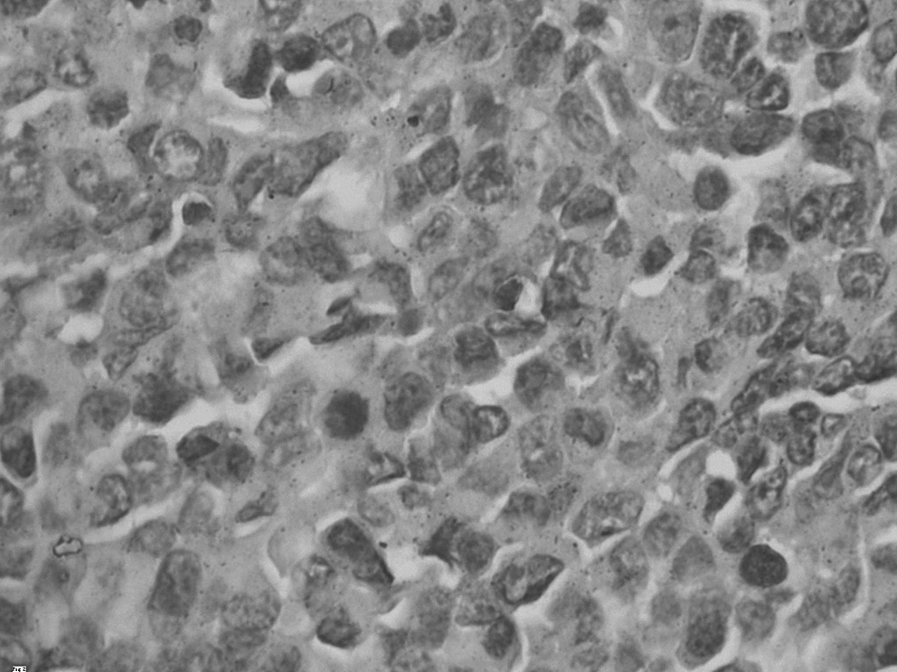
Control group: cell apoptosis with no disposal (hematoxylin stain, × 200).

IR group: tumor cell apoptosis when dealt with IR (hematoxylin stain, × 200). IR, irradiation.
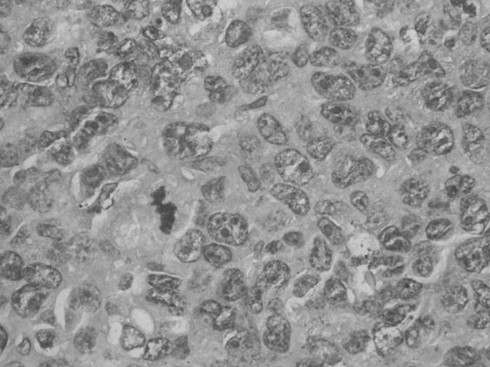
CpG ODN group: tumor cell apoptosis when dealt with CpG ODN 1826 (hematoxylin stain, × 200). ODN, oligodeoxynucleotide.
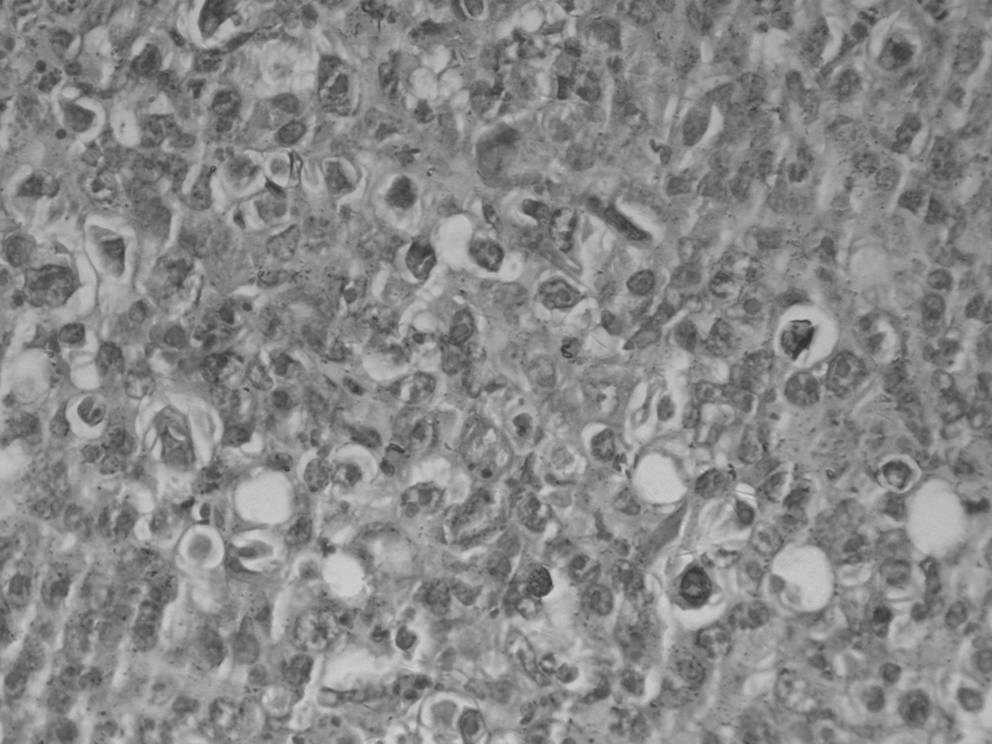
CpG ODN + IR group: tumor cell apoptosis when dealt with CpG ODN 1826 and IR (hematoxylin stain, × 200). IR, irradiation; ODN, oligodeoxynucleotide.
The results represent percentage of total mice in every group. Data are expressed as mean ± standard deviation (n = 8).
Significantly different from control at p < 0.01.
Significantly different from control at p < 0.001.
Significantly different from IR at p < 0.001.
IR, irradiation; ODN, oligodeoxynucleotides.
The release of IL-10 and TNF-α
As shown in Table 4, compared with the control group, the level of IL-10 decreased dramatically when the mice were treated with CpG ODN 1826 (p < 0.01); on the other hand, the level of TNF-α increased dramatically (p < 0.01).
Effect of CpG ODN1826 on IL-10 and tumor necrosis factor-α in serum. Values represent mean ± standard deviation (n = 8) of total mice in every group.
Not statistically different from control at p > 0.05.
Significantly different from control at p < 0.01.
Significantly different from IR at p < 0.05.
IR, irradiation; ODN, oligodeoxynucleotide.
Compared with the control group, the level of IL-10 decreased and the level of TNF-α increased when the mice were treated with X-ray irradiation, but the differences were of no statistical significance (p > 0.05). But, the group treated with CpG ODN 1826 and X-ray irradiation showed a significantly lower amount of IL-10 and a significantly higher amount of TNF-α than the X-ray irradiation group.
Spleen exponent
In addition, the spleen exponent of mice was assessed. As shown in Table 5, the spleen exponent of the group treated with X-ray irradiation was remarkably smaller than that of the control group (p < 0.05) and remarkably smaller than that of the group treated with CpG ODN 1826 and X-ray irradiation (p < 0.01). And, the exponent of the group treated with CpG ODN 1826 was dramatically larger than that of the control group (p < 0.05).
Values represent mean ± standard deviation (n = 8) of total mice in every group.
Significantly different from control at p < 0.01.
Significantly different from control at p < 0.05.
Significantly different from IR at p < 0.01.
IR, irradiation; ODN, oligodeoxynucleotide.
Discussion
CpG-ODNs, which contain unmethylated CpG dinucleotides in the context of particular base sequences, have demonstrated substantial potential as monotherapy or combination therapies for the treatment of cancer. In both animal and human experiments in vivo, researchers have found that CpG ODN, combined with conventional therapies, could increase the apoptosis of tumor cells to achieve the purpose of improving cancer cure rates through the activation of TH1-dominated specific immune response and NK cell-participated innate immune response.
The results of the present study showed that treatment of mice bearing large established tumors with CpG ODN 1826 resulted in remarkably enhanced response of these tumors to fractionated radiotherapy. Enhancement of tumor treatment response was demonstrated by a strong prolongation in tumor growth delay, which was a significant treatment end point in this study, and a higher rate of apoptosis than that from tumor radiotherapy alone was also observed. Also, a more powerful immune function, which was reflected by determining the concentration of IL-10 and TNF-α in serum and the spleen exponent, than that from X-ray irradiation alone was observed.
In this experiment, every mouse was treated with CpG ODN 1826 with a fractional dose of 0.05 mg, the total dose being 0.3 mg. The results showed that tumor growth could be delayed when mice were treated with CpG ODN 1826 or X-ray irradiation alone: the ATGD value was 2.0 ± 0.5 and 2.2 ± 0.4, respectively, and the SER of CpG ODN 1826 was 2.4. Generally speaking, a drug has clinical significance if it could improve the sensitivity of anoxic cells by 30% to irradiation (SER>1.3). So, it has the possibility of clinical application. The total dose of CpG ODN 1826 used in this study was safe; toxic events did not happen, such as allergies, dermatitis, and induration; and no mice died. At present, the CpG ODN dose range applied in clinical trials is between 0.0025 and 0.8100 mg/kg and no obvious side-effects have been reported. So, the CpG ODN dose explored in this study is safe and effective, providing some experimental basis for future clinical trials of CpG ODN. In contrast, Sparsser et al.
20
observed that high-dose CpG ODN could evoke toxic effect, like LPS, and when combined with
The levels of cytokines IL-10 and TNF-α in serum and the spleen exponent were measured to study the influence of CpG ODN 1826 on the immune function of mice. The immunomodulation function of IL-10 mainly included two aspects: inhibits macrophage from secreting certain cytokines, such as TNF, interleukin-12, and chemotaxis factors, and inhibits the other functions of macrophage in activating T cells. In brief, IL-10 downregulates the immune function of organism. TNF-α is a cytokine having a multitude of actions: activates neutronphilic granulocytes and macrophages, excites monocytes, and regulates specific and nonspecific immunity by the method of regulating T and B leucocytes. Also, TNF-α inhibits the growth of some cancer cells directly. Spleen is an important immune organ, and the spleen exponent reflects immune function to some degree. So, elevation in the weight of spleen was considered as one index of raising immune function.
By animal experiments, it was observed that, compared with the X-ray irradiation group, CpG ODN 1826 decreased the level of IL-10 and increased the level of TNF-α in serum. And, it also raised the spleen exponent.
Nowadays, apoptosis study is popular in oncology research, and it provides new ideas for human screening of antitumor drugs and for studying their mechanisms. CpG ODNs have a feasible basis of inducing apoptosis. CpG ODNs have immunostimulatory activity, that is, they activate a multitude of immunocytes to generate a multitude of cytokines, such as IL-2, IL-6, IL-12, IL-18, IL-1β, and TNF-α, and then cause apoptosis and tumor growth inhibition.
In this experiment, tumor tissues were fixed, blades were embedded with mineral wax, cut sheets were dyed, and the influence of CpG ODN 1826 on apoptosis of Lewis lung cancer in vivo was observed. The results showed that, compared with the control group, the apoptosis index of mice treated with CpG ODN 1826 or X-ray irradiation alone was obviously bigger, and the significances were of statistical value (p < 0.001). Moreover, the apoptosis index of the CpG group was statistically higher than that of the X-ray irradiation group (p < 0.001). When mice received both the treatments, the apoptosis index was statistically higher than that of either treatment alone. The results disclosed that CpG ODN 1826 could induce apoptosis, and further, it aggravated the effect of X-ray irradiation, inducing apoptosis. The present study cued that CpG ODN 1826 could produce a marked effect by inducing apoptosis and this was consistent with previous reports. Jahrsdörfer et al. 21 demonstrated that in contrast to the classic understanding of CpG ODN as inhibitors of B-cell apoptosis, IS ODNs including CpG ODNs induce apoptosis of B-CLL cells. El Andaloussi et al. 22 reported that CpG ODNs could prolong the life time of GL261 glioma tumor-bearing mice and can induce apoptosis of glioma in vivo and vitro.
In the present study, the effect of tumor growth delay effect and, perhaps, the mechanism of CpG ODN 1826 combined with X-ray irradiation in Lewis lung cancer treatment have been approached. The results have preliminarily verified that CpG ODNs had produced antitumor effect by activating the immune system, elevating immune function, and enhancing the action of radiation-induced apoptosis.
Existing investigations have shown that CpG ODNs were of tempting applied perspectives. The outcomes have provided a spacious perspective for people in the prevention and cure of malignant tumors. The present study researched only the antitumor and sensitive effects of CpG ODN 1826. The interactions between the immune system and radiation resulting in improvement of tumor control are multiple and complex and have been reviewed recently. 23 The concrete mechanism, optimal application channel, and dose of CpG ODN have not yet been found. So, further studies will be required to solve these questions.
Footnotes
Disclosure Statement
No conflicts of interest exist.