
Abstract
The radioprotective effect of hydroalcholic Zataria multiflora (Avishan–e shirazi) extract was investigated against genotoxicity induced by γ irradiation in human lymphocytes. Peripheral blood samples were collected from human volunteers and incubated with Z. multiflora extract at different concentrations (5, 10, and 50 μg/mL) for 1 hour. At each dose point, the whole blood was exposed in vitro to 150 cGy of cobalt-60 γ irradiation, and then the lymphocytes were cultured with mitogenic stimulation to determine number of the micronuclei in cytokinesis-blocked binucleated cells. The treatment of lymphocytes with extract showed a significant decrease in the incidence of micronuclei binucleated cells, compared with similarly irradiated lymphocytes without extract against γ irradiation. The maximum protection and decrease in frequency of micronuclei was observed at 50 μg/mL of Zataria extract by 32% reduction. High-performance liquid chromatography analysis of extract showed that it contains high amounts of thymol. Zataria extract exhibited concentration-dependent radical-scavenging activity on 1,1-diphenyl-2-picryl hydrazyl free radicals. These data have an important application for the protection of human lymphocyte from the genetic damage and side-effects induced by γ irradiation in personnel exposed to radiation.
Introduction
Free radicals are main toxic substances produced by ionizing irradiation. These hazardous compounds can induce damage to critical macromolecules in cells, such as DNA. The cellular DNA damage leads to mutation and cancer. 1 Exposure to high doses of γ irradiation can cause mortality in mammals. With respect to radiation damage to humans, it is important to protect biological systems from radiation-induced genotoxicity. One of the main radioprotective classes is thiol-based synthetic compounds such as amifostine. Amifostine is a powerful radioprotective agent compared with other agents, but this drug has limited usage in clinical practice because of its side-effects and toxicity. 2,3 The search for less-toxic radioprotectors has spurred interest in the development of natural products. It has been reported that citrus extract protects bone marrow cells against γ irradiation in mice. 4 Citrus extract contains high amounts of flavonoids. 5 Although several herbal medicines have been studied for a radioprotective effect in animals, there were not assessed in human samples. Recently, it was shown that hawthorn has powerful protection effects on DNA damage induced by γ irradiation on human lymphocytes. 6 This medicinal herb has phenolic and flavonoids compounds with antioxidant activity. 7
Zataria multiflora is a potential medicinal plant grown extensively in several countries such as Iran, Pakistan, and Afghanistan; it is called Avishan–e shirazi in Iran. This herbal medicine is used extensively in Iran. Several pharmacological properties including antimicrobial, antinoceptive, anti-inflammatory effect, and antioxidant activities have been reported for this herb. 8 –11
Thus, the important evaluation of Zataria extract is warranted on the genotoxic effect induced by γ rays in human volunteer's blood lymphocytes with in vitro method. The micronucleus assays have emerged as one of the preferred methods for assessing chromosome damage. The cytokinesis-block micronucleus method is now applied for population monitoring of genetic damage induced by ionizing radiation. In this study, the in vitro radioprotective activity of Z. multiflora was investigated using in vitro radiation-induced genetic damage in human volunteers' blood lymphocytes.
Materials and Methods
Plant material
Z. multiflora Boiss (Lamiaceae) tops were collected from plants growing in Fars Province. Aliquots of 200 g of the dried powder of the plant were extracted with 2000 mL of ethanol (75%) for 72 hours. After evaporation of solvent under reduced vacuum at a temperature below 50°C, 23.5 g of dried powder was obtained.
HPLC analysis
The content of thymol in Zataria extract was analyzed by high-performance liquid chromatography (HPLC) method. The HPLC system consists of a model K-1001 solvent delivery system equipped with an autosampler injector (100 μL injection volume; MIDAS type 830) and a photodiode array detector model K-2800 set at 274 nm (all from Knauer Assoc.) with Chromgate software (Version 3.1.7). Analysis was performed using an ODS-C18 column (250 × 4.6 mm i.d., 5 μm particle size; MZ-analysentechnik GmbH) and the corresponding guard column (5 × 4.6 mm i.d., 5 μm particle size). All solvents were filtered and degassed before entering the column. The mobile phase was methanol–water–acetic acid (70-40-2). The mobile phase flow rate was 1.0 mL/minute, and all the measurements were done at ambient temperature.
Measurement of free radical-scavenging activity
The free radical-scavenging capacity of methanolic extracts were determined as bleaching of the stable 1,1-diphenyl-2-picryl hydrazyl radical (DPPH). 12 The different concentrations of Zataria extract (0.05–0.8 mg/mL) were added, at 1 mL, to 3 mL methanol solution of DPPH (10 mg/250 mL). After 15 minutes at room temperature, the absorbance was recorded at 517 nm. The experiment was performed in triplicate; butylated hydroxytoluene (BHT) was used a standard antioxidant agent. The percentage of scavenging was calculated using the following formula: ([Control − Test]/Control) × 100.
Blood treatment
After obtaining permission from the medical ethics committee of the university, this study was performed. Informed consent was obtained from 5 healthy, nonsmoking human volunteers, who were men aged between 25 and 35 years. Ten milliliters of whole blood was collected in heparinized tubes and divided in centrifuge tubes at 2 mL. Blood samples were treated with 50 μmL solution of Zataria extract at concentrations 5, 10, or 50 μL/mL (final concentration). These samples were incubated for 1 hour at 37°C.
γ irradiation and culture setup to determine micronuclei
At each concentration, for each volunteer, an aliquot of heparinized whole blood was divided into two tubes of 1 mL. One tube was control sample and another tube was irradiated at 37°C with cobalt-60 source (Theratron 780, Canada) at a dose of 1.5 Gy. The samples were kept at 37°C for 1 hour. Subsequently, 0.5 mL of each sample (control and irradiated samples) was added to 4.5 mL of RPMI 1640 culture medium (Gibco) containing 20% fetal calf serum, 20 μL/mL phytohemagglutinin (Gibco), 50 U/mL penicillin, 50 μg/mL streptomycin, and 2 mM glutamine (sigma). All cultures were set up in duplicate. All cultures were incubated at 37°C ± 1°C in a humidified atmosphere of 5% CO2/95% air. Cytochalasin B (Fluka; final concentration: 6 μL/mL) was added after 44 hours of culture. At the end of 72 hours of incubation, the cells were collected by centrifugation and resuspended in 0.075 M cold potassium chloride for 8 minutes, at 1000 rpm; they were immediately fixed in a fixative solution (methanol:acetic acid, 6:1) for three times. Fixed cells were dropped onto clean microscopic slides, air dried, and stained with a Giemsa solution. All slides were coded by an individual other than the score and evaluated at 40 × magnification for the frequency of micronuclei in cytokinesis-blocked binucleated cells with well-preserved cytoplasm. To be scored as micronuclei, candidates had to have a diameter between 1/16th and 1/3rd of main nuclei, be nonrefractile, and not be linked to or overlap with the main nuclei. 13 At each extract concentration, for each volunteer, from irradiated and control cultures, a total of 2000 binucleate cells (1000 cells each from duplicate cultures) were examined to record the frequency on micronuclei.
Statistical analysis
For each volunteer, at each extract concentration, the incidence of radiation-induced micronuclei was recorded. Data were analyzed with Student's t-test. A probability value of 0.05 was accepted to denote significance.
Results
In this study, an isocratic elution of methanol in acidic aqueous was used to achieve complete separation of thymol in extract. The separation profile of thymol is shown in Figure 1. Thymol had a typical retention time of 13–14 minutes. Comparison of its retention time with that of thymol identified the peak of the analytes in the Zataria extract. The photodiode array detector was applied for purity peak of thymol in extract. The Zataria extract was standardized based on thymol by HPLC method. The calibration curve of thymol was linear over the range 10–100 μg/mL. The thymol content was 9.99 ± 0.128 mg/100 mg of extract powder.

High-performance liquid chromatography profile of thymol and Zataria multiflora total extract analyzed at 274 nm.
The mean values of micronucleated binucleated cells in control samples were 6.6 ± 1.1, 7.8 ± 2.6, 7.2 ± 2.3, and 7.4 ± 1.8 at control and at 5, 10, and 50 μg/mL without irradiation samples per 1000 binucleated lymphocytes (Table 1). There were no significant differences between these concentrations of Zataria extract and control groups. The data presented in Table 1 show a significant difference in the percentage of micronucleated binucleated cells in irradiated lymphocytes, compared with those in control cells. γ irradiation increased the frequency of micronuclei in binucleated lymphocytes to 126 ± 13.5. The frequencies of micronuclei found in the Zataria-treated cell groups were 85 ± 9.6, 101 ± 16.9, and 107 ± 31.4 at irradiated alone sample and at 50, 10, and 5 μg/mL with irradiation, respectively. Zataria extract at a concentration of 50 μg/mL was significantly lower than the irradiation control sample (p < 0.01). The total micronucleated binucleated cells values were 32%, 20%, and 15% less in the 50, 10, and 5 μg/mL concentrations of Zataria extract (Table 1). It was usually lower at 50 μg/mL, compared with those at 5 and 10 μg/mL concentrations (Fig. 2).

In vitro protection by Z. multiflora extract against radiation-induced genetic damage in cultured blood lymphocytes. Data represent the average ± standard deviation of 5 human volunteers. p < 0.01: irradiated sample without extract compared with similarly irradiated lymphocytes from the blood sample collected with extract at a concentration of 50 μg/mL.
One thousand binucleated cells were examined in each sample.
Excellent scavenging effect was observed with Avishan-e shirazi extract. The scavenging effects of methanolic extracts from Zataria on DPPH radicals increased with increasing concentrations; it was 94% at 0.8 mg/mL (Fig. 3).
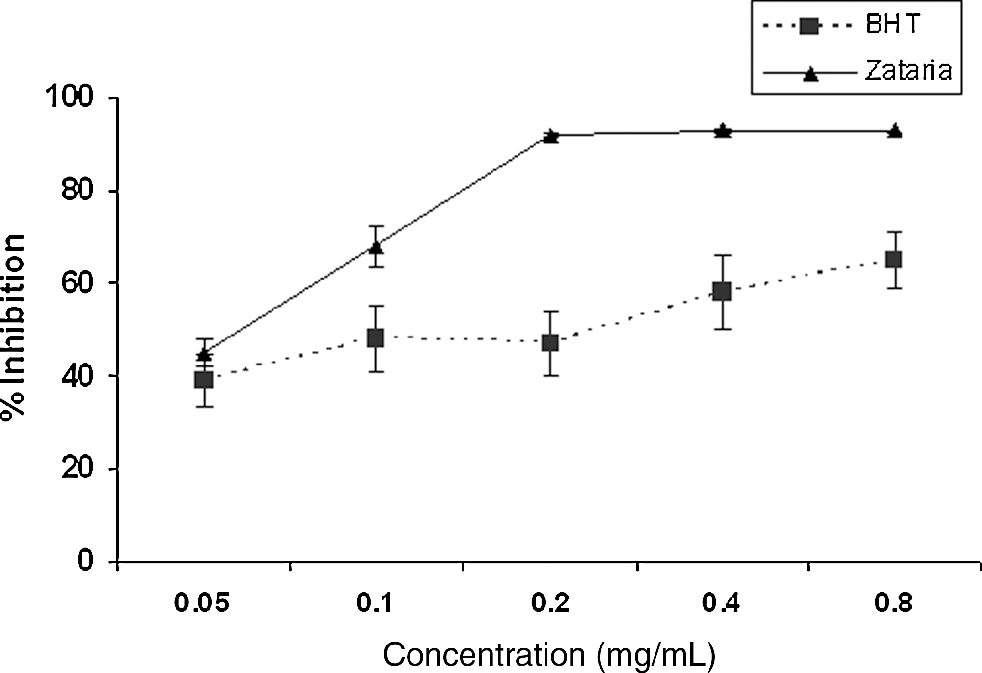
The scavenging effect of different concentrations of Z. multiflora (▴) and butylated hydroxytoluene (▪) on 1,1-diphenyl-2-picrylhydrazyl free radical at 517 nm.
Discussion
This study demonstrated that Z. multiflora has potent radioprotective effects against the genotoxicity induced by γ irradiation in human lymphocytes. Zataria extract reduced the frequency of micronuclei in binucleated lymphocytes that were affected by ionizing irradiation. Zataria showed excellent antioxidant activity. With respect to side-effects induced by ionizing radiation in patients undergoing radiotherapy, the radioprotectors have an important role for tolerance and increasing survival rate in patients.
Natural compounds including flavonoids and phenolic compounds may play a role in scavenging free radicals such as hydroxyl radicals generated by chemical hazardous agents. Increase in the intracellular level of reactive oxygen species, frequently referred to as oxidative stress, represents a potentially toxic insult, which interacts with macromolecules to induce DNA damage. In this study, Z. multiflora had radioprotective effects in reducing micronuclei induced by γ irradiation. Treatment of whole blood with Z. multiflora for 1 hour at a dose of 50 μg/mL before irradiation reduced the frequency of micronuclei by almost 32%. The chemical composition of Zataria showed that it has several phenolic compounds such as thymol, hydroxyl benzoic acid, and cymene. 14,15 HPLC analysis showed that Zataria extract contain high amounts of thymol (up to 10% of powdered extract). It has been previously shown that administration of hawthorn, an herbal medicine, protected human lymphocytes against the genotoxicity induced by γ irradiation. 6 Hawthorn extract, which has strong antioxidant activity, may affect the scavenging of free radicals such as hydroxyl radicals generated by γ-rays in cells. 7 Several studies have shown that thymol has chemoprotective and radioprotective effects against toxicity and genotoxicity induced by chemical agents and ionizing radiation. 16 –19 Thymol exhibited a protective effect against radiation-induced oxidative stress and lipid peroxidation. 19 This protective effect is mainly companied by its antioxidant activity. 17
Zataria extract showed a potent radical-scavenging activity against free radicals by DPPH method, which shows that it has more antioxidant activity than BHT, a standard antioxidant. As phenolic compounds exert excellent antioxidant activity, thymol is the main compound for the antioxidant activity of Zataria. The main characteristic of antioxidants is an ability to trap free radicals. Highly reactive free radicals and oxygen species are present in biological systems from a wide variety of sources. The free radicals may oxidize nucleic acids, proteins, lipid, and DNA and can initiate degenerative diseases. 20 Antioxidant compounds such as phenolic acids, polyphenols, and flavanoids scavenge free radicals such as peroxide and lipid peroxyl and thus inhibit the oxidative mechanisms that lead to degenerative diseases. 21
Conclusions
This study shows that Z. multiflora with antioxidant properties can probably contribute to reduce the genotoxicity induced by γ irradiation in human lymphocytes, as well as its use as herbal medicine for several diseases can help the body's defense against the side-effects induced by γ irradiation in patients during radiotherapy.
Footnotes
Acknowledgments
This work was supported by a grant from Mazandaran University of Medical Sciences, Sari, Iran.
Disclosure Statement
The authors declare that there are no conflicts of interest.